论著

感染性肺炎与免疫相关间质性肺病中淋巴细胞亚群的表达

  • 赵豆豆 1, 2, * ,
  • 齐晅 1, 3, * ,
  • 黄博 1 ,
  • 高伟波 4 ,
  • 裴源源 4 ,
  • 金月波 1 ,
  • 邵苗 1 ,
  • 何菁 , 1, *
展开
  • 1. 北京大学人民医院风湿免疫科, 北京 100044
  • 2. 晋城市人民医院风湿免疫科, 山西晋城 048000
  • 3. 河北医科大学第二医院风湿免疫科, 石家庄 050000
  • 4. 北京大学人民医院急诊科, 北京 100044

* These authors contributed to this work equally

收稿日期: 2024-07-26

  网络出版日期: 2026-04-09

版权

版权所有,未经授权,不得转载。

Expression and clinical significance of lymphocyte subsets in infectious pneumonia and immune-related interstitial lung disease

  • Doudou ZHAO 1, 2 ,
  • Xuan QI 1, 3 ,
  • Bo HUANG 1 ,
  • Weibo GAO 4 ,
  • Yuanyuan PEI 4 ,
  • Yuebo JIN 1 ,
  • Miao SHAO 1 ,
  • Jing HE , 1, *
Expand
  • 1. Peking University People's Hospital, Rheumatology and Immunology Department, Beijing 100044, China
  • 2. Jincheng People's Hospital, Rheumatology and Immunology Department, Jincheng 048000, Shanxi, China
  • 3. The Second Hospital of Hebei Medical University, Rheumatology and Immunology Department, Shijiazhuang 050000, China
  • 4. Peking University People's Hospital, Emergency Department, Beijing 100044, China
HE Jing, e-mail,

Received date: 2024-07-26

  Online published: 2026-04-09

Copyright

All rights reserved. Unauthorized reproduction is prohibited.

摘要

目的: 比较感染性肺炎和免疫相关间质性肺病(immune-related interstitial lung disease, IRILD)患者淋巴细胞亚群表达的差异,评估不同肺炎患者的免疫状态,从免疫学的角度为有效鉴别不同肺炎提供新的实验室指标。方法: 连续纳入2023年1月至2024年3月就诊于北京大学人民医院风湿免疫科的感染性肺炎患者27例、单纯IRILD患者27例以及IRILD合并感染患者24例,并选择同期进行体检的50名健康志愿者作为对照组。采用流式细胞术检测各组淋巴细胞亚群水平。结果: 病例组与对照组间年龄差异无统计学意义(P>0.05)。在实验室指标方面,与IRILD组相比,感染性肺炎组患者中性粒细胞、C反应蛋白、降钙素原(procalcitonin,PCT)、红细胞沉降率(erythrocyte sedimentation rate,ESR)、D-二聚体、纤维蛋白原水平增高(P<0.05),IRILD合并感染组患者PCT、ESR、免疫球蛋白G水平增高(P<0.05)。在淋巴细胞亚群水平方面,与对照组相比,感染性肺炎组患者的T、B、自然杀伤(natural killer,NK)、CD4+T细胞数,CD4+/CD8+比值明显降低(P<0.05),IRILD组患者的T、B,CD4+T细胞数、CD4+/CD8+比值明显降低(P<0.05),IRILD合并感染组患者T、B、NK、CD4+T、CD8+T细胞数明显降低(P<0.05)。与IRILD组患者相比,感染性肺炎组患者NK细胞数明显下降(P<0.05),IRILD合并感染组患者NK、CD8+T细胞数明显降低(P<0.05)。结论: 不同肺炎淋巴细胞亚群水平存在一定差异,为有效鉴别不同肺炎提供了新的思路。

本文引用格式

赵豆豆 , 齐晅 , 黄博 , 高伟波 , 裴源源 , 金月波 , 邵苗 , 何菁 . 感染性肺炎与免疫相关间质性肺病中淋巴细胞亚群的表达[J]. 北京大学学报(医学版), 2026 , 58(3) : 624 -630 . DOI: 10.19723/j.issn.1671-167X.2026.03.024

Abstract

Objective: To delineate differences in lymphocyte subset expression between patients with infectious pneumonia and immune-related interstitial lung disease (IRILD), to assess the immune status across distinct pneumonia types, and to provide novel laboratory markers through immunological profiling to effectively differentiate pneumonias. Methods: A total of 78 patients sequentially enrolled from Peking University People' s Hospital between January 2023 and March 2024 were stratified into three groups: 27 with infectious pneumonia, 27 with isolated immune-related interstitial lung disease (IRILD), and 24 with IRILD complicated by infection. Fifty age-matched healthy individuals undergoing routine physical examinations during the same period served as controls. Peripheral lymphocyte subsets were quantified in all subjects by multiparametric flow cytometry. Results: There was no statistically significant difference in age between the pneumonia patients and the healthy control group in this study (P>0.05). Analysis of laboratory parameters revealed that compared with the IRILD group, the infectious pneumonia group demonstrated significantly elevated levels of neutrophils, C-reactive protein, procalcitonin (PCT), erythrocyte sedimentation rate (ESR), D-dimer, and fibrinogen (P < 0.05), while the IRILD with superimposed infection group exhibited significantly increased levels of PCT, ESR, and immunoglobulin G (P < 0.05). At the lymphocyte subset level, compared with the control group, the infectious pneumonia group exhibited significantly decreased counts of T cells, B cells, natural killer (NK) cells, CD4+T cells, and the CD4+/CD8+ ratio (P < 0.05). Similarly, compared with the control group, the IRILD group showed significantly reduced counts of T cells, B cells, CD4+T cells, and the CD4+/CD8+ ratio (P < 0.05). Furthermore, compared with the control group, the IRILD-with-infection group demonstrated significantly lower counts of T cells, B cells, NK cells, CD4+T cells, and CD8+T cells (P < 0.05). Compared with the IRILD group, the infectious pneumonia group had a significantly lower NK cell count (P < 0.05). Additionally, compared with the IRILD group, the IRILD-with-infection group displayed significantly reduced counts of NK cells and CD8+T cells (P < 0.05). Conclusion: The study revealed significant variations in lymphocyte subset profiles among the distinct pneumonia groups, providing novel insights with potential diagnostic value for differentiating between pneumonia types.

肺炎是一种常见的呼吸系统疾病,其发病机制和临床表现复杂,可能由多种因素引起(如环境污染物、自身免疫性疾病和感染等)[1],其中,较为常见的是感染和自身免疫性疾病所引起的肺部炎症。感染性肺炎是由各种病原体(如细菌、病毒或真菌等)引起的肺部感染,患者常出现咳嗽、发热、呼吸困难等症状,严重时可导致呼吸衰竭甚至死亡,免疫系统发育不全的幼儿和继发于衰老、免疫功能低下或慢性肺病的免疫屏障受损的成人更容易发生感染性肺炎[2]。免疫相关间质性肺病(immune-related interstitial lung disease, IRILD)是一种多因素相关的复杂疾病,其中自身免疫病[如系统性硬化症(systemic sclerosis, SSc)、类风湿性关节炎(rheumatoid arthritis, RA)、特发性炎性肌病(idiopathic inflammatory myopathy, IIM)和干燥综合征(Sjögren syndrome, SS)]常累及肺部可表现为IRILD,除此之外新近提出的一类具有自身免疫特征的间质性肺炎(interstitial pneumonia with autoimmune features,IPAF)也属于IRILD,IPAF的特点是患者仅有血清免疫学异常,但临床上没有表现出特定结缔组织病(connective tissue disease,CTD)的全身性症状[3]。当肺部影像学出现肺炎表现时,临床医生鉴别肺炎是感染所致还是非感染所致至关重要[2]。先天和获得性免疫应答的激活参与了炎症性自身免疫性疾病和感染性疾病的发病,在感染性肺炎和IRILD中均可导致免疫失衡,而作为适应性免疫重要组成部分的淋巴细胞亚群也会出现异常分布[4]。本研究将对感染性肺炎、IRILD及其合并感染患者的淋巴细胞亚群表达水平进行分析,旨在探索不同肺炎免疫状态的潜在变化,为临床有效鉴别不同肺炎提供新的实验室指标。

1 资料与方法

1.1 研究对象

本研究为回顾性研究,连续纳入2023年1月至2024年3月就诊于北京大学人民医院的感染性肺炎患者27例、单纯IRILD患者27例,以及IRILD合并感染患者24例,并选取同期健康体检人群志愿者50例作为正常对照组。本研究开始前已经北京大学人民医院伦理委员会审查批准(2023PHB093-001)。

1.2 诊断及排除标准

1.2.1 感染性肺炎

纳入标准:所有感染性肺炎患者均符合《内科学》(第9版)中肺炎的诊断标准[5];年龄≥18岁;入院后根据临床症状、呼吸道病原微生物结果阳性及影像学检查确诊为感染性肺炎。排除标准:排除既往诊断为IRILD及其他肺部疾病患者。

1.2.2 IRILD

纳入标准:患者于北京大学人民医院排除感染并明确诊断为结缔组织病相关间质性肺炎(CTD-ILD)及IPAF,将CTD-ILD及IPAF统称为IRILD[3];呼吸道病原微生物结果阴性患者。排除标准:排除其他原因(如感染、环境、药物、职业暴露等)导致的ILD患者。

1.2.3 IRILD合并感染

纳入标准:患者既往在排除感染的情况下已明确诊断为CTD-ILD或IPAF;本次呼吸道病原微生物结果阳性。排除标准:排除其他原因(如感染、环境、药物、职业暴露等)导致的ILD患者。

1.3 淋巴细胞亚群检测方法

采集患者和健康志愿者静脉血2~4 mL,用乙二胺四乙酸(ethylene diamine tetraacetic acid,EDTA)抗凝,各取50 μL全血加入2个流式管内,用配套荧光标记抗体(深圳迈瑞公司),混匀,室温避光孵育15 min,加人500 μL配比好的溶血素(美国BD公司),室温避光孵育15 min,上机检测(BriCyteE6流式仪,深圳迈瑞公司),收集细胞。

1.4 临床资料收集

记录患者主要临床表现(发热、咳嗽、呼吸困难),初始用药情况、合并症情况等,并记录患者实验室检查[血常规、肝肾功能、免疫球蛋白(immunoglobulin, Ig)、补体、红细胞沉降率(erythrocyte sedimentation rate, ESR)、C反应蛋白(C-reactive protein, CRP)、降钙素原(procalcitonin, PCT)、D-二聚体(d-dimer)、纤维蛋白原(fibrinogen, FIB)等]结果。

1.5 统计学分析

应用SPSS 22.0软件,计量资料首先通过Shapiro-Wilk正态性检验,服从正态分布的计量资料以$\bar x \pm s$表示,组间比较采用单因素方差分析(ANOVA),两两比较采用Hodges-Lehmann估计法;不服从正态分布的计量资料以M(P25P75)表示,组间比较用Kruskal-Wallis检验;计数资料以n(%)表示,组间比较采用卡方检验或Fisher精确概率法,P<0.05认为差异有统计学意义。

2 结果

2.1 一般临床资料

研究最终纳入了感染性肺炎患者27例、单纯IRILD患者27例、IRILD合并感染患者24例和对照组50例(表 1)。
表1 患者基线资料比较

Table 1 Comparison of baseline data of patients

Items Control (n=50) Infectious pneumonia (n=27) IRILD (n=27) IRILD with infection (n=24) F/χ2 P
Male 17 (34.0) 19 (70.4) 8 (29.6) 9 (37.5) χ2=11.82 P<0.05
Age/years 55.3±9.4 60.3±16.9 60.5±13.9 60.0±12.8 F=1.50 P>0.05
Infection type χ2=2.06 P>0.05
  Virus 10 (37.0) 6 (25.0)
  Bacteria 14 (51.9) 17 (70.8)
  Bacteria + virus 2 (7.4)
  Fungus 1 (3.7) 1 (4.2)
IRILD etiology χ2=5.56 P>0.05
  IIM 12 (44.4) 12 (50.0)
  RA 2 (7.4) 3 (12.5)
  SS 2 (7.4) 3 (12.5)
  SSc 2 (8.3)
  AAV 1 (4.2)
  IPAF 11 (40.8) 3 (12.5)

Data are $\bar x \pm s$ or n (%). IRILD, immune-related interstitial lung disease; IIM, idiopathic interstitial pneumonia; RA, rheumatoid arthritis; SS, Sjögren syndrome; SSc, systemic sclerosis; AAV, anti-neutrophil cytoplasmic antibody-associated vasculitis; IPAF, interstitial pneumonia with autoimmune features.

感染性肺炎组:男性19例,女性8例,平均年龄(60.3±16.9)岁;其中病毒性肺炎10例,细菌性肺炎14例,混合感染2例,真菌性肺炎1例。
IRILD组:男性8例,女性19例,平均年龄(60.5±13.9)岁;CTD-ILD 16例(IIM相关ILD 12例,RA相关ILD 2例,SS相关ILD 2例),IPAF 11例。
IRILD合并感染组:男性9例,女性15例,平均年龄(60.0±12.8)岁;其中CTD-ILD 21例(IIM相关ILD 12例,RA相关ILD 3例,SS相关ILD 3例,SSc相关ILD 2例,AAV相关ILD 1例),IPAF 3例;根据感染的病原体类型进行分类,其中合并细菌感染17例,病毒感染6例,真菌感染1例。
对照组:男性17例,女性33例,平均年龄(55.3±9.4)岁。
病例组与对照组间年龄差异无统计学意义(P>0.05)。与感染性肺炎组相比,IRILD组及IRILD合并感染组女性多见,差异有统计学意义(P<0.05)。感染性肺炎组与IRILD合并感染组在感染的病原体类型上差异无统计学意义(P>0.05);IRILD组与IRILD合并感染组在疾病组成上差异无统计学意义(P>0.05)。
病例组患者常见症状中(表 2),感染性肺炎组发热比例较IRILD组和IRILD合并感染组高(P<0.05),IRILD合并感染组发热比例较IRILD组高(P<0.05);感染性肺炎组和IRILD合并感染组咳嗽比例较IRILD组高(P<0.05),感染性肺炎组和IRILD合并感染组咳嗽比例差异无统计学意义(P>0.05);各亚组间呼吸困难比例差异均无统计学意义。
表2 患者组间临床特征比较

Table 2 Comparison of clinical characteristics among patient groups

Characteristics Infectious pneumonia group(n=27) IRILD group(n=27) IRILD with infection group(n=24) F/x2/H P value
Common symptoms
  Fever 24 (88.9) 2 (7.4)a 13 (54.2)a,b χ2=36.09 P<0.001
  Cough 18 (66.7) 8 (29.6)a,c 19 (79.2) χ2=14.14 P<0.001
  Dyspnea 5 (18.5) 3 (11.1) 7 (29.2) χ2=2.59 0.321#
Co-existing complications
  Other 11 (40.7) 17 (63.0) 14 (58.3) χ2=2.96 0.222
  Hypertension 14 (51.9) 8 (29.6) 6 (25.0) χ2=4.69 0.101
  Diabetes 11 (40.7) 6 (22.2) 4 (16.7) χ2=4.96 0.20#
  CVD 2 (7.4) 2 (7.4) 4 (16.7) χ2=1.45 0.577#
Biochemical indexes
  WBC/(×109/L) 8.73 (5.61, 10.75) 7.80 (6.39, 8.30) 7.09 (5.15, 9.63) H=3.59 0.166
  LYM/(×109/L) 1.32±0.77 1.73±0.91 1.28±0.64 F=2.61 0.080
  NEU/(×109/L) 6.72 (4.42, 8.30)b 4.85 (3.80, 5.58) 5.17 (3.68, 7.27) H=6.57 0.038
  HB/(g/L) 110.40±21.78 129.89±16.90a 122.25±16.24a F=7.55 0.001
  PLT/(×109/L) 234.48±127.90 230.89±76.54 262.38±101.67 F=0.68 0.509
  ALT/(U/L) 22.00 (12.00, 36.00) 18.00 (13.00, 27.00) 15.00 (11.25, 49.75) H=0.38 0.828
  AST/(U/L) 21.00 (15.00, 28.00) 21.00 (16.00, 24.00) 26.00 (18.00, 37.75) H=2.13 0.346
  Scr/(μmol/L) 77.00 (60.00, 138.96) 65.00 (55.00, 79.00) 72.00 (56.25, 83.00) H=3.46 0.177
  CRP/(mg/L) 55.40 (14.20, 91.80) 1.10 (0.50, 3.90)a 5.90 (1.30, 24.70)a H=28.95 P<0.001
  PCT/(μg/L) 0.15 (0.10, 0.61)b 0.04 (0.03, 0.07) 0.20 (0.06, 0.52)b H=21.42 P<0.001
  ESR/(mm/h) 47.32 (39.00, 53.00)b 13.00 (7.00, 18.00) 33.50 (11.00, 70.00)b H=28.95 P<0.001
  D-dimer/(μg/L) 477.00 (203.00, 1 146.00)b 112.00 (75.00, 322.80) 254.00 (92.75, 496.25) H=14.67 P<0.001
  FIB/(mg/dL) 417.50±105.09 330.58±54.89a 377.33±115.00 F=5.72 0.005
  IgG/(g/L) 9.40±3.66c 11.49±3.66c 15.81±4.47 F=17.37 P<0.001
  IgA/(g/L) 2.00 (1.48, 2.26) 2.37 (1.63, 2.79) 2.65 (1.94, 3.36) H=5.79 0.055
  IgM/(g/L) 0.91 (0.43, 1.07) 1.05 (0.67, 1.52) 1.14 (0.77, 2.07) H=5.34 0.069
  C3/(g/L) 1.29±0.29c 1.34±0.18c 1.14±0.29 F=3.73 0.028
  C4/(g/L) 0.38 (0.36, 0.41)c 0.34 (0.27, 0.41) 0.26 (1.94, 3.36) H=11.18 0.004
Initial treatment
  GC therapy 0 (0) 23 (85.2)a 21 (87.5)a χ2=61.00 P<0.001#
  csDMARDs 0 (0) 23 (85.2)a 22 (91.7)a χ2=64.43 P<0.001#
  tDMARDs 0 (0) 1 (3.7) 5 (20.8)a χ2=7.02 0.012#

Data are M(P25, P75), $\bar x \pm s$ or n (%). IRILD, immune-related interstitial lung disease; CVD, cardiovascular diseases; WBC, white blood cell; LYM, lymphocyte; NEU, neutrophil; HB, hemoglobin; PLT, platelet; ALT, alanine aminotransferase; AST, aspartate aminotransferase; Scr, serum creatinine; CRP, C-reactive protein; PCT, procalcitonin; ESR, erythrocyte sedimentation rate; FIB, fibrinogen; IgG, immunoglobulin G; IgA, immunoglobulin A; IgM, immunoglobulin M; C3, complement component 3; C4, complement component 4; GC, glucocorticoid; csDMARDs, conventional synthetic disease-modifying anti-rheumatic drugs; tDMARDs, targeted synthetic disease-modifying anti-rheumatic drugs. a, P<0.05, compared with the infectious pneumonia group. b, P<0.05, compared with the IRILD group. c, P<0.05, compared with the IRILD with infection group. # Fisher ’ s exact test.

三组间糖尿病、高血压、心血管疾病及其他合并症发生比例差异均无统计学意义(P>0.05)。
初始用药:感染性肺炎组未接受激素、传统合成改善病情抗风湿药(conventional synthetic disease-modifying anti-Rheumatic drugs, csDMARDs)和靶向合成改善病情抗风湿药(targeted synthetic disease-modifying anti-Rheumatic drugs, tDMARDs)治疗,与IRILD组及IRILD合并感染组相比,激素和csDMARDs使用上差异有统计学意义(P<0.05)。同时,与IRILD合并感染组相比,tDMARDs使用上差异有统计学意义(P<0.05)。而IRILD组与IRILD合并感染组在激素、csDMARDs和tDMARDs的使用比例上差异无统计学意义(P>0.05)。
实验室检查:感染性肺炎组中性粒细胞(neutrophil, NEU)、CRP、PCT、ESR、D-二聚体、FIB水平高于IRILD组(P<0.05),血红蛋白(hemoglobin, HB)低于IRILD组(P<0.05),感染性肺炎组中CRP、C4水平高于IRILD合并感染组(P<0.05),HB、IgG水平低于IRILD合并感染组(P<0.05)。IRILD合并感染组中PCT、ESR、IgG水平高于IRILD组(P<0.05),C3水平低于IRILD组(P<0.05)。各亚组间白细胞(white blood cell, WBC)、淋巴细胞(lymphocyte, LYM)、血小板(platelet, PLT)、丙氨酸氨基转移酶(alanine aminotransferase, ALT)、天冬氨酸氨基转移酶(aspartate aminotransferase, AST)、肌酐(serum creatinine, Scr)、IgA、IgM水平差异均无统计学意义(P>0.05)。

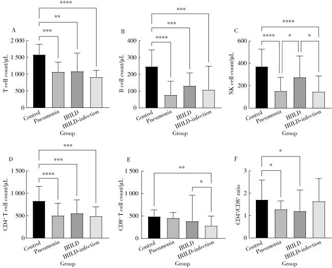
2.2 各组间T淋巴细胞亚群表达水平比较(图 1)

图1 淋巴细胞亚群表达水平对比

Figure 1 Comparative expression levels of lymphocyte subsets

A, T lymphocyte count comparison; B, B lymphocyte count comparison; C, NK count comparison; D, CD4+ cell count comparison; E, CD8+ cell count comparison; F, CD4/CD8 ratio comparison. Control, health control group; Pneumonia, infectious pneumonia group; IRILD, immune-related interstitial lung disease group; IRILD-infection, immune-related interstitial lung disease with concurrent infection group.* P < 0.05, * * P < 0.01, * * * P < 0.001, * * * * P < 0.000 1.

2.2.1 对照组与病例组比较

与对照组相比,感染性肺炎组的T细胞数、B细胞数、自然杀伤(natural killer,NK)细胞数、CD4+T细胞数、CD4+/CD8+比值降低,差异均有统计学意义(P<0.01);CD8+T细胞数降低,但差异无统计学意义(P>0.05)。与对照组相比,IRILD组的T细胞数、B细胞数、CD4+T细胞数、CD4+/CD8+比值降低,差异均有统计学意义(P<0.01);NK细胞数、CD8+T细胞数降低,但差异无统计学意义(P>0.05)。与对照组相比,IRILD合并感染组T细胞数、B细胞数、NK细胞数、CD4+T细胞数、CD8+T细胞数降低,差异均有统计学意义(P<0.05);CD4+/CD8+比值升高,但差异无统计学意义(P>0.05)。

2.2.2 IRILD组与感染性肺炎组比较

与IRILD组相比,感染性肺炎组NK细胞数降低,差异有统计学意义(P<0.05);T细胞数、B细胞数、CD4+T细胞数降低,CD8+T细胞数、CD4+/CD8+比值升高,但差异无统计学意义(P>0.05)。

2.2.3 IRILD组与IRILD合并感染组比较

与IRILD组相比,IRILD合并感染组NK细胞数、CD8+T细胞数降低,差异均有统计学意义(P<0.05);T细胞数、B细胞数、CD4+T细胞数降低,CD4+/CD8+比值升高,但差异均无统计学意义(P>0.05)。

2.2.4 感染性肺炎组与IRILD合并感染组比较

与感染性肺炎组相比,T细胞数降低,B细胞数升高,NK细胞数降低,CD4+T细胞数升高,CD8+T细胞数降低,CD4+/CD8+比值升高,但差异均无统计学意义(P>0.05)。

3 讨论

淋巴细胞亚群中的T细胞、B细胞和NK细胞是人体重要的免疫细胞,是机体免疫系统的重要组成部分,T淋巴细胞进一步分为辅助/诱导T细胞(CD3+CD4+T细胞)即CD4+T细胞与抑制/细胞毒性T细胞(CD3+CD8+T细胞)即CD8+T淋巴细胞;B淋巴细胞介导体液免疫;NK细胞与非特异免疫(即固有免疫)有关[6]。本研究发现感染性肺炎、IRILD及IRILD合并感染患者的淋巴细胞亚群表达水平是不同的。
本研究结果表明,与正常对照组相比,感染性肺炎患者淋巴细胞亚群的数量呈现出显著的下降趋势,包括T、B、NK、CD4+T细胞以及CD4+/CD8+比值。这些研究结果与现有文献的研究结论基本一致,即无论是由细菌还是病毒引起的肺部感染,淋巴细胞的减少均是一个普遍现象。Wang等[7]的研究支持了这一观点,他们在重症社区获得性肺炎(severe community-acquired pneumonia, SCAP)患者中发现CD3+T细胞、CD4+T细胞和CD8+T细胞的数量普遍较低,研究结果强调了监测CD4+T细胞在识别和管理SCAP患者中的重要性,并提示适当的免疫支持疗法可能有助于控制病情,改善临床结局。Qin等[8]在COVID-19患者的研究中也发现,无论疾病的严重程度如何,淋巴细胞的减少是该疾病的一个显著特征,特别是CD8+T细胞、CD4+T细胞和NK细胞的数量减少对整体淋巴细胞计数的降低起到了关键作用。当感染发生时,淋巴细胞的减少可能与一系列复杂的生物学机制有关。Doughty[9]发现,淋巴细胞凋亡、抗原反应性的降低、细胞因子表达的异常、免疫检查点分子的上调,以及调节性T细胞(Treg)群体的扩张等因素,共同导致了T细胞功能的广泛抑制。另外, Finfer等[10]还发现,除了淋巴细胞凋亡外,免疫细胞向感染部位的迁移也会导致外周血中淋巴细胞数量的减少。
在IRILD发病机制中,免疫系统紊乱扮演了至关重要的角色[11]。本研究结果发现, IRILD患者T、B、CD4+T细胞以及CD4+/CD8+比值均较健康对照组显著降低, CD8+T细胞数保持相对稳定。然而,在IRILD合并感染的患者中,NK细胞、CD8+T细胞数较IRILD组显著下降,这些结果再次强调了感染对淋巴细胞亚群的影响。既往研究也支持本研究的结果[12-13],在IIM-ILD的患者中发现淋巴细胞数量和CD4+T细胞数量较非ILD患者明显减少,ILD外周血中CD3+T细胞和CD8+T细胞数量显著降低。Shi等[11]研究发现,SS-ILD患者外周血NK细胞数量低于健康对照者,Th2细胞的绝对计数和百分比均低于无ILD的SS患者。也有研究证实[13-15],Treg在CTD-ILD的发展中起着重要作用,其数量减少和功能受损均与肺纤维化密切相关。Treg、Th1、Th2均为CD4+T细胞的亚群,它们的共同作用可能导致CD4+T细胞的减少。遗憾的是本研究未能对CD4+ T细胞亚群进一步分析。左艳华等[16]发现外周血中CD4+和CD8+T细胞水平与CTD-ILD患者病情严重程度呈负相关,表明CTD-ILD病情越严重,机体免疫功能受损越严重。本研究中,与对照组相比,IRILD组CD8+T细胞数并未下降趋势,这与上述研究结果不一致。但IRILD合并感染的情况下,CD8+T细胞数与对照组相比明显降低,这与张晶晶等[17]在IIM-ILD合并肺部感染的患者发现一致;耿陶然等[18]也发现重症流感患者中CD8+T细胞计数显著下降的趋势与患者较高的死亡风险相关。这提示,在IRILD合并感染的情况下,免疫功能的损害可能进一步加重,而CD8+T细胞的减少可能是患者感染风险增加的主要原因,也可能与患者较高的死亡风险相关[19]。因此,对这类患者应尽早进行免疫功能评估并给与积极的临床干预。IRILD患者淋巴细胞亚群减少的可能原因包括:(1)循环中活化的淋巴细胞被募集到靶器官,如外分泌腺、肺组织等,在CTD-ILD患者的肺组织中发现大量免疫细胞浸润,尤其是CD4+、CD8+T细胞[11, 15, 20-21];(2)原发病活动和各种病原微生物(如细菌、病毒等)感染引起了外周血中免疫细胞过度消耗;(3)免疫抑制剂和激素药物的使用可能直接或间接影响淋巴细胞的数量和功能。
本研究存在一定的局限性,首先,本研究样本量较少并且为单中心研究,今后需要多中心的前瞻性队列进行进一步分析;其次,本研究未对细菌、真菌和病毒等不同病原体进行进一步分层,这可能会引入潜在的混杂因素。未来,本课题组将继续分析不同病原微生物对感染性肺炎及IRILD合并感染患者淋巴细胞亚群表达水平的影响,并探索对感染性疾病的临床免疫治疗。
综上所述,本研究通过对比感染性肺炎、IRILD以及IRILD合并感染患者的淋巴细胞亚群表达水平,揭示了不同肺炎类型之间存在显著的免疫特征差异。这些差异不仅反映了不同肺炎状态下免疫系统的特异性改变,也为临床鉴别提供了潜在的实验室免疫学指标。

利益冲突  所有作者均声明不存在利益冲突。

作者贡献声明  赵豆豆、齐晅:统计数据,撰写论文;黄博、高伟波、裴源源:收集数据;金月波:设计研究方案,审定论文;邵苗:审定论文;何菁:提出研究思路,设计研究方案,总体把关和审定论文。所有作者均参与论文修改,并对最终文稿进行审读和确认。

1
Gopallawa I , Dehinwal R , Bhatia V , et al. A four-part guide to lung immunology: Invasion, inflammation, immunity, and intervention[J]. Front Immunol, 2023, 14, 1119564.

DOI

2
Lletí MS , Cabañero Navalón MD , Pitarch MT , et al. Etiology, diagnosis, and management of pneumonia in immunosuppressed patients[J]. Rev Esp Quimioter, 2022, 35 (Suppl 1): 89- 96.

3
Sverzellati N , Lynch DA , Hansell DM , et al. American thoracic society-European respiratory society classification of the idiopathic interstitial pneumonias: Advances in knowledge since 2002[J]. Radiographics, 2015, 35 (7): 1849- 1871.

DOI

4
Spagnolo P , Cordier JF , Cottin V . Connective tissue diseases, multimorbidity and the ageing lung[J]. Eur Respir J, 2016, 47 (5): 1535- 1558.

DOI

5
葛均波, 徐永健, 王辰. 内科学[M]. 9版. 北京: 人民卫生出版社, 2018: 41- 44.

6
中华医学会健康管理学分会. TBNK淋巴细胞检测在健康管理中的应用专家共识[J]. 中华健康管理学杂志, 2023, 17 (2): 85- 95.

7
Wang J , Pei L , Zhao T , et al. CD4+ T cells related to disease severity in elderly and frailty community-acquired pneumonia patients: A retrospective cohort study[J]. Immun Inflamm Dis, 2023, 11 (9): e1009.

DOI

8
Qin L , Duan X , Dong JZ , et al. The unreversible reduced but persistent activated NK and CD8+ T cells in severe/critical COVID-19 during omicron pandemic in China[J]. Emerg Microbes Infect, 2023, 12 (1): 2208679.

DOI

9
Doughty L . Adaptive immune function in critical illness[J]. Curr Opin Pediatr, 2016, 28 (3): 274- 280.

DOI

10
Finfer S , Venkatesh B , Hotchkiss RS , et al. Lymphopenia in sepsis: An acquired immunodeficiency[J]. Immunol Cell Biol, 2023, 101 (6): 535- 544.

DOI

11
Shi L , Wang J , Guo HX , et al. Circulating Th2 cell reduction and Th1/Th2 imbalance are correlated with primary Sjögren ' s syndrome-associated interstitial lung disease[J]. Arthritis Res Ther, 2022, 24 (1): 121.

DOI

12
Cheng L , Li Y , Luo Y , et al. Decreased Th1 cells and Increased Th2 cells in peripheral blood are associated with Idiopathic Inflammatory myopathies patients with Interstitial lung disease[J]. Inflammation, 2023, 46 (1): 468- 479.

DOI

13
Feng M , Guo H , Zhang C , et al. Absolute reduction of regulatory T cells and regulatory effect of short-term and low-dose IL-2 in polymyositis or dermatomyositis[J]. Int Immunopharmacol, 2019, 77, 105912.

DOI

14
Kotsianidis I , Nakou E , Bouchliou I , et al. Global impairment of CD4+CD25+FOXP3+ regulatory T cells in idiopathic pulmonary fibrosis[J]. Am J Respir Crit Care Med, 2009, 179 (12): 1121- 1130.

DOI

15
Moyé S , Bormann T , Maus R , et al. Regulatory T cells limit pneumococcus-induced exacerbation of lung fibrosis in mice[J]. J Immunol, 2020, 204 (9): 2429- 2438.

DOI

16
左艳华, 刘靓, 许化恒, 等. 结缔组织病相关性间质性肺炎患者涎液化糖链抗原与T细胞亚群水平变化及其临床意义[J]. 临床军医杂志, 2021, 49 (3): 336- 337.

17
张晶晶, 陈智勇, 王红, 等. 特发性炎性肌病间质性肺炎患者并发肺部感染的临床特征[J]. 中华风湿病学杂志, 2020, 24 (4): 253- 257.

18
耿陶然, 韩扬, 邱志峰, 等. 重症流行性感冒患者T淋巴细胞与预后研究[J]. 中华内科杂志, 2020, 59 (3): 200- 206.

19
Deng Y , Ou YY , Mo CJ , et al. Characteristics and clustering analysis of peripheral blood lymphocyte subsets in children with systemic lupus erythematosus complicated with clinical infection[J]. Clin Rheumatol, 2023, 42 (12): 3299- 3309.

DOI

20
Luzina IG , Todd NW , Iacono AT , et al. Roles of T lymphocytes in pulmonary fibrosis[J]. J Leukoc Biol, 2008, 83 (2): 237- 244.

21
Qin L , Wang WZ , Liu HR , et al. CD4+ and CD8+ T lymphocytes in lung tissue of NSIP: Correlation with T lymphocytes in BALF[J]. Respir Med, 2013, 107 (1): 120- 127.

DOI

文章导航

/