
肾血管平滑肌脂肪瘤(renal angiomyolipoma, RAML)是肾脏常见的良性肿瘤,50%~70%为散发性,30%~50%与遗传综合征相关。对于较小的RAML(<4 cm),可以基于主动监测进行保守治疗[1-2];而对于较大的RAML,因可能存在并发症(自发破裂、腹膜后出血、高血压等)或相关症状(腹部肿块、腰痛、血尿等),通常需要积极手术治疗,其中,自发性破裂出血是泌尿外科中常见的危急重症,可能引发失血性休克,严重时可危及患者生命,约14%的散发性RAML患者会出现这一并发症[3-5]。保留肾单位手术(nephron-sparing surgery, NSS)可在完整切除RAML的前提下最大程度地保留肾功能[6],但对于巨大(≥8 cm)RAML,由于存在肿物体积大、易出血且腹腔镜操作空间有限等难点,手术有一定困难,需要术者具备更好的经验与手术技巧。本研究回顾性分析2012年1月至2025年9月北京大学第三医院收治的45例巨大(≥8 cm)RAML行腹腔镜NSS治疗患者的临床资料,探讨该术式的疗效及安全性。
1 资料与方法
1.1 一般资料
本组共纳入45例患者,其中男11例,女34例,年龄20~74岁,平均(40.9±14.1)岁,体重指数(23.7±4.0) kg/m2,其中9例体重指数>25.0 kg/m2,5例手术前曾有自发性出血。所有病例术前均行肾脏CT或MRI检查(图 1),提示肿瘤呈外生性,边界清晰,肿瘤最大直径8.0~15.4 cm,平均(10.3±2.2) cm。2例合并妊娠,2例术前行介入栓塞治疗,1例诊断为结节性硬化病。术前血肌酐45~101 μmol/L,平均(66.1±11.8) μmol/L。
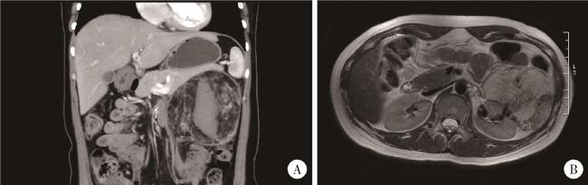
图1 巨大肾血管平滑肌脂肪瘤的术前影像学检查Figure 1 Preoperative imaging of a giant renal angiomyolipoma A, CTU examination shows a mass with mixed density anterior to the left kidney, containing fat and soft tissue densities, measuring approximately 12.8 cm×8.1 cm×12.3 cm. Contrast-enhanced scan reveals inhomogeneous enhancement. B, MRI examination shows a roundish mixed-signal mass in the left kidney, measuring approximately 8.9 cm×6.9 cm×10.8 cm, predominantly characterized by short T1 and long T2 signals. CTU, computed tomography urography; MRI, magnetic resonance imaging. |
45例患者均行腹腔镜NSS,其中6例采用达芬奇机器人手术系统辅助腹腔镜手术,32例(71.1%)为经后腹腔入路,13例(28.9%)为经腹腔入路。
根据术后组织病理结果,参考《2022年世界卫生组织肿瘤分类:泌尿系与男性生殖系统肿瘤病理》[7],对RAML和上皮样血管平滑肌脂肪瘤进行鉴别,(1)RAML:光镜下可见存在厚壁血管,存在比例不等的肌样成分和/或脂肪样成分,免疫组织化学检测提示黑色素细胞标志物(Melan-A、HMB45)阳性;(2)上皮样血管平滑肌脂肪瘤:光镜下呈癌样外观和/或呈弥漫性生长的上皮样细胞及丰满的梭形细胞,同时,超过80%的肿瘤细胞为上皮样细胞,免疫组织化学检测提示黑色素细胞标志物(如Melan-A)和组织蛋白酶K呈阳性。
1.2 手术方法
以后腹腔入路腹腔镜手术为例。气管插管、全身麻醉成功后,患者取完全健侧卧位,升高腰桥。制备气腹并放置套管,常规在第12肋下腋后线处、肋缘下腋前线处、髂棘上腋中线处三点建立通道,以手指及气囊扩张法制备腹膜后操作空间,气腹压力为12 mmHg。游离肾脏及肾蒂血管,顺肿瘤与肾包膜之间的层面向深方游离寻找并显露肿瘤基底部。显露困难时可通过旋转肾蒂来改善手术视野和操作空间,处理肾下极或肾门部RAML时注意保护输尿管。分离并阻断肾动脉,在距肿瘤基底部约0.5 cm处用剪刀楔形切除肿物及部分肾组织,完整切除肿瘤,缝合肾实质,恢复肾脏血供。若肿瘤与脂肪等周围结构粘连严重,可在解除动脉阻断后再游离粘连。确切止血后将肿物装入标本袋,经腋后线切口将肿瘤取出。放置引流,关闭切口。
1.3 观察指标
主要观察指标包括手术时长、术中出血量、肾动脉阻断时长、术后血肌酐变化、术后引流管拔管时长、术后住院时长、手术并发症、组织病理结果、术后复发情况等。
1.4 数据分析
采用SPSS 21.0软件,正态型连续性变量以均数±标准差表示,非正态型连续性变量以中位数(范围)表示,分类型变量以n (%)表示。
2 结果
45例患者的手术均顺利完成,5例中转开放手术。手术时长86~312 min,平均(180.6±55.7) min;术中出血量10~1 300 mL,中位值100 mL;术后住院3~14 d,平均(6.2±2.6) d;术后拔管时间2~14 d,平均(5.1±2.2) d;肾动脉阻断时长6~40 min,平均(21.9±9.8) min,其中36例≤30 min,6例>30 min。3例患者因考虑肿瘤位置较表浅、未侵及集合系统,经术者评估后采用无肾动脉阻断技术。
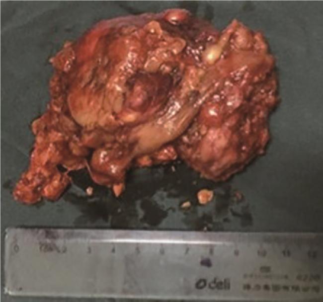
术后组织病理均为RAML,切缘均为阴性。术后血肌酐38~325 μmol/L,平均(70.3±42.9) μmol/L,1例在术后出现急性肾功能不全,1例术后出现肺部感染,经抗感染治疗后好转出院,其余患者无明确并发症。
表1 45例巨大RAML患者的临床资料及观察指标Table 1 Clinical data and observational indicators of 45 patients with giant RAML |
| Characteristic | Value |
| Age/years | 40.9±14.1 |
| Gender | |
| Male | 11 (24.4) |
| Female | 34 (75.6) |
| Body mass index/(kg/m2) | 23.7±4.0 |
| Tumor diameter/cm | 10.3±2.2 |
| Surgical side | |
| Left | 23 (51.1) |
| Right | 22 (48.9) |
| Preoperative imaging suggestive of contralateral RAML | |
| No | 38 (84.4) |
| Yes | 7 (15.6) |
| Preoperative estimated residual renal volume/mL | 85.4±23.6 |
| Surgical method | |
| Laparoscopic | 39 (86.7) |
| Robotic | 6 (13.3) |
| Surgical approach | |
| Transperitoneal | 13 (28.9) |
| Retroperitoneal | 32 (71.1) |
| Operative time/min | 180.6±55.7 |
| Renal artery clamping time/min | 21.9±9.8 |
| Estimated blood loss/mL | 100 (10, 1 300) |
| Postoperative hospital stay/d | 6.2±2.6 |
| Time to drainage removal/d | 5.1±2.2 |
Data are presented as ${\bar x}$±s, n (%) or median (minimum, maximum). RAML, renal angiomyolipoma. |
3 讨论
RAML起源于间充质,由不同比例的异常增生血管、平滑肌和脂肪组织构成,绝大多数为良性表现,组织病理学上可大致分为经典型和上皮样型。现有证据表明,RAML好发于女性,可能与雌激素相关[10]。RAML一般无典型临床表现,大约80%的病例为偶然发现,在约40%的病例中可存在腹部肿块、腰痛和血尿的经典三联征,其诊断依赖于MRI或CT[11]。破裂出血是RAML的严重并发症,常伴有严重的疼痛、贫血,可导致失血性休克,其风险严重程度通常与瘤体大小、血管分布、位置、生长速度等有关,直径越大的RAML发生破裂出血的可能性越高[12]。关于RAML患者的手术治疗指征目前尚无定论,一般包括破裂出血风险高、症状明显。
本研究遵循既有研究[13]所报道的,将巨大RAML定义为最大直径≥8 cm的病例,也有其他研究以7~10 cm为界[12, 14-16],肿瘤体积大小与手术难度、出血风险、预后、恶变风险有相关性。对于巨大RAML病例,非药物治疗方法通常包括选择性动脉栓塞术(selective artery embolization, SAE)、NSS、全肾切除术以及介入栓塞与手术联合治疗[6, 17]。NSS的目的在于完整切除肿瘤的同时,尽可能多地为患者保留肾功能以改善预后,通常适用于肿瘤呈外生性、边界清晰的病例,但由于肿瘤体积、质地、位置等因素,巨大RAML病例在施行NSS时,需要更高的手术技巧及医疗条件[18]。本文所报道45例接受NSS治疗的巨大RAML病例均取得了较为满意的效果。
在面对难以整块切除的肿瘤时,可以考虑采用分区切除技术,可将巨大的瘤体分步进行减容、游离和切除,以突破空间瓶颈,减少热缺血时间。使用该项技术需要考虑肿瘤的供血分支、肿瘤与集合系统的关系,例如,面对遮蔽肾门的巨大瘤体,可首先在远离正常肾实质的肿瘤“外周区”进行切除,为寻找肾门血管和暴露正常肾实质边界创造空间。既往有研究报道了类似技巧,RAML基底与肾脏粘连、且粘连组织内血供不丰富时,将附着于肾脏的RAML完整切除后其基底便能良好显露。RAML往往滋养血管丰富且缺乏正常血管结构,对于不清晰的条索状结构,可使用Hem-o-lock夹以减少撕扯血管造成的出血[20]。切除肿瘤和重建过程中,若出现突然的喷射性出血,首选电凝出血点,若效果不满意,则用双极钳夹闭出血点并以缝线结扎。对于多极供血的巨大肿瘤,可考虑将选择性分支动脉阻断技术与分区切除技术相结合。此外,早期肿瘤基底部离断、逆向游离技术也是一项可以应用于巨大RAML行NSS的技巧。进入手术空间后,可不急于游离巨大的瘤体表面,而是先暴露肾门血管以及肿瘤与正常肾实质交界的基底区域,在肿瘤基底部沿假包膜层面切开与分离,肿瘤脱离肾脏后即刻重建创面,之后再从周围组织中逐步剥离瘤体。该项技术使瘤体游离步骤移到恢复肾脏供血之后,有利于保护患肾功能,且可减少瘤体碎裂风险。
RAML出血后3周左右的粘连严重,手术风险较大。一般认为,肾脏热缺血时间的安全阈值应限制在25~30 min内,本研究中,6例肾热缺血时间较长的患者主要为肿瘤总体积及肾实质内体积大、粘连严重、游离肿瘤过程中需要控制出血所致,也进一步导致了手术时间的延长。术前手术方案的设计、术中的充分游离及有序操作可缩短肾脏缺血时间,亦可根据出血量变化实时收紧或放松止血带以缩短阻断时长,必要时可结合使用冰盐水、冰屑覆盖等冷缺血手段保护患肾功能。处理左侧RAML时,如肿瘤周边及肾蒂处严重粘连,可顺主动脉侧方向上分离,找到并游离左肾静脉及左肾动脉,将肾脏腹侧、背侧及下极进一步游离。经腹腔入路具有视野开阔、解剖显露难度较低、大血管损伤处理方便等优点;而经后腹腔入路对腹腔脏器干扰小,其有效性也已得到肯定,且随着术前影像评估技术的发展其安全性进一步提高[5, 13]。
综上所述,对于巨大RAML病例施行NSS,可在完整切除肿瘤、保证切缘阴性、避免并发症的基础上,实现有效保留肾功能。采用机器人手术系统辅助,在缩短手术时间、减少并发症方面可能具有一定优势。我们也将在未来持续随访,关注患者的远期功能及肿瘤学结局。



