
1 病例资料
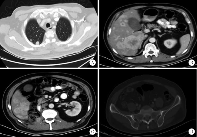
图1 治疗前基线胸部及腹盆CTFigure 1 Baseline chest and abdominopelvic CT before treatment A, multiple bilateral pulmonary metastases (largest 14 mm×13 mm); B, multiple hepatic lesions with enhancement, partially confluent (largest 64 mm×56 mm); C, right psoas metastasis (21 mm×13 mm); D, osteolytic lesion in the right iliac bone, consistent with metastasis. |
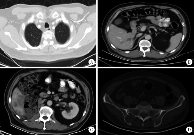
图2 治疗6周后胸部及腹盆CTFigure 2 Chest and abdominopelvic CT at 6 weeks after treatment A, bilateral pulmonary metastases decreased in size (largest 11 mm ×9 mm); B, hepatic lesions reduced in size with decreased enhancement (largest 58 mm×54 mm); C, right psoas metastasis decreased (16 mm×8 mm); D, osteolytic lesion in the right iliac bone increased in size. |
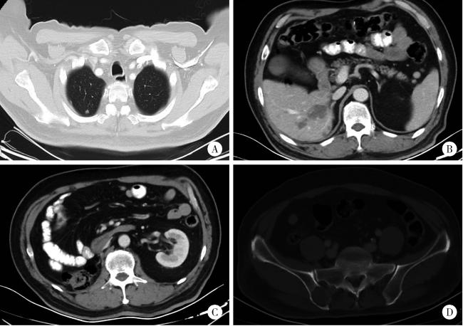
图3 治疗12周后复查胸部及腹盆CTFigure 3 Chest and abdominopelvic CT at 12 weeks after treatment A, bilateral pulmonary metastases further decreased (largest 6 mm×5 mm); B, hepatic lesions further reduced with decreased enhancement (largest 33 mm×23 mm); C, right psoas-adjacent lesion measuring 11 mm×6 mm; D, osteolytic lesion in the right iliac bone remained stable. |
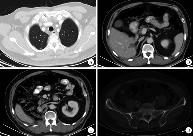
图4 治疗24周后复查胸部及腹盆CTFigure 4 Chest and abdominopelvic CT at 24 weeks after treatment A, bilateral pulmonary metastases remained stable, with the largest 6 mm×5 mm; B, hepatic lesions partly stable, with the largest 33 mm×23 mm, with some lesions further decreased from 18 mm×14 mm to 16 mm×9 mm; C, right psoas-adjacent lesion remained stable; D, osteolytic lesion in the right iliac bone remained stable. |
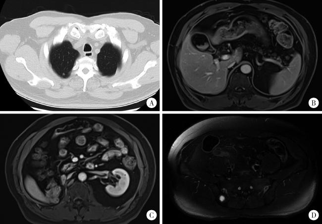
图5 2023年10月末次复查胸部CT及腹盆磁共振成像,维持PR状态Figure 5 Chest CT and abdominopelvic MRI at last follow-up in October 2023, the patient maintained PR A, bilateral pulmonary metastases remained stable, with the largest lesion measuring 6 mm × 5 mm; B, hepatic lesions remained stable, with the largest lesion measuring 33 mm × 23 mm; C, right psoas-adjacent lesion remained stable; D, osteolytic lesion in the right iliac bone remained stable. PR, partial response. |



