病例报告

不同膀胱异物的手术方式:2例报告

  • 王甡 1 ,
  • 宋欣怡 1 ,
  • 孙晓娜 2 ,
  • 郑冀鲁 , 1, * ,
  • 吕吉 , 1, *
展开
  • 1. 康复大学青岛中心医院泌尿外科,山东青岛 266042
  • 2. 康复大学青岛中心医院护理部,山东青岛 266042

收稿日期: 2026-01-31

  网络出版日期: 2026-06-30

版权

版权所有,未经授权,不得转载。

Surgical approaches to different foreign bodies in the urinary bladder: A report of 2 cases

  • Shen WANG 1 ,
  • Xinyi SONG 1 ,
  • Xiaona SUN 2 ,
  • Jilu ZHENG , 1, * ,
  • Ji LV , 1, *
Expand
  • 1. Department of Urology, Qingdao Central Hospital, University of Health and Rehabilitation Sciences, Qingdao 266042, Shandong, China
  • 2. Nursing Department, Qingdao Central Hospital, University of Health and Rehabilitation Sciences, Qingdao 266042, Shandong, China
ZHENG Jilu,

Received date: 2026-01-31

  Online published: 2026-06-30

Copyright

All rights reserved. Unauthorized reproduction is prohibited.

摘要

回顾2例膀胱异物的患者临床资料。由于异物的大小和类型不同,采用了不同的手术方式。病例1为21岁的男性患者,因尿痛于康复大学青岛中心医院就诊,患者自述2天前自行将一异物通过尿道插入体内,查体提示耻骨上疼痛,以膀胱异物收入泌尿外科。盆腔CT检查显示膀胱内有一长4.2 cm的长条状金属异物,经过评估膀胱异物的形状和体积,考虑行膀胱镜检查以取出异物。患者于全身麻醉状态下,取截石位,直视下硬质膀胱镜进入膀胱,观察到膀胱内一金属管状结构异物。尝试使用异物夹将其取出,调整异物的长轴与膀胱镜平行。由于金属异物表面光滑,经过多次尝试后终于成功将异物取出。病例2为42岁男性患者,因排尿困难和尿痛于我院门诊就诊,并被收入泌尿外科。患者自述1天前自行将异物经尿道插入体内。泌尿系统CT检查提示膀胱内有一根金属管状结构异物,长度为21.5 cm。考虑到异物较长(从髂嵴顶部至会阴部),且两端尖锐,不能排除膀胱穿孔可能;加之异物表面光滑,通过膀胱镜取出难度较大,且会增加医源性膀胱穿孔的风险,最终决定采用腹腔镜手术探查是否存在膀胱穿孔并取出异物。在全身麻醉下,患者取仰卧位,于脐上做1 cm切口,使用气腹针建立气腹,维持二氧化碳气腹压力12 mmHg;于脐下进入腹腔后,插入10 mm Trocar和腹腔镜,在左腹直肌外侧缘脐下2 cm处插入5 mm Trocar,在右腹直肌对称位置插入12 mm Trocar;于腹腔镜下可见异物将膀胱顶部顶入腹腔,切开膀胱壁暴露异物,从会阴部向头端推挤异物,并通过脐上Trocar取出异物。

关键词: 异物; 膀胱; 腹腔镜; 膀胱镜

本文引用格式

王甡 , 宋欣怡 , 孙晓娜 , 郑冀鲁 , 吕吉 . 不同膀胱异物的手术方式:2例报告[J]. 北京大学学报(医学版), 2026 , 58(4) : 877 -880 . DOI: 10.19723/j.issn.1671-167X.2026.04.028

Abstract

We reported the clinical records of 2 patients each with a foreign body in the urinary bladder admitted to Qingdao Central Hospital, University of Health and Rehabilitation Sciences. The size and shape of each foreign body in the urinary bladder were different, thus we chose different surgical approaches to remove them. Case 1 was a 21-year-old male patient. He presented to the outpatient service with urodynia. The patient currently complained of urodynia, and narrated that a foreign body was self-inserted via the urethra 2 days before. Physical examination revealed suprapubic pain. Ultimately he was admitted to the Department of Urology with a diagnosis of "bladder foreign body". A computed tomography (CT) scan of the pelvis was performed and the results demonstrated a linear metallic structure, mea-suring 4.2 cm in length, within the urinary bladder. Under general anesthesia, the patient was placed in lithotomy position and a rigid cystoscope was inserted into the bladder via urethra. The foreign body was a metal tubular structure and was located within the urinary bladder. Removal by foreign body forceps was attempted and long axis of the foreign body should be parallel to cystoscope. Removal was carefully performed after several attempts due to the smooth surface of the metal foreign body. Case 2 was a 42-year-old male patient. He presented to the outpatient service with dysuria and urodynia and was admitted to the Department of Urology. The patient narrated that a foreign body was self-inserted via the urethra 1 day before, and currently complained of dysuria, urodynia, and slight hematuria. A CT scan of the urologic system was performed and the results demonstrated a metal tubular structure, measuring 21.5 cm in length. The length of the foreign body was long (from top of iliac crest to perineum) and both ends were sharp, thus bladder perforation could not be excluded. Besides, with smooth metal surface, it was difficult to remove via cystoscopy and the risk of iatrogenic bladder perforation would increase simultaneously. Therefore, laparoscopic surgery was chosen to detect whether there would be a bladder perforation and could remove the foreign body. Under general anesthesia, the patient was placed in supine position and supraumbilical incision of 1 cm was performed. Pneumoperitoneum needle was used to establish a pneumoperitoneum and insufflation CO2 pressure was maintained at 12 mmHg. The peritoneum was accessed and a 10-mm trocar and laparoscopic camera were inserted. A 5-mm trocar was inserted at 2 cm below the umbilical level of lateral margin of left rectus abdominis muscle. While a 12-mm trocar was inserted at symmetrical site of his right rectus abdominis muscle. Under laparoscope, dome of the bladder was lifted into the abdominal cavity by the foreign body, then the bladder wall was incised to expose the foreign body. We pushed the bottom of the foreign body from the perineum extracorporally and removed the foreign body from the supraumbilicus trocar.

膀胱异物在临床上并不罕见,但对此类病例的恰当治疗仍是泌尿外科医生面临的挑战。异物通常由患者通过尿道自行置入,目的往往出于性刺激或好奇,或因患有精神疾病;另外,异物也可能由医源性因素所导致,比如遗留或移位的医疗器具[1]。目前的报道中膀胱异物的种类多样,包括金属丝、铅笔、圆珠笔、钉子、发夹、塑料管、橡皮擦、磁珠、棉签、体温计、尿道探子等[2-5], 由于每个膀胱异物的大小和形状各不相同,因此,选择合适的器械和手术方式至关重要。本文回顾了2023年9月至2024年3月期间康复大学青岛中心医院收治的2例膀胱异物患者的临床资料,通过不同的手术方式取出异物,现报告如下。

1 病例资料

病例1为21岁男性,因尿痛于我院门诊就诊,患者自述2天前自行将一异物通过尿道插入体内,查体提示耻骨上疼痛,以膀胱异物收入泌尿外科。盆腔CT检查提示膀胱内有一长约4.2 cm的长条状金属异物(图 1A),对膀胱异物的形状和体积进行评估后,考虑通过膀胱镜取出异物。全身麻醉状态下,患者取截石位,在直视下将26F(1F≈0.33 mm)硬质膀胱镜(Karl Storz,Tuttlingen,Germany)经尿道置入膀胱,观察到该异物为一金属管状结构,位于膀胱腔内。尝试使用异物夹将其取出,调整异物的长轴与膀胱镜平行,但由于金属异物表面光滑,经过多次尝试后才成功将异物取出(图 1B)。术后为患者提供了性心理教育,术后第2天患者出院,术后1个月随访时患者排尿顺畅,尿流率检测正常(25 mL/s)。
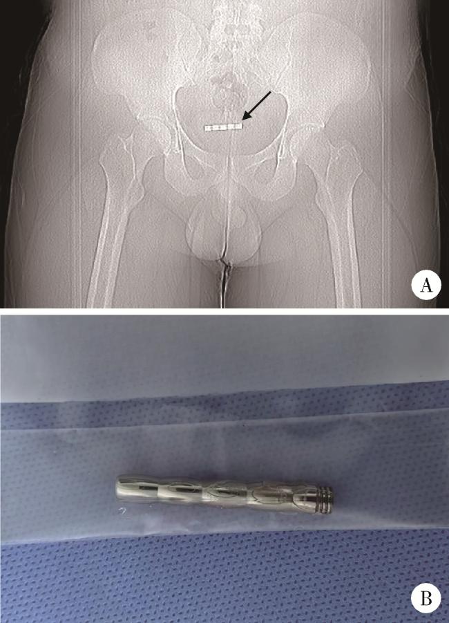
图1 病例1的盆腔CT图像和取出的异物

Figure 1 The pelvic CT image of Case 1 and the removed foreign body

A, CT scan of the pelvis, indicating a metal tubular structure within the urinary bladder (arrow); B, extracted foreign body (a metal tube).

病例2为42岁男性,因排尿困难和尿痛就诊于我院,并被收入泌尿外科。患者自述1天前自行将异物经尿道插入体内。泌尿系CT提示膀胱内有一根金属管状结构,长度约21.5 cm(图 2A)。由于异物较长(从髂嵴顶部至会阴部)且两端尖锐,不能排除膀胱穿孔可能;此外,考虑到金属异物表面光滑,膀胱镜取出难度较大,且会增加医源性膀胱穿孔的风险,因此决定采用腹腔镜手术检查是否有膀胱穿孔并取出异物。全身麻醉下,患者取仰卧位,于脐上做1 cm切口,使用气腹针建立气腹,维持二氧化碳气腹压力12 mmHg。于脐下进入腹腔,置入10 mm Trocar和腹腔镜; 在左腹直肌外侧缘脐下2 cm处置入5 mm Trocar;在右腹直肌对称位置置入12 mm Trocar。腹腔镜下可见异物将膀胱顶部顶入腹腔,遂切开膀胱壁以暴露异物,并从会阴部向头端推挤异物,最终通过脐上Trocar取出异物(图 2B)。异物成功取出后,使用2-0型Ⅴ-loc缝线(Ethicon,Cincinnati,OH,USA)连续缝合膀胱壁,使用3-0型Ⅴ-loc缝线缝合腹部切口。术后即刻通过三腔导尿管对膀胱进行冲洗,术后第2天拔除腹腔引流管,术后第3天出院。术后1个月随访,患者自主排尿顺畅,尿流率为20 mL/s。
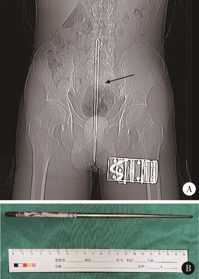
图2 病例2的泌尿系统CT图像及取出的异物

Figure 2 CT image of the urinary system in Case 2 and the removed foreign body

A, CT scan of the urologic system, indicating a metal tubular structure(arrow) in the urinary bladder (bladder perforation can not be excluded); B, extracted foreign body (a metal chopstick).

2 分析与讨论

膀胱异物的典型症状包括尿路感染、排尿困难、下腹部疼痛或轻度肉眼血尿(伴或不伴有疼痛)[5]。性自慰行为或精神疾病所导致的膀胱异物,症状可能并不典型,因此会延误诊断,全面的影像学检查有助于减少漏诊、误诊[6]。异物残留可能会导致慢性尿路感染、膀胱刺激症状、膀胱穿孔、腹膜后脓肿、肠道瘘、阴道瘘以及结石形成[7-8]。因此,影像学诊断、详细的病史资料采集以及及时的手术取出是治疗膀胱异物的有效方法。
关于膀胱异物的来源,除患者自行置入外,也可能是医源性的,或者从邻近器官移位而来。据报道,膀胱异物种类较多,包括缝线、纱布、体温计、金属管等,其最佳的处理方式取决于异物的性质、位置、外科医生的专业水平以及所在医院可用的器械[9-11]。因此,不同的异物以及不同的情况需要采用不同的手术方式。
通过在PubMed上以“bladder foreign body”为关键词检索文献(年代设定为2000—2023年),我们对其中20篇有代表性的文献进行回顾,总结了取出膀胱异物的5种常见手术方式:(1)膀胱镜(包括气膀胱镜),此为最常见的手术方式,文献中通过该手术方式取出的异物通常为体积较小且孤立于膀胱内的异物,包括医源性异物,如气囊导管的碎片、双J管导管碎片[2],以及非医源性异物,如铅笔(长9 cm)[8]、管状金属结构(长10 cm)[12]、卫生棉条、回形针、钢笔帽、钉子、橡皮、注射器、圆珠笔[2]、管状塑料异物、弹簧样金属物质[13]、发卡、电线(金属)[14]、磁力球[15]。(2)耻骨上膀胱切开术,通过该手术方式取出的异物有金属碎片(3.5 cm×2.0 cm)[16]、外科敷料[17]、膀胱结石(7.0 cm×5.2 cm,起初膀胱异物为人造钓鱼虫,后续以此为核心形成膀胱结石)[18]、骨水泥[2]和寄生虫[14]。(3)膀胱镜-腹腔镜手术(在腹腔镜下松解粘连,再通过膀胱镜取出异物),有研究报道通过此手术方式取出一枚嵌入膀胱壁内的宫内节育器[19]。(4)剖腹探查手术,有文献报道1例通过此种手术方式取出一枚长8.2 cm钉子的病例,该例患者CT检查提示膀胱破裂,且生命体征及实验室检查提示低血容量性休克,因此,在急症下推荐采取此手术方式。(5)腹腔镜手术,本文中病例2采用了该方法。
此外,我们也回顾了我院自2015年至今收治的6例膀胱异物患者的临床资料。其中,1例通过腹腔镜取出,5例使用膀胱镜取出,所有患者均康复并出院。在术后3个月的随访期间内,患者均未出现下尿路不适症状,也无血尿或尿路感染复发的情况。上述对既往文献报道和本科室曾经收治病例的汇总分析提示,膀胱镜检查是创伤较小的首选的膀胱异物治疗方法;只有当异物为锐器、与膀胱壁分离并可能穿破膀胱壁,或异物体积较大,引起严重并发症甚至危及生命时,才需要考虑通过有创的方式取出,比如耻骨上膀胱切开术、膀胱镜-腹腔镜手术、剖腹探查手术或腹腔镜手术。
另外,通过文献的阅读,我们认为性别不会对手术方式、手术效果存在影响, 在本研究检索到的病例中[1-19],男女比例为87 ∶107,因此,性别不是影响手术方式的主要因素。异物的性质、是否突破膀胱、是否造成严重且紧急的并发症,以及当地医院的设备,是影响手术方式的主要因素。膀胱异物是否伴有感染、瘘口等并发症是影响手术效果的关键因素。
总之,对于膀胱异物,应尽早进行取出手术,以减轻不必要的疼痛和并发症。手术方式需根据异物的性质及并发症情况来制定,并应尽量减少对膀胱和尿道的损伤。

利益冲突  所有作者均声明不存在利益冲突。

作者贡献声明  王甡:撰写论文;宋欣怡:设计研究方案;孙晓娜:收集、分析、整理数据;郑冀鲁:提出研究思路和修改论文;吕吉:总体把关和审定论文。所有作者均参与论文修改,并对最终文稿进行审读和确认。

1
Winot S , Hill AC , Simon EL . A case report you can't make up: A bladder foreign body[J]. J Emerg Med, 2021, 61 (1): 73- 75.

DOI

2
Kochakarn W , Pummanagura W . Foreign bodies in the female urinary bladder: 20-year experience in ramathibodi hospital[J]. Asian J Surg, 2008, 31 (3): 130- 133.

DOI

3
Bansal A , Yadav P , Kumar M , et al. Foreign bodies in the urinary bladder and their management: A single-centre experience from north India[J]. Int Neurourol J, 2016, 20 (3): 260- 269.

DOI

4
Al-Zubaidi M , McCombie S , Bangash H , et al. A novel technique to remove a urinary bladder foreign body endoscopically using an Endoloop[J]. Urol Case Rep, 2022, 40, 101899.

5
Zhang K , Zhang Y , Zhang Y , et al. Pneumovesicoscopy: An available technique for the retrieval of a rare foreign body in the urinary bladder[J]. Asian J Surg, 2022, 45 (5): 1180- 1181.

DOI

6
Gibson E , Glaser Z , Joseph D , et al. Previously banned magnets as foreign bodies in the lower urinary system: A single-institution case series and review of the literature[J]. Clin Pediatr (Phila), 2019, 58 (1): 110- 113.

DOI

7
Walsh P , Moustafa M . Retention of urethrovesical foreign bodies: Case report and literature review[J]. J Emerg Med, 2000, 19 (3): 241- 243.

DOI

8
Ortogǧlu F , Gürlen G , Altunkol A , et al. A rare foreign material in the bladder: Piece of pencil[J]. Arch Iran Med, 2015, 18 (9): 616- 617.

9
Lindsay JS . The practice of 'urethral sounding' complicated by retained magnetic beads within the bladder and urethra: Diagnosis and review of management[J]. Am J Case Rep, 2019, 20, 1841- 1844.

DOI

10
Liu ZH , Zhu XF , Zhou N . Retrieval of 159 magnetic balls from urinary bladder: A case report and literature review[J]. Urol Case Rep, 2019, 26, 100975.

11
Ogbetere F , Irekpita E . A self-insertion of an uncommon urethrovesical foreign body for autoerotism[J]. Niger J Surg, 2021, 27 (1): 81- 83.

DOI

12
Fabià Mayans A , Segarra Tomás J , et al. Bladder foreign body[J]. Arch Esp Urol, 2018, 71 (2): 224.

13
Huang HH , Huang YH , Chang HC , et al. Foreign bodies in the urinary bladder and urethra: A case series[J]. Asian J Surg, 2023, 46 (4): 1649- 1650.

DOI

14
Datta B , Ghosh M , Biswas S . Foreign bodies in urinary bladders[J]. Saudi J Kidney Dis Transpl, 2011, 22 (2): 302- 305.

15
Graziottin TM , de Freitas G Soares D , Da Ros CT , et al. Magne-tic spheres as foreign body into the bladder[J]. J Sex Med, 2013, 10 (10): 2590- 2592.

DOI

16
Klingenberg C , Koehler T , Due J . A foreign body in the urinary bladder[J]. Tidsskr Nor Laegeforen, 2019, 139 (2)

17
Cardinale M , Scheiwe C , Boubotte-Salmon F , et al. A rare foreign body in the bladder: A surgical dressing as a bladder stone[J]. Med Sante Trop, 2019, 29 (2): 222- 224.

18
Schmitt BH , Feder MT , Rokke DL , et al. An unusual foreign body in the urinary bladder mimicking a parasitic worm[J]. J Clin Microbiol, 2012, 50 (7): 2520- 2522.

DOI

19
Jin C , Fan Y , Zhang Q , et al. Removal of foreign bodies embedded in the urinary bladder wall by a combination of laparoscopy and carbon dioxide cystoscopic assistance: Case report and literature review[J]. Investig Clin Urol, 2016, 57 (6): 449- 452.

DOI

文章导航

/