病例报告

妊娠合并原发性甲状旁腺功能亢进致高钙危象及急性胰腺炎1例

  • 谢晓琪 ,
  • 张秀英 ,
  • 王相清 ,
  • 陈玲 ,
  • 张放 ,
  • 张瑞 ,
  • 纪立农 ,
  • 韩学尧 ,
  • 刘蔚 , *
展开
  • 北京大学人民医院内分泌科,北京 100044

收稿日期: 2024-07-25

  网络出版日期: 2026-05-22

版权

版权所有,未经授权,不得转载。

Primary hyperparathyroidism in pregnancy resulting in hypercalcemic crisis and acute pancreatitis: A case report

  • Xiaoqi XIE ,
  • Xiuying ZHANG ,
  • Xiangqing WANG ,
  • Ling CHEN ,
  • Fang ZHANG ,
  • Rui ZHANG ,
  • Linong JI ,
  • Xueyao HAN ,
  • Wei LIU , *
Expand
  • Department of Endocrinology, Peking University People's Hospital, Beijing 100044, China

Received date: 2024-07-25

  Online published: 2026-05-22

Copyright

All rights reserved. Unauthorized reproduction is prohibited.

摘要

原发性甲状旁腺功能亢进症(primary hyperparathyroidism,PHPT) 是由于甲状旁腺产生过多的甲状旁腺激素所致,妊娠期间首次发现的PHPT十分罕见,且对母体和胎儿有严重不良影响。本文报告了一例妊娠合并PHPT、高钙危象、急性胰腺炎的病例,患者行急诊剖宫产术,产后第47天行甲状旁腺腺瘤切除术,所娩出胎儿在分娩后曾有数次低钙抽搐。产后9个月随访时患者恢复良好,婴儿生长发育良好。本例患者为30岁妊娠期女性,孕32周时因腹痛、呕吐就诊,实验室检查提示血钙3.56 mmol/L、甲状旁腺激素(parathyroid hormone,PTH)388.6 ng/L,影像学检查证实急性胰腺炎及甲状旁腺占位。值得注意的是,患者分娩后PTH水平较产前明显下降(48.91~53.08 ng/L),结合术后病理所见腺瘤内出血及含铁血黄素沉积,分析可能与急性胰腺炎期间有效循环血容量减少及瘤体部分缺血坏死有关。临床医师应提高对于妊娠合并PHPT的警惕性,特别是孕前检查及孕早期应当注意血钙水平,并尽早采取预防措施以避免并发症及不良妊娠结局的发生。

本文引用格式

谢晓琪 , 张秀英 , 王相清 , 陈玲 , 张放 , 张瑞 , 纪立农 , 韩学尧 , 刘蔚 . 妊娠合并原发性甲状旁腺功能亢进致高钙危象及急性胰腺炎1例[J]. 北京大学学报(医学版), 2026 , 58(4) : 885 -888 . DOI: 10.19723/j.issn.1671-167X.2026.04.030

Abstract

Primary hyperparathyroidism (PHPT) is caused by excessive production of parathyroid hormone by the parathyroid gland. PHPT discovered for the first time during pregnancy is extremely rare and has serious adverse effects on both the mother and the fetus. This article reports a case of pregnancy complicated by PHPT, hypercalcemic crisis, and acute pancreatitis. The patient was a 30-year-old woman at 32 weeks of gestation who presented with a four-day history of progressive abdominal pain and vomiting. Laboratory evaluation on admission revealed a markedly elevated serum total calcium of 3.56 mmol/L and a parathyroid hormone (PTH) level of 388.6 ng/L, with the normal reference range being 15 to 65 ng/L. Imaging studies confirmed the presence of acute pancreatitis, left nephrolithiasis, right hydronephrosis, and a suspicious cystic-solid mass at the lower pole of the right thyroid lobe measuring 2.1 cm × 2.0 cm × 1.2 cm. Due to rapid clinical deterioration and the development of a hypercalcemic crisis, an emergency cesarean section was performed, resulting in the delivery of a live premature male infant with a birth weight of 2 400 g and a length of 45 cm. The neonate's initial serum calcium was 3.08 mmol/L, and several episodes of hypocalcemic seizures occurred shortly after birth, necessitating admission to the neonatal intensive care unit. Postpartum, the mother was managed with aggressive intravenous fluid resuscitation, subcutaneous calcitonin, and intravenous bisphosphonate therapy. Notably, her serum PTH level decreased substantially after delivery, ranging from 48.91 to 53.08 ng/L. This spontaneous decline was likely attributable to reduced effective circulating blood volume during the episode of acute pancreatitis and partial ischemic necrosis of the parathyroid adenoma. At 47 days postpartum, the patient underwent successful surgical resection of a right inferior parathyroid adenoma measuring approximately 3 cm in diameter. Pathological examination of the resected specimen confirmed the diagnosis and revealed focal vascular proliferation and congestion, interstitial fibrosis with hyaline degeneration, and hemosiderin deposition. Following parathyroidectomy, her serum calcium and PTH levels normalized rapidly and remained stable. At the 9-month follow-up visit, the mother was completely asymptomatic with normal biochemical parameters, and the infant demonstrated appropriate growth and development. Clinicians should be vigilant about the presence of PHPT during pregnancy, especially during preconception and early pregnancy, monitoring blood calcium levels and taking preventive measures early to avoid complications and adverse pregnancy outcomes.

原发性甲状旁腺功能亢进症(primary hyperparathyroidism,PHPT) 是由于甲状旁腺产生过多的甲状旁腺激素所致[1]。普通人群中PHPT的发病率为0.3%,是高钙血症最常见的病因。女性发病率是男性的两倍[2],在20~40岁的女性中,PHPT的患病率为0.05%[3],但在妊娠期间PHPT的真实患病率尚不明确[4]。PHPT的病因包括甲状旁腺增生、腺瘤或腺癌,也可能作为多发性内分泌腺瘤、罕见的遗传或代谢综合征的部分临床表现[1]。已有PHPT导致急性胰腺炎的病例报道, 但在妊娠期间出现者极为罕见[5]。本文报告一例32周初产妇中的PHPT导致高钙危象及急性胰腺炎病例,并分析其临床特征、治疗及母儿预后随访。

1 病例资料

1.1 患者一般情况

女,30岁,孕32周,2017年10月1日因“腹痛,血钙升高4天”就诊于外院。患者4天前进食油腻食物后出现剑突下疼痛,进行性加重,伴呕吐数次就诊。孕22周诊断妊娠期糖尿病,饮食及运动控制,血糖基本达标。孕期曾口服乳酸钙2片(含钙量200 mg/片),1次/d;复合维生素片1片,1次/d;二十二碳六烯酸1粒,1次/d,不规律口服B族维生素及补血颗粒,无明确大量维生素A、D及钙片摄入史。否认高钙血症、肾结石、肿瘤家族史; 否认高血压、糖尿病家族史。
体格检查:体温37.5 ℃,脉搏102次/min,呼吸:18次/min,血压122/74 mmHg。痛苦表情,斜侧位被动体位,腹部膨隆,上腹部剑突下压痛,无肌紧张,无反跳痛,肾区叩痛(+),甲状腺Ⅰ度,未及结节。查血清淀粉酶549 IU/L↑,血清脂肪酶995 IU/L↑,血钙3.56 mmol/L↑、甲状旁腺素(parathyroid hormone,PTH) 388.6 ng/L↑(正常为15~65 ng/L)。腹部彩超提示胰腺水肿,考虑胰腺炎。左肾结石,右肾肾盂积水,右输尿管上段扩张。甲状腺超声提示甲状腺右叶下极囊实性占位,2.1 cm×2.0 cm×1.2 cm,形态规则,边界清,内部低回声为主,可见粗大钙化,内部可见较丰富血流信号,甲状旁腺来源可能性大。给予禁食、扩容补液、抗感染治疗,4 d后腹痛加重,复查血清淀粉酶较前升高至990 IU/L,于2017年10月5日转入北京大学人民医院,诊断妊娠合并急性胰腺炎、甲状旁腺功能亢进症、高钙血症、肾积水、肾结石。

1.2 手术治疗及病理检查

患者入院后全身麻醉下行急诊剖宫产术,胎儿取出后宫腔出血汹涌,双侧子宫动脉上行支结扎,同时行膀胱镜下双侧输尿管支架植入术。分娩一早产男活婴,体重2 400 g,身长45 cm,出生后哭声弱,呼吸不规则,立即胸外按压、气管插管正压通气转入儿科重症监护病房,出生后即刻查血钙3.08 mmol/L,之后波动在2.00~2.46 mmol/L,曾有数次“抽搐”发作。
给予患者补液、利尿、降钙素皮下注射、双膦酸盐静脉输液治疗,完善甲状旁腺断层显像提示,甲状腺右叶下极结节,1.2 cm×1.0 cm×1.8 cm,99mTc-甲氧基异丁基异腈(99mTc-methoxyisobutylisonitrile, MIBI)摄取增高,考虑为甲状旁腺腺瘤(图 1A)。复查血钙波动在2.75~3.10 mmol/L,PTH水平降低(48.91~53.08 ng/L)。产后第47天于北京大学人民医院行甲状旁腺探查术+喉返神经探查术+甲状旁腺病损切除术。探查甲状旁腺发现右侧下甲状旁腺肿大约有2~3 cm,质地较硬,手术切除,术后标本最大经为3 cm,边界清,右侧上旁腺未触及明显肿大(图 1B)。送病理检查,回报为甲状旁腺腺瘤,局灶血管增生及充血,间质纤维化伴玻璃样变性,含铁血黄素沉积(图 1C~F)。患者术后无口周及四肢麻木、刺痛,无四肢搐搦,无呼吸困难,无声音嘶哑及饮水呛咳。术后第1天复查血钙2.25 mmol/L,PTH 2.76 ng/L。
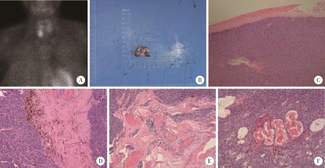
图1 患者术前影像学及术中病理切除标本(HE染色×40)

Figure 1 Preoperative imaging and intraoperative pathological resection specimen of the patient (HE staining ×40)

A, parathyroid single photon emission computed tomography imaging suggests a nodule at the lower pole of the right thyroid lobe; B, intraoperative resection specimen has a diameter of 3 cm; C, postoperative pathology indicates a parathyroid adenoma; D to F, focal vascular proliferation and congestion, interstitial fibrosis with hyaline degeneration, and deposition of hemosiderin in the parathyroid adenoma.

1.3 术后随访和结局

术后随访9个月,监测患者血钙水平2.19~2.42 mmol/L,PTH在13.58~82.41 ng/L。随访期间进一步结合患者病史、家族史及相关内分泌评估对多发性内分泌腺瘤综合征进行了筛查,未见支持多发性内分泌腺瘤综合征的证据。新生儿于重症监护病房治疗24 d后出院,期间曾并发新生儿败血症、应激性溃疡,第9个月时随访,生长发育情况良好,血钙2.21 mmol/L。

2 讨论

妊娠期间首次发现的PHPT是一种罕见的临床情况,可能对母体和胎儿产生严重的负面影响[6-7]。据报道,妊娠期间PHPT相关的母体并发症发生率高达67%[7-8],但最常见的是妊娠剧烈呕吐和肾结石,仅有极为罕见的病例报告危及生命的并发症,如急性胰腺炎和高钙危象[9-11]。由PHPT引起的高钙血症会导致细胞内钙稳态的破坏和胰腺腺泡细胞胰蛋白酶原等的过早激活[12],进而诱发急性胰腺炎的发生,妊娠期胰腺炎与子痫、早产和宫内胎儿死亡直接相关[13]。值得注意的是,非胆源性胰腺炎可能导致更为严重的并发症和较差的临床预后,与急性胰腺炎相关的母体死亡亦偶有报道[14-15]。另外,未经有效治疗的PHPT母亲所分娩的新生儿可能会由于子宫内甲状旁腺受到抑制而出现新生儿低钙血症和手足搐搦症[16]。既往个案报告显示,婴儿在出生后不久常出现短暂性低钙血症[9]
尽管恶心和呕吐在早孕期很常见,但伴随腹痛的情况却极为罕见[17]。临床需要考虑的鉴别诊断包括胃肠炎、阑尾炎、胆囊炎、消化性溃疡、肠梗阻、泌尿系统感染和滋养细胞肿瘤性疾病。对于有剧烈呕吐的孕妇,当出现腹痛或感染迹象,尤其症状在第一孕期后开始时,临床应引起警惕并进行进一步检查以明确原因。
本例患者在分娩后PTH水平较前明显降低,一方面可能与分娩前急性胰腺炎期间有效循环血容量较低有关[18],另一方面,术后病理提示腺瘤内部存在间质纤维化、玻璃样变性以及含铁血黄素沉积,可能与急症及手术期间瘤体血供减少和部分缺血坏死有关。对于PHPT患者由于瘤体缺血坏死而出现短暂的自发缓解的个案既往亦有报告[19],我国学者近期亦报道一例高钙危象后双侧甲状旁腺腺瘤内部囊内出血的病例[20],并认为应该结合患者血钙水平、PTH水平以及相关影像学检查评估合适的手术时机。本例患者在术后仍存在高钙血症,且核素显像提示MIBI摄取增高,接受手术治疗后证实为甲状旁腺腺瘤。
尽管妊娠期合并PHPT较为罕见,然而一旦合并高钙血症及急性胰腺炎则严重威胁母儿安全。由于PHPT较易发生于女性,临床医师应提高对妊娠期合并PHPT的警惕性。备孕期及孕早期检查时应关注有无高钙血症;对于妊娠期出现不明原因恶心、呕吐、腹痛、泌尿系结石或肾积水者,亦应及时筛查血钙,并在发现高钙血症后进一步检测PTH、结合颈部超声评估甲状旁腺病变,以期尽早识别并干预,避免高钙危象、急性胰腺炎及不良母儿结局的发生。

利益冲突  所有作者均声明不存在利益冲突。

作者贡献声明  谢晓琪、王相清、刘蔚:收集数据,撰写论文;张秀英、陈玲、张放、张瑞:收集数据;纪立农、韩学尧、刘蔚:总体把关和审定论文。所有作者均参与论文修改,并对最终文稿进行审读和确认。

1
MacKenzie-Feder J , Sirrs S , Anderson D , et al. Primary hyperparathyroidism: An overview[J]. Int J Endocrinol, 2011, 2011, 251410.

2
Wermers RA , Khosla S , Atkinson EJ , et al. Incidence of primary hyperparathyroidism in Rochester, Minnesota, 1993-2001:An update on the changing epidemiology of the disease[J]. J Bone Miner Res, 2006, 21 (1): 171- 177.

DOI

3
Hirsch D , Kopel V , Nadler V , et al. Pregnancy outcomes in women with primary hyperparathyroidism[J]. J Clin Endocrinol Metab, 2015, 100 (5): 2115- 2122.

DOI

4
DiMarco A , Christakis I , Constantinides V , et al. Undiagnosed primary hyperparathyroidism and recurrent miscarriage: The first prospective pilot study[J]. World J Surg, 2018, 42 (3): 639- 645.

DOI

5
Horjus C , Groot I , Telting D , et al. Cinacalcet for hyperparathyroidism in pregnancy and puerperium[J]. J Pediatr Endocrinol Metab, 2009, 22 (8): 741- 749.

6
Schnatz PF , Curry SL . Primary hyperparathyroidism in pregnancy: Evidence-based management[J]. Obstet Gynecol Surv, 2002, 57 (6): 365- 376.

DOI

7
Kort KC , Schiller HJ , Numann PJ . Hyperparathyroidism and pregnancy[J]. Am J Surg, 1999, 177 (1): 66- 68.

DOI

8
García MA , Acosta Feria M , Soto Moreno A , et al. Primary hyperparathyroidism in pregnancy[J]. Gynecol Endocrinol, 2004, 19 (2): 111- 114.

DOI

9
Hong MK , Hsieh CT , Chen BH , et al. Primary hyperpara-thyroidism and acute pancreatitis during the third trimester of pregnancy[J]. J Matern Fetal Neonatal Med, 2001, 10 (3): 214- 218.

DOI

10
Iqbal N , Aldasouqi S , Peacock M , et al. Life-threatening hypercalcemia associated with primary hyperparathyroidism during pregnancy: Case report and review of literature[J]. Endocr Pract, 1999, 5 (6): 337- 342.

DOI

11
Cherry TAD , Kauffman RP , Myles TD . Primary hyperpara-thyroidism, hypercalcemic crisis and subsequent seizures occurring during pregnancy: A case report[J]. J Matern Fetal Neonatal Med, 2002, 12 (5): 349- 352.

DOI

12
Frick TW . The role of calcium in acute pancreatitis[J]. Surgery, 2012, 152 (3 Suppl 1): S157- S163.

13
Hacker FM , Whalen PS , Lee VR , et al. Maternal and fetal outcomes of pancreatitis in pregnancy[J]. Am J Obstet Gynecol, 2015, 213 (4): 568.e1- 568.e5.

DOI

14
Kondo Y , Nagai H , Kasahara K , et al. Primary hyperpara-thyroidism and acute pancreatitis during pregnancy[J]. Int J Pancreatol, 1998, 24 (1): 43- 47.

DOI

15
Yang J , Dong MJ , Chen F . A rare lethal case of severe acute necrotizing pancreatitis due to a parathyroid adenoma in a third-trimester pregnant woman[J]. BMC Endocr Disord, 2019, 19 (1): 82.

DOI

16
Diaz-Soto G , Linglart A , Sénat MV , et al. Primary hyperparathyroidism in pregnancy[J]. Endocrine, 2013, 44 (3): 591- 597.

DOI

17
Richa CG , Saad KJ , Chaaban AK , et al. A rare case of hypercalcemia-induced pancreatitis in a first trimester pregnant woman[J]. Endocrinol Diabetes Metab Case Rep, 2018, 2018, 17- 0175.

18
Horton JW , Burnweit CA . Hemodynamic function in acute pancreatitis[J]. Surgery, 1988, 103 (5): 538- 546.

19
Pereira FA , Brandão DF , Elias J , et al. Parathyroid adenoma apoplexy as a temporary solution of primary hyperparathyroidism: A case report[J]. J Med Case Rep, 2007, 1 (1): 139.

DOI

20
Liu Y , Li J , Liu H , et al. Spontaneous remission after a hypercalcemic crisis caused by an intracystic hemorrhage of bilateral parathyroid adenomas: A case report and literature review[J]. Front Endocrinol, 2021, 12, 766234.

DOI

文章导航

/