Journal of Peking University(Health Sciences) ›› 2019, Vol. 51 ›› Issue (5): 954-958. doi: 10.19723/j.issn.1671-167X.2019.05.027
Previous Articles Next Articles
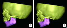
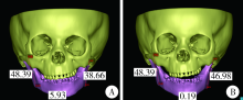
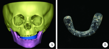
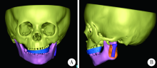
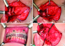
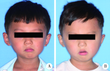
Application of computer-aided virtual mandibular position in the simultaneous treatment of children with temporomandibular joint ankylosis and jaw deformity
Shuo CHEN,Yang HE,Jin-gang AN,Yi ZHANG( )
)
- Department of Oral and Maxillofacial Surgery,Peking University School and Hospital of Stomatology & National Clinical Research Center for Oral Diseases & National Engineering Laboratory for Digital and Material Technology of Stomatology & Beijing Key Laboratory of Digital Stomatology, Beijing 100081, China
CLC Number:
- R782.6


| [1] | Zhang X, Chen M, Wu Y , et al. Management of temporomandi-bular joint ankylosis associated with mandibular asymmetry in infancy[J]. J Craniofac Surg, 2011,22(4):1316-1319. |
| [2] | Kaban LB, Perrott DH, Fisher K . A protocol for management of temporomandibular joint ankylosis[J]. J Oral Maxillofac Surg, 1990,48(11):1145-1151. |
| [3] | Kaban LB, Bouchard C, Troulis MJ . A protocol for management of temporomandibular joint ankylosis in children[J]. J Oral Maxillofac Surg, 2009,67(9):1966-1978. |
| [4] | Perrott DH, Umeda H, Kaban LB . Costochondral graft construction/reconstruction of the ramus/condyle unit: long-term follow-up[J]. Int J Oral Maxillofac Surg, 1994,23(6 Pt 1):321-328. |
| [5] | Padwa BL, Mulliken JB, Maghen A , et al. Midfacial growth after costochondral graft construction of the mandibular ramus in hemifacial microsomia[J]. J Oral Maxillofac Surg, 1998,56(2):122-127. |
| [6] | Zhu S, Li J, Luo E , et al. Two-stage treatment protocol for management of temporomandibular joint ankylosis with secondary deformities in adults: our institution’s experience[J]. J Oral Maxillofac Surg, 2011,69(12):e565-572. |
| [7] | Lu C, Huang D, He D , et al. Digital occlusal splint for condylar reconstruction in children with temporomandibular joint ankylosis[J]. J Oral Maxillofac Surg, 2014,72(8):1585-1593. |
| [8] | Sawhney CP . Bony ankylosis of the temporomandibular joint: follow-up of 70 patients treated with arthroplasty and acrylic spacer interposition[J]. Plast Reconstr Surg, 1986,77(1):29-40. |
| [9] | 孙力, 郭亚娟, 赵妍 , 等. 根据CT扫描数据及印模三维重建牙列数字模型精度的初步研究[J]. 口腔颌面修复学杂志, 2011,12(1):6-9. |
| [10] | Besl PJ, Mckay ND . A method for registration of 3-D shapes[J]. IEEE T Pattern Anal, 1992,14(2):239-256. |
| [11] | 赵一姣, 原福松, 谢晓艳 . 牙颌模型激光扫描数据与锥形束CT数据配准方法的精度比较[J]. 中华口腔医学杂志, 2013,48(3):173-176. |
| [12] | Gateno J, Xia JJ, Teichgraeber JF , et al. Clinical feasibility of computer-aided surgical simulation (CASS) in the treatment of complex cranio-maxillofacial deformities[J]. J Oral Maxillofac Surg, 2007,65(4):728-734. |
| [13] | Zhao J, He D, Yang C , et al. 3-D computed tomography measurement of mandibular growth after costochondral grafting in growing children with temporomandibular joint ankylosis and jaw deformity[J]. Oral Surg Oral Med Oral Pathol Oral Radiol, 2017,124(4):333-338. |
| [14] | Kaban LB, Padwa BL, Mulliken JB . Surgical correction of mandibular hypoplasia in hemifacial microsomia: the case for treatment in early childhood[J]. J Oral Maxillofac Surg, 1998,56(5):628-638. |
| [1] | Xinyu XU,Ling WU,Fengqi SONG,Zili LI,Yi ZHANG,Xiaojing LIU. Mandibular condyle localization in orthognathic surgery based on mandibular movement trajectory and its preliminary accuracy verification [J]. Journal of Peking University (Health Sciences), 2024, 56(1): 57-65. |
| [2] | Sui LI,Wenjie MA,Shimin WANG,Qian DING,Yao SUN,Lei ZHANG. Trueness of different digital design methods for incisal guidance of maxillary anterior implant-supported single crowns [J]. Journal of Peking University (Health Sciences), 2024, 56(1): 81-87. |
| [3] | FENG Sha-wei,GUO Hui,WANG Yong,ZHAO Yi-jiao,LIU He. Initial establishment of digital reference standardized crown models of the primary teeth [J]. Journal of Peking University (Health Sciences), 2022, 54(2): 327-334. |
| [4] | LI Yi,WONG Lai U,LIU Xiao-qiang,ZHOU Ti,LYU Ji-zhe,TAN Jian-guo. Marginal features of CAD/CAM laminate veneers with different materials and thicknesses [J]. Journal of Peking University (Health Sciences), 2022, 54(1): 140-145. |
| [5] | QIU Shu-ting,ZHU Yu-jia,WANG Shi-min,WANG Fei-long,YE Hong-qiang,ZHAO Yi-jiao,LIU Yun-song,WANG Yong,ZHOU Yong-sheng. Preliminary clinical application verification of complete digital workflow of design lips symmetry reference plane based on posed smile [J]. Journal of Peking University (Health Sciences), 2022, 54(1): 193-199. |
| [6] | XU Xiao-xiang,CAO Ye,ZHAO Yi-jiao,JIA Lu,XIE Qiu-fei. In vitro evaluation of the application of digital individual tooth tray in the impression making of mandibular full-arch crown abutments [J]. Journal of Peking University (Health Sciences), 2021, 53(1): 54-61. |
| [7] | YUE Zhao-guo,ZHANG Hai-dong,YANG Jing-wen,HOU Jian-xia. Comparison of residual cement between CAD/CAM customized abutments and stock abutments via digital measurement in vitro [J]. Journal of Peking University (Health Sciences), 2021, 53(1): 69-75. |
| [8] | LI Zheng,LIU Yu-shu,WANG Shi-min,ZHANG Rui,JIA Lu,YE Hong-qiang,HU Wen-jie,ZHAO Wen-yan,LIU Yun-song,ZHOU Yong-sheng. Application of biocopy function of temporary crown occlusal morphology in patients with severe attrition [J]. Journal of Peking University (Health Sciences), 2021, 53(1): 62-68. |
| [9] | FANG Shuo-bo,YANG Guang-ju,KANG Yan-feng,SUN Yu-chun,XIE Qiu-fei. Method and accuracy of determining the jaw position of repositioning splint with the aid of digital technique [J]. Journal of Peking University (Health Sciences), 2021, 53(1): 76-82. |
| [10] | Lang YOU,Ke-hui DENG,Wei-wei LI,Yi-jiao ZHAO,Yu-chun SUN,Yong-sheng ZHOU. Visual sensitivity threshold of lateral view of nasolabial Angle changes in edentulous jaw patients [J]. Journal of Peking University(Health Sciences), 2020, 52(1): 107-112. |
| [11] | Kuan-paul WANG,Hong-qiang YE,Hu CHEN,Yong WANG,Yu-chun SUN,Yong-sheng ZHOU. Establishment and preliminary clinical evaluation of edentulous custom trays designed and fabricated by chair-side CAD and 3D printing systems [J]. Journal of Peking University(Health Sciences), 2019, 51(2): 349-355. |
| [12] | Xin-xin LI,Yu-shu LIU,Yu-chun SUN,Hu CHEN,Hong-qiang YE,Yong-sheng ZHOU. Evaluation of one-piece polyetheretherketone removable partial denture fabricated by computer-aided design and computer-aided manufacturing [J]. Journal of Peking University(Health Sciences), 2019, 51(2): 335-339. |
| [13] | Shi-min WANG,Zheng LI,Guan-bo WANG,Hong-qiang YE,Yun-song LIU,Dai TONG,Wen-hui GAO,Yong-sheng ZHOU. Preliminary clinical application of complete digital workflow of design and manufacturing occlusal splint for sleep bruxism [J]. Journal of Peking University(Health Sciences), 2019, 51(1): 105-110. |
| [14] | PENG Li, WANG Zu-hua, SUN Yu-chun, QU Wei,HAN Yang, LIANG Yu-hong. Computer aided design and three-dimensional printing forapicoectomy guide template [J]. Journal of Peking University(Health Sciences), 2018, 50(5): 905-910. |
| [15] | CHAI Jin-you, LIU Jian-zhang, WANG Bing, QU Jian, SUN Zhen, GAO Wen-hui, GUO Tian-hao, FENG Hai-lan, PAN Shao-xia. Evaluation of the fabrication deviation of a kind of milling digital implant surgical guides#br# [J]. Journal of Peking University(Health Sciences), 2018, 50(5): 892-898. |
|
||