Journal of Peking University (Health Sciences) ›› 2022, Vol. 54 ›› Issue (1): 54-61. doi: 10.19723/j.issn.1671-167X.2022.01.009

Previous Articles     Next Articles

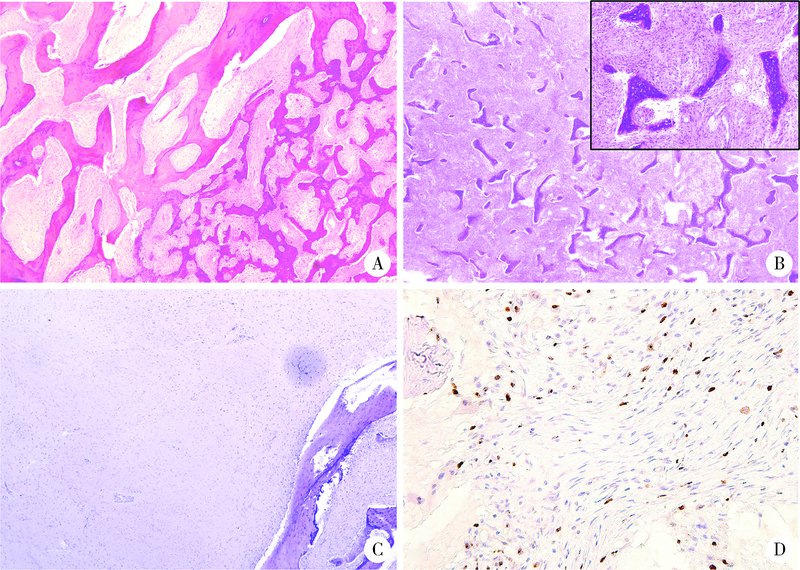
Clinicopathological analysis of 105 patients with fibrous dysplasia of cranio-maxillofacial region

XUE Jiang1,ZHANG Jian-yun1,SHI Rui-rui2,XIE Xiao-yan3,BAI Jia-ying1,LI Tie-jun1,()   

  1. 1. Department of Oral Pathology, Peking University School and Hospital of Stomatology, Beijing 100081, China
    2. Central Laboratory, Peking University School and Hospital of Stomatology, Beijing 100081, China
    3. Department of Oral and Maxillofacial Radiology, Peking University School and Hospital of Stomatology & National Center of Stomatology & National Clinical Research Center for Oral Diseases & National Engineering Laboratory for Digital and Material Technology of Stomatology & Beijing Key Laboratory of Digital Stomatology, Beijing 100081, China
  • Received:2021-09-03 Online:2022-02-18 Published:2022-02-21
  • Contact: Tie-jun LI E-mail:litiejun22@vip.sina.com
  • Supported by:
    Research Unit of Precision Pathologic Diagnosis in Tumors of the Oral and Maxillofacial Regions, Chinese Academy of Medical Sciences(2019RU034);Education and Teaching Research Project Fund of Peking University School of Stomatology(YS030120)

Abstract:

Objective: To compare the clinicopathologic features and prognosis of the different types of fibrous dysplasia (FD) of cranio-maxillofacial region, so as to provide a new reference for clinicians to treat these patients and make prognostic judgement. Methods: Clinical records, radiographic data and pathological information of 105 patients diagnosed with FD or McCune-Albright syndrome (MAS) at the Department of Oral Pathology, Peking University Hospital of Stomatology from January 2013 to December 2020 were collected. The patients were divided into 4 groups: monostotic FDs, polyostotic FDs, MAS and a specific type called craniofacial fibrous dysplasia (CFD) limited in the craniofacial region. The clinicopathological characteristics, treatment and follow-up data of each type were analyzed. Results: Of all the 105 patients, 46 were males and 59 were females, with a male-to-female ratio of 1 ∶1.3. The onset age ranged from 0 to 56 years and the median age was 12 years. On the basis of different involvement conditions, 4 types were divided. The most common type was monostotic FDs (43 cases, 40.95%), including maxilla (29 cases), mandibular (12 cases) and zygoma (2 cases). 32 cases (30.48%) were diagnosed with polyostotic FDs, 7 cases (6.67%) were MAS, and 23 cases (21.90%) were CFDs confirmed by computed tomography (CT) analysis. CFD was clearly distinct from other types of FD, such as the patient gender and the serum alkaline phosphatase level in peripheral blood before operative surgery. The pathologic findings of various types FD were quite similar, whilst the predominant fibrous tissue hyperplasia could be observed in polyostotic FDs and MAS types. Conclusion: The clinicopathologic features of FD in the cranio-maxillofacial region are different from the FD lesions in other parts of the body. The clinicopathological features of CFD are significantly different from those of monostotic and polyostotic FDs in the cranio-maxillofacial region. Therefore, the clinicians should pay attention to distinguish CFD in clinic, imaging and pathology aspects, so as to further clarify its features in clinic management and prognosis.

Key words: Fibrous dysplasia of bone, Facial bones, Pathology, clinical, Alkaline phosphatase

CLC Number: 

  • R739.8

Figure 1

A case of McCune-Albright syndrome A and B, a typical Café-au-lait skin pigmentation on the back of the neck (A), and lower back (B) of a 37-year-old woman with McCune-Albright syndrome which demonstrates jagged “coast of Maine” borders; C-E, CT images demonstrate a ground-glass ill-defined pattern in the cranio-maxillofacial region; F, bilateral clavicle and scapula are involved with chest deformity; G and H, bilateral tibia and femur are involved with bone deformity."

Table 1

Clinical characteristics and follow-up data of the different types of FD patients"

Items MFD (n=43) PFD (n=32) MAS (n=7) CFD (n=23) Total (n=105)
Onset age/years 15.00 (13.00, 30.00) 9.50 (5.50, 12.75) 8.57±2.37 10.00 (6.00, 14.00) 12.00 (8.00, 17.00)
Onset age group/years
≤10 7 (16.28) 21 (65.63) 5 (71.43) 13 (56.52) 46 (43.81)
11-20 23 (53.49) 7 (21.88) 2 (28.57) 7 (30.43) 39 (37.14)
21-30 4 (9.30) 2 (6.25) 0 (0) 2 (8.70) 8 (7.62)
31-40 6 (13.95) 1 (3.13) 0 (0) 1 (4.35) 8 (7.62)
41-50 1 (2.32) 1 (3.13) 0 (0) 0 (0) 2 (1.90)
51-60 2 (4.65) 0 (0) 0 (0) 0 (0) 2 (1.90)
Gender
Male 20 (46.51) 12 (37.50) 1 (14.29) 13 (56.52) 46 (43.81)
Female 23 (53.49) 20 (62.50) 6 (85.71) 10 (43.48) 59 (56.19)
Clinical sign
Facial deformity 39 (90.70) 32 (100.00) 7 (100.00) 23 (100.00) 101 (96.19)
Pain 8 (18.60) 2 (6.25) 1 (14.29) 1 (4.35) 12 (11.43)
Treatment
Conservative surgery 41 (95.35) 30 (93.75) 6 (85.71) 21 (91.30) 98 (93.33)
Radical surgery with reconstruction 2 (4.65) 2 (6.25) 1 (14.29) 2 (8.70) 7 (6.67)
Number of conservative operations 1.0 (1.0, 1.5) 2.0 (1.0, 3.0) 2.0 (1.0, 3.5) 1.0 (1.0, 1.5) 1.0 (1.0, 2.0)
Course 8.78±7.86 17.09±10.27 18.86±9.56 10.39±6.77 12.34±9.34
Serum ALP concentration
Elevation 5 (11.63) 22 (68.75) 6 (85.71) 11 (47.83) 44 (41.90)
Non-elevation 36 (83.72) 10 (31.25) 1 (14.29) 11 (47.83) 58 (55.24)
NA 2 (4.65) 0 (0) 0 (0) 1 (4.35) 3 (2.86)
Follow-up
No regrowth 28 (65.12) 18 (56.25) 3 (42.86) 16 (69.57) 65 (61.90)
Slow regrowth 3 (6.98) 3 (9.38) 1 (14.29) 1 (4.35) 8 (7.62)
Marked regrowth 2 (4.65) 3 (9.38) 0 (0) 2 (8.70) 7 (6.67)
Death 0 (0) 0 (0) 1 (14.29) 0 (0) 1 (0.95)
Loss to follow-up 10 (23.26) 8 (25.00) 2 (28.57) 4 (17.39) 24 (22.86)

Figure 2

The age, gender and clinical characteristics of the different types of FD patients A, the differential onset age distribution of four types of FD; B, CFD was more present in men than in women; C, the median onset age of CFD was significantly lower than MFD; D, the proportion of CFD patients with elevated serum ALP before operation was significantly higher than MFD; E, the average course of CFD was significantly shorter than PFD; F, the median number of operations of CFD was significantly less than PFD. * P<0.05, ** P<0.01, *** P<0.001. Abbreviations as in Table 1."

Figure 3

Radiologic characteristics of FD Single bone was involved in monostotic fibrous dysplasia in A (left mandible) and C (right maxilla), and clear zygomaticomaxillary suture was shown in C and D (white arrow). Multiple bones were involved in B (right mandible and left maxilla). The right maxilla and zygoma were involved crossing the zygomaticomaxillary suture (white arrow) in CFD (D, case 5 in Table 2), while the bone sutures were indistinct in E (case 20 in Table 2). FD, fibrous dysplasia; CFD, craniofacial fibrous dysplasia."

Table 2

Clinical characteristics and follow-up data of CFD patients"

No. Gender Last treatment Onset age/
years
Number of the
operation
Age at
operation/
years
Age at
follow-up/
years
Bone involved Current status
1 Male Conservative surgery 11 1 19 20 Left zygoma and left sphenoid bone Loss to follow-up
2 Female Conservative surgery 6 1 16 17 Left maxilla, left ethmoid bone, left nasal bone, left inferior nasal concha, left temporal bone, left parietal bone, bilateral frontal bone and bilateral sphenoid bone No regrowth
3 Female Conservative surgery 23 1 26 28 Left sphenoid bone, left ethmoid bone, left nasal bone, left temporal bone, bilateral frontal bone No regrowth
4 Male Conservative surgery 11 1 17 20 Bilateral occipital bone, bilateral frontal bone, right temporal bone, right maxilla, right sphenoid bone, right zygoma, right ethmoid bone Marked regrowth
5 Female Conservative surgery 10 2* 10, 17 20 Right maxilla, right sphenoid bone, right zygoma, right temporal bone No regrowth
6 Female Extended resection with
flap reconstruction
6 2& 9, 10 13 Right maxilla, right sphenoid bone Loss to follow-up
7 Female Conservative surgery 16 2* 16, 31 33 Left maxilla, left sphenoid bone No regrowth
8 Female Conservative surgery 11 1 18 21 Left maxilla, left sphenoid bone, left zygoma, left ethmoid bone, left occipital bone, left frontal bone No regrowth
9 Female Conservative surgery 35 1 35 38 Left maxilla, left sphenoid bone, left zygoma, left ethmoid bone, left occipital bone, left frontal bone No regrowth
10 Female Conservative surgery 10 1 25 28 Right maxilla, right ethmoid bone, right zygoma Loss to follow-up
11 Male Conservative surgery 6 1 7 11 Right maxilla, right sphenoid bone, right temporal bone No regrowth
12 Male Conservative surgery 13 1 43 47 Right maxilla, right zygoma No regrowth
13 Male Conservative surgery 9 1 19 23 Left maxilla, left sphenoid bone, left zygoma Slow regrowth
14 Male Conservative surgery 15 1 21 25 Right maxilla, right sphenoid bone No regrowth
15 Female Conservative surgery 9 1 15 18 Left maxilla, left sphenoid bone, left zygoma No regrowth
16 Male Conservative surgery 14 1 21 25 Left maxilla, left sphenoid bone, left temporal bone No regrowth
17 Male Conservative surgery 4 2* 14, 18 22 Right maxilla, right sphenoid bone, right zygoma Loss to follow-up
18 Male Conservative surgery 6 2* 12, 23 27 Left maxilla, left sphenoid bone, left temporal bone Marked regrowth
19 Male Conservative surgery 7 2* 7, 18 22 Right maxilla, right sphenoid bone, right zygoma, right temporal bone, right ethmoid bone No regrowth
20 Male Extended resection with
flap reconstruction
22 4# 22, 25,
40, 47
53 Left zygoma, bilateral maxilla No regrowth
21 Male Conservative surgery 9 1 22 27 Left maxilla, left sphenoid bone, left zygoma No regrowth
22 Female Conservative surgery 8 1 22 28 Left maxilla, left zygoma No regrowth
23 Male Conservative surgery 4 1 19 27 Right maxilla, right sphenoid bone, right zygoma, right temporal bone No regrowth

Figure 4

Histopathologic characteristics of FD A, the lesion is ill-defined in continuity with normal bone (HE ×40); B, typical histological features of FD show thin, irregular, and disconnected trabeculae and fibrous tissue replaces the normal bone structures (HE ×40, HE ×400 in the upper right box); C, the predominant of fibrous tissue (HE ×40); D, the low expression of Ki-67 in FD lesions (HE ×400). FD, fibrous dysplasia."

[1] Dumitrescu CE, Collins MT. McCune-Albright syndrome[J]. Orphanet J Rare Dis, 2008, 3:12.
doi: 10.1186/1750-1172-3-12 pmid: 18489744
[2] 叶为民, 竺涵光, 郑家伟, 等. 46例颌面部骨纤维异常增殖症临床分析[J]. 中国口腔颌面外科杂志, 2008, 6(3):170-173.
[3] El-Naggar AK, Chan JK, Grandis JR, et al. WHO classification of head and neck tumours [M]. Lyon: IARC Press, 2017: 253-254.
[4] 张壁, 韩其滨, 赵吉宏, 等. 30例颌面部骨纤维异常增殖症诊治的临床分析[J]. 口腔医学研究, 2010, 26(5):713-715.
[5] Sweeney K, Kaban LB. Natural history and progression of craniofacial fibrous dysplasia: A retrospective evaluation of 114 patients from Massachusetts General Hospital[J]. J Oral Maxillofac Surg, 2020, 78(11):1966-1980.
doi: 10.1016/j.joms.2020.05.036
[6] Cheng J, Wang Y, Yu H, et al. An epidemiological and clinical analysis of craniomaxillofacial fibrous dysplasia in a Chinese population[J]. Orphanet J Rare Dis, 2012, 7:80.
doi: 10.1186/1750-1172-7-80 pmid: 23074969
[7] Javaid MK, Boyce A, Appelman-Dijkstra N, et al. Best practice management guidelines for fibrous dysplasia/McCune-Albright syndrome: A consensus statement from the FD/MAS international consortium[J]. Orphanet J Rare Dis, 2019, 14(1):139.
doi: 10.1186/s13023-019-1102-9
[8] World Health Organization. WHO classification of tumours of soft tissue and bone [M]. 5th ed. Lyon: IARC Press, 2020: 472-474.
[9] Ma J, Liang L, Gu B, et al. A retrospective study on craniofacial fibrous dysplasia: Preoperative serum alkaline phosphatase as a prognostic marker?[J]. J Craniomaxillofac Surg, 2013, 41(7):644-647.
doi: 10.1016/j.jcms.2012.12.007
[10] Chen YR, Wong FH, Hsueh C, et al. Computed tomography characteristics of non-syndromic craniofacial fibrous dysplasia[J]. Chang Gung Med J, 2002, 25(1):1-8.
[11] Burke AB, Collins MT, Boyce AM. Fibrous dysplasia of bone: Craniofacial and dental implications[J]. Oral Dis, 2017, 23(6):697-708.
doi: 10.1111/odi.12563 pmid: 27493082
[12] Akintoye SO, Lee JS, Feimster T, et al. Dental characteristics of fibrous dysplasia and McCune-Albright syndrome[J]. Oral Surg Oral Med Oral Pathol Oral Radiol Endod, 2003, 96(3):275-282.
doi: 10.1016/S1079-2104(03)00225-7
[13] Waldron CA. Fibro-osseous lesions of the jaws[J]. J Oral Maxillofac Surg, 1993, 51(8):828-835.
doi: 10.1016/S0278-2391(10)80097-7
[14] Gupta D, Garg P, Mittal A. Computed tomography in craniofacial fibrous dysplasia: A case series with review of literature and classification update[J]. Open Dent J, 2017, 11:384-403.
doi: 10.2174/1874210601711010384
[15] 李铁军. 口腔组织学与病理学 [M]. 3版. 北京: 北京大学医学出版社, 2020: 360-361.
[16] Slootweg PJ. Maxillofacial fibro-osseous lesions: Classification and differential diagnosis[J]. Semin Diagn Pathol, 1996, 13(2):104-112.
pmid: 8734416
[17] Riminucci M, Liu B, Corsi A, et al. The histopathology of fibrous dysplasia of bone in patients with activating mutations of the Gs alpha gene: Site-specific patterns and recurrent histological hallmarks[J]. J Pathol, 1999, 187(2):249-258.
pmid: 10365102
[18] Sissons HA, Steiner GC, Dorfman HD. Calcified spherules in fibro-osseous lesions of bone[J]. Arch Pathol Lab Med, 1993, 117(3):284-290.
pmid: 8442673
[19] Dorfman HD. New knowledge of fibro-osseous lesions of bone[J]. Int J Surg Pathol, 2010, 18(3 Suppl):62S-65S.
[20] Sargolzaei S, Ghelejkhani A, Baghban AA. Diagnostic and bio-logical significance of immunohistochemical expression of osteopontin and Ki67 in fibro-osseous lesions of jaws[J]. J Islam Dent Assoc IRAN, 2017, 29(2):70-78.
doi: 10.30699/jidai.29.2.70
[21] Shi RR, Li XF, Zhang R, et al. GNAS mutational analysis in differentiating fibrous dysplasia and ossifying fibroma of the jaw[J]. Mod Pathol, 2013, 26(8):1023-1031.
doi: 10.1038/modpathol.2013.31
[22] Li Z, Raynald, Wang Z, et al. Malignant transformation of craniofacial fibrous dysplasia: A systematic review of overall survival[J]. Neurosurg Rev, 2020, 43(3):911-921.
doi: 10.1007/s10143-019-01089-1
[1] Hui WEI, Luo-zeng, Ci-dan-yang-zong, Bai-ma-yang-jin. Analysis of clinical characteristics of Henoch-Schonlein purpura patients from different altitudes in plateau areas [J]. Journal of Peking University (Health Sciences), 2021, 53(6): 1072-1077.
[2] Zhi-bo SONG,Yan GENG,Xue-rong DENG,Xiao-hui ZHANG,Zhuo-li ZHANG. Benefit of ultrasound in the phenotype recognition of psoriatic arthritis [J]. Journal of Peking University (Health Sciences), 2021, 53(6): 1061-1066.
[3] Mei-ge LIU,Pu FANG,Yan WANG,Lu CONG,Yang-yi FAN,Yuan YUAN,Yan XU,Jun ZHANG,Dao-jun HONG. Clinical, pathological and genetic characteristics of 8 patients with distal hereditary motor neuropathy [J]. Journal of Peking University (Health Sciences), 2021, 53(5): 957-963.
[4] XIA Fang-fang,LU Fu-ai,LV Hui-min,YANG Guo-an,LIU Yuan. Clinical characteristics and related factors of systemic lupus erythematosus with interstitial pneumonia [J]. Journal of Peking University (Health Sciences), 2021, 53(2): 266-272.
[5] GAO Yang-xu,SUN Qing,LI Hui,XIE Yao,YAO Hong-xin,ZHAO Wei-hong. Therapeutic effect and clinical cost of multi-disciplinary team model of hepatoblastoma in children [J]. Journal of Peking University (Health Sciences), 2021, 53(1): 200-203.
[6] ZHANG Sheng-nan,AN Na,OUYANG Xiang-ying,LIU Ying-jun,WANG Xue-kui. Role of growth arrest-specific protein 6 in migration and osteogenic differentiation of human periodontal ligament cells [J]. Journal of Peking University (Health Sciences), 2021, 53(1): 9-15.
[7] Yu-zhou GAN,Yu-hui LI,Li-hua ZHANG,Lin MA,Wen-wen HE,Yue-bo JIN,Yuan AN,Zhan-guo LI,Hua YE. Comparison of clinical and immunological features between clinically amyopathic dermatomyositis and typical dermatomyositis [J]. Journal of Peking University (Health Sciences), 2020, 52(6): 1001-1008.
[8] Shi-xiong WEI,Shu-jia LI,Yi LIU. Clinical characteristics and biological treatment of adult patient with juvenile idiopathic arthritis [J]. Journal of Peking University (Health Sciences), 2020, 52(6): 1014-1022.
[9] Gong CHENG,Xia ZHANG,Fei YANG,Jia-yu CHENG,Yan-ying LIU. Angioimmunoblastic T-cell lymphoma with fever, arthritis and skin pigmentation: A case report [J]. Journal of Peking University (Health Sciences), 2020, 52(6): 1150-1152.
[10] Zhi-feng XU,Yun-peng LING,Zhong-qi CUI,hong ZHAO,Yi-chen GONG,Yuan-hao FU,Hang YANG,Feng WAN. Feasibility and safety of minimally invasive cardiac coronary artery bypass grafting surgery for patients with multivessel coronary artery disease: Early outcome and short-mid-term follow up results [J]. Journal of Peking University (Health Sciences), 2020, 52(5): 863-869.
[11] Yi LIU,Zhi-jian LIU,Qi SHEN,Jing-yun WU,Yu FAN,De-run LI,Wei YU,Zhi-song HE. A clinical analysis of 14 cases of prostatic stromal tumor of uncertain malignant potential [J]. Journal of Peking University (Health Sciences), 2020, 52(4): 621-624.
[12] Yi-chang HAO,Ye YAN,Fan ZHANG,Min QIU,Lang ZHOU,Ke LIU,Jian LU,Chun-lei XIAO,Yi HUANG,Cheng LIU,Lu-lin MA. Surgical strategy selection and experience summary of prostate cancer with positive single needle biopsy [J]. Journal of Peking University (Health Sciences), 2020, 52(4): 625-631.
[13] Ling WEI,Dong ZOU,Hu CHEN,Shao-xia PAN,Yu-chun SUN,Yong-sheng ZHOU. Evaluation of clinical efficacy of a kind of digital complete denture [J]. Journal of Peking University (Health Sciences), 2020, 52(4): 762-770.
[14] Hang-ci ZHENG,Xiao-tong LI,Peng MEN,Xiang MA,Qiang WANG,Yao-long CHEN,Suo-di ZHAI. Selecting and defining the clinical questions and outcomes of Guideline for the Emergency Treatment of Anaphylaxis [J]. Journal of Peking University (Health Sciences), 2020, 52(4): 715-718.
[15] Ru MA,Xin-bao LI,Feng-cai YAN,Yu-lin LIN,Yan LI. Clinical evaluation of tumor-stroma ratio in pseudomyxoma peritonei from the appendix [J]. Journal of Peking University (Health Sciences), 2020, 52(2): 240-246.
Viewed
Full text


Abstract

Cited

  Shared   
  Discussed   
[1] . [J]. Journal of Peking University(Health Sciences), 2009, 41(4): 456 -458 .
[2] . [J]. Journal of Peking University(Health Sciences), 2009, 41(2): 125 -128 .
[3] . [J]. Journal of Peking University(Health Sciences), 2009, 41(1): 52 -55 .
[4] . [J]. Journal of Peking University(Health Sciences), 2009, 41(5): 599 -601 .
[5] . [J]. Journal of Peking University(Health Sciences), 2009, 41(5): 516 -520 .
[6] . [J]. Journal of Peking University(Health Sciences), 2007, 39(3): 310 -314 .
[7] . [J]. Journal of Peking University(Health Sciences), 2007, 39(3): 12 .
[8] . [J]. Journal of Peking University(Health Sciences), 2007, 39(4): 342 .
[9] . [J]. Journal of Peking University(Health Sciences), 2007, 39(4): 365 -368 .
[10] . [J]. Journal of Peking University(Health Sciences), 2007, 39(4): 388 -393 .