Journal of Peking University (Health Sciences) ›› 2025, Vol. 57 ›› Issue (5): 947-955. doi: 10.19723/j.issn.1671-167X.2025.05.020

Previous Articles     Next Articles

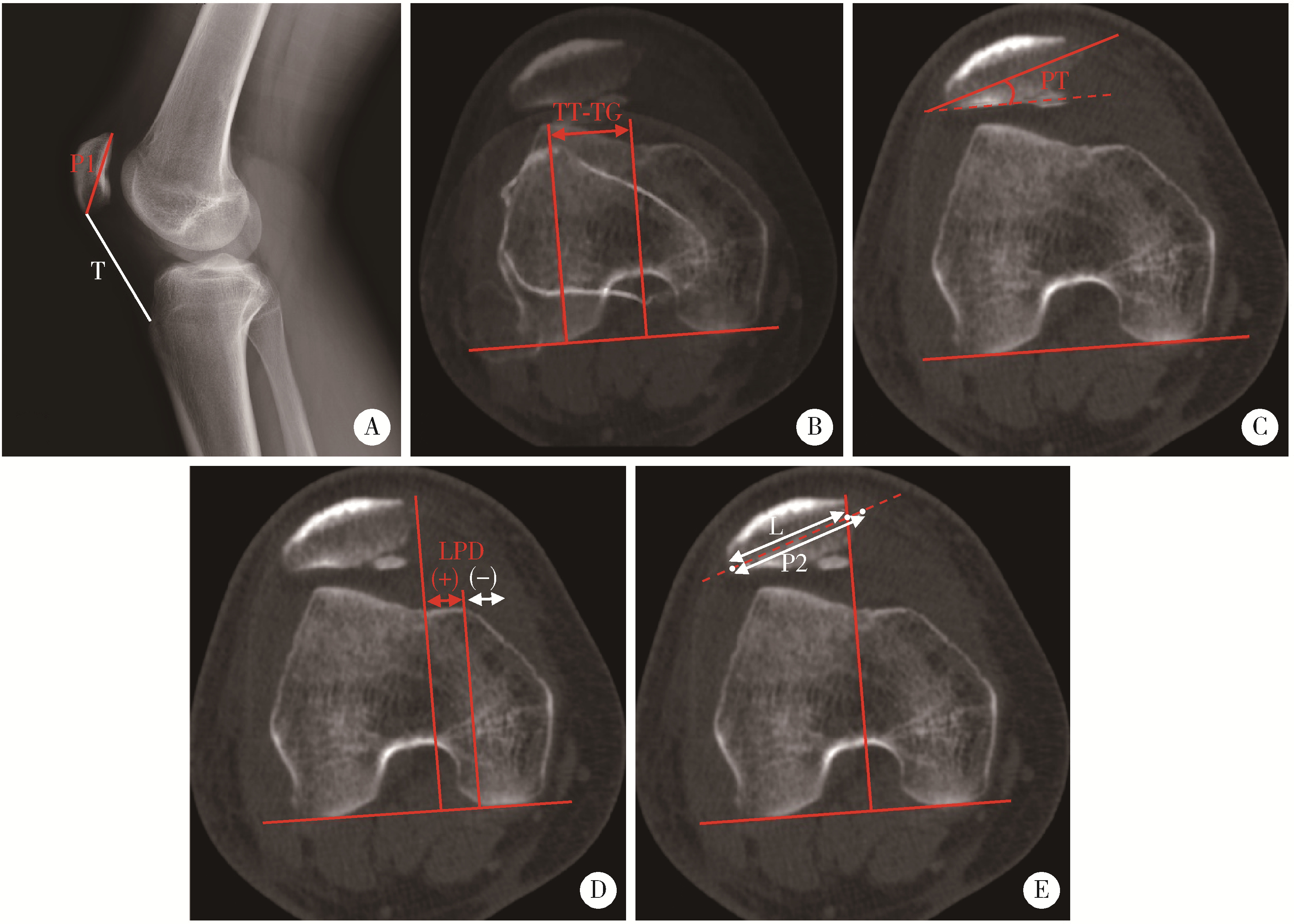
Clinical efficacy of arthroscopic medial patellofemoral complex reconstruction for recurrent patellar dislocation with high-grade trochlear dysplasia

Fengyi HU, Qingyang MENG, Nayun CHEN, Jianing WANG, Zhenlong LIU, Yong MA, Yuping YANG, Xi GONG, Cheng WANG, Ping LIU*(), Weili SHI*()   

  1. Department of Sports Medicine, Peking University Third Hospital; Institute of Sports Medicine of Peking University; Beijing Key Laboratory of Sports Injuries, Beijing 100191, China
  • Received:2023-02-06 Online:2025-10-18 Published:2024-05-07
  • Contact: Ping LIU, Weili SHI
  • Supported by:
    Peking University Third Hospital Clinical Key Program(BYSYZD2022011); Interdisciplinary Cooperation Project of Beijing Nova Program of Beijing Municipal Science & Technology Commission(Z211100002121015)

RICH HTML


Abstract: Objective: To investigate the midterm clinical efficacy of medial patellofemoral complex (MPFC) reconstruction for recurrent patellar dislocation with high-grade trochlear dysplasia. Methods: A retrospective analysis was carried out among adult patients who underwent arthroscopically assisted MPFC reconstruction between January 2014 and December 2020. Dejour classification was evaluated to grade trochlear dysplasia; tibial tubercle-trochlear groove (TT-TG) distance and Insall-Salvati index were measured. Preoperative and postoperative patient-reported outcome measures (PROMs) were compared, including International Knee Documentation Committee (IKDC) score, Kujala score, Lysholm score and Tegner score. Information regarding returning-to-sport rate, re-instability events and complications was collected. Patellar tilt (PT), lateral patellar displacement (LPD) and bisect offset (BSO) ratio were measured based on axial computed tomography before and after surgery to assess the patellofemoral congruence. Results: A total of 46 MPFC reconstructions in 43 patients were enrolled, including 16 male and 27 female. Mean age at surgery was (22.2±7.6) years (range: 14-44 years). Mean follow-up was (49.9±22.6) months (range: 18-102 months). The percentages of Dejour B, C and D dysplasia were 37.0% (17/46), 43.5% (20/46), and 19.6% (9/46), respectively. Mean Insall-Salvati index was 1.2±0.2 (range: 0.85-1.44), and mean TT-TG distance was (19.6±3.5) mm (range: 10.6-28.7 mm). At latest follow-up, there were significant improvements in all PROMs (P < 0.001): IKDC score, from 56.3±15.1 to 86.2±8.1; Kujala score, from 58.9±15.6 to 92.6±5.4; Lysholm score, from 63.7±15.0 to 94.0±5.7; Tegner score, from 3.1±1.4 to 4.7±1.4, and there were no significant differences in the improvements of the scores between the patients with Dejour B, C and D dysplasia. Overall, ninety percent of the patients returned to their preoperative sports level. One patient reported a postoperative subluxation, while no cases of infection, limited range of motion or patella fracture were observed. PT, LPD and BSO ratio were all significant altered (P < 0.001) after MPFC reconstruction. Conclusion: Arthroscopically assisted MPFC reconstruction yielded satisfactory midterm clinical results for recurrent patellar dislocation with high-grade trochlear dysplasia. No significant differences of improvements in knee function were observed among the three types of high-grade trochlear dysplasia.

Key words: Patellar dislocation, Recurrence, Medial patellofemoral complex reconstruction, Femoral trochlear dysplasia, Dejour classification

CLC Number: 

  • R684.76

Figure 1

Arthroscopically assisted medial patellofemoral complex reconstruction A, a patellar tunnel was made at the upper third point of the medial patella with a 4.5 mm drill; B, guide sutures were introduced through the patellar tunnel with an eyelet pin; C, a quadricep-tendon tunnel was created at the midpoint of the superior patella using a blunt cannula; D, guide sutures were pulled out through the medial femoral incision with a grasper; E, the autograft was introduced into the patellar tunnel (arrow 1) and the quadricep-tendon tunnel (arrow 2) with guide sutures; F, intraarticular view of the double-bundle (arrow 3 and 4) reconstructed graft."

Figure 2

Examples of 4-grade Dejour classification based on axial CT A, a 38-year-old male was diagnosed with Dejour A trochlear dysplasia, whose CT scan indicated a trochlear groove angle of >145 degree and symmetric trochlear facets; B, a 21-year-old male was diagnosed with Dejour B trochlear dysplasia, whose CT scan indicated a flat trochlea (arrow); C, a 16-year-old female was diagnosed with Dejour C trochlear dysplasia, whose CT scan indicated asymmetric trochlear facets and hypoplastic medial facet (arrow); D, a 17-year-old female was diagnosed with Dejour D trochlear dysplasia, whose CT scan indicated a positive cliff pattern (arrow). CT, computed tomography."

Figure 3

Measurements of radiographic parameters on lateral plain radiographs and axial CT A, Insall-Salvati index is the ratio of the patellar tendon length (T) to the patellar height (P1, distance from the superior pole to the inferior pole) on lateral plain radiographs; B, TT-TG distance is calculated by the transverse length between the trochlear groove and the tibial tuberosity on axial images; C, PT is the angle between the transverse axis of the patella and the posterior femoral condylar line; D, LPD describes the distance between the medial margin of the patella and the medial trochlear margin measured by lines perpendicular to the posterior femoral condylar line; E, bisect offset is determined by the ratio at which the patella width was bisected from its lateral aspect to the transverse width of the patella (L/P2). CT, computed tomography; LPD, lateral patellar displacement; PT, patellar tilt; TT-TG, tibial tubercle-trochlear groove."

Table 1

Anatomical risk factors and chondral lesion"

Parameters Data
Dejour classification
  Type B 17 (37.0)
  Type C 20 (43.5)
  Type D 9 (19.6)
Patellar height (Insall-Salvati index) 1.2±0.2
   < 1.2 21 (45.7)
  ≥ 1.2 25 (54.3)
TT-TG distance 19.6±3.5
   < 20 mm 24 (52.2)
  ≥ 20 mm 22 (47.8)
Patellar tilt/(°) 30.9±10.5
Chondral lesion
  Patella > ICRS Ⅱ 23 (50.0)
  Trochlea > ICRS Ⅱ 1 (2.2)
  Lateral femoral condyle > ICRS Ⅱ 8 (17.3)

Table 2

Comparison of anatomical risk factors among Dejour type B, C and D dysplasia"

Parameters Type B (n=17) Type C (n=20) Type D (n=9) F P
Insall-Salvati index 1.1±0.2 1.2±0.2 1.3±0.1 2.641 0.083
TT-TG distance/mm 19.0±3.3 19.4±3.9 21.2±2.5 1.232 0.302
Patellar tilt/(°) 30.2±9.1 31.2±12.7 31.9±8.7 0.066 0.936

Table 3

Comparison of patient-reported outcome measures before and after surgery"

Outcome measures Preoperative score Postoperative score Z P
IKDC score 56.3±15.1 86.2±8.1 -5.896 < 0.001
Kujala score 58.9±15.6 92.6±5.4 -5.906 < 0.001
Lysholm score 63.7±15.0 94.0±5.7 -5.906 < 0.001
Tegner score 3.1±1.4 4.7±1.4 -4.867 < 0.001
Subjective satisfaction 9.1±1.0

Table 4

Comparison of improvements in patient-reported outcome measures among Dejour type B-D dysplasia"

Outcome measures Type B (n=17) Type C (n=20) Type D (n=9) F P
ΔIKDC score 27.2±17.7 29.1±15.2 37.0±13.7 1.173 0.319
ΔKujala score 27.5±12.8 30.8±13.9 34.4±13.5 1.378 0.263
ΔLysholm score 31.6±14.8 32.1±14.9 41.0±14.6 0.815 0.449
ΔTegner score 1.4±1.5 1.5±1.7 2.2±1.4 0.896 0.416

Table 5

Relationship between postoperative symptomatic knee pain and stages of chondral lesion"

Chondral lesion No lesion ICRS Ⅰ ICRS Ⅱ ICRS Ⅲ ICRS Ⅳ P
Patella 0.869
  Symptomatic knee pain (-) 3 2 13 9 8
  Symptomatic knee pain (+) 2 0 3 3 3
Trochlea facet 0.287
  Symptomatic knee pain (-) 12 2 21 0 0
  Symptomatic knee pain (+) 5 0 5 1 0
Lateral femoral condyle >0.995
  Symptomatic knee pain (-) 26 1 1 2 5
  Symptomatic knee pain (+) 10 0 0 0 1

Table 6

Comparison between preoperative and postoperative CT parameters of the patellofemoral joint"

Parameters Preoperative value Postoperative value Z P
Patellar tilt/(°) 30.9±10.5 9.8±6.0 -5.443 < 0.001
Lateral patellar displacement/mm 10.4±8.1 -8.7±7.1 -5.442 < 0.001
Bisect offset ratio 1.1±0.3 0.6±0.2 -5.442 < 0.001

Figure 4

Comparison of axial CT scans of the patellofemoral joint before and after MPFC reconstruction Patient 1 was a 17-year-old male diagnosed with recurrent patellar dislocation of the right knee. Preoperative CT scan showed significant patellar tilt and lateral displacement, which were both corrected on postoperative scan. Patient 2 was a 16-year-old male diagnosed with recurrent patellar dislocation of the right knee. Preoperative CT scan demonstrated a dislocated patella with remarkable tilt and lateral translation, while the patellofemoral congruence was restored on postoperative scan. CT, computed tomography; MPFC, medial patellofemoral complex reconstruction."

1
Sanders TL , Pareek A , Hewett TE , et al. Incidence of first-time lateral patellar dislocation: A 21-year population-based study[J]. Sports Health, 2018, 10 (2): 146- 151.

doi: 10.1177/1941738117725055
2
Liu JN , Steinhaus ME , Kalbian IL , et al. Patellar instability ma-nagement: A survey of the International Patellofemoral Study Group[J]. Am J Sports Med, 2018, 46 (13): 3299- 3306.

doi: 10.1177/0363546517732045
3
Nomura E , Inoue M , Osada N . Anatomical analysis of the medial patellofemoral ligament of the knee, especially the femoral attachment[J]. Knee Surg Sports Traumatol Arthrosc, 2005, 13 (7): 510- 515.

doi: 10.1007/s00167-004-0607-4
4
Fulkerson JP , Edgar C . Medial quadriceps tendon-femoral ligament: Surgical anatomy and reconstruction technique to prevent patella instability[J]. Arthrosc Tech, 2013, 2 (2): e125- e128.

doi: 10.1016/j.eats.2013.01.002
5
Tanaka MJ , Voss A , Fulkerson JP . The anatomic midpoint of the attachment of the medial patellofemoral complex[J]. J Bone Joint Surg Am, 2016, 98 (14): 1199- 1205.

doi: 10.2106/JBJS.15.01182
6
Huddleston HP , Campbell KJ , Madden BT , et al. The quadriceps insertion of the medial patellofemoral complex demonstrates the greatest anisometry through flexion[J]. Knee Surg Sports Traumatol Arthrosc, 2021, 29 (3): 757- 763.

doi: 10.1007/s00167-020-05999-2
7
Christian DR , Redondo ML , Cancienne JM , et al. Differential contributions of the quadriceps and patellar attachments of the proximal medial patellar restraints to resisting lateral patellar translation[J]. Arthroscopy, 2020, 36 (6): 1670- 1676.

doi: 10.1016/j.arthro.2020.01.058
8
印钰, 陈临新, 梅宇, 等. 内侧股四头肌腱股骨韧带重建联合单骨道内侧髌股韧带重建治疗复发性髌骨脱位的临床疗效[J]. 中国运动医学杂志, 2021, 40 (12): 936- 941.
9
Spang RC , Tepolt FA , Paschos NK , et al. Combined reconstruction of the medial patellofemoral ligament (MPFL) and medial quadriceps tendon-femoral ligament (MQTFL) for patellar instability in children and adolescents: Surgical technique and outcomes[J]. J Pediatr Orthop, 2019, 39 (1): e54- e61.

doi: 10.1097/BPO.0000000000001259
10
Temponi EF , Saithna A , Gonçalves MBJ , et al. Combined reconstruction of the medial patellofemoral ligament and medial quadriceps tendon-femoral ligament[J]. Arthrosc Tech, 2021, 10 (1): e193- e198.

doi: 10.1016/j.eats.2020.09.026
11
Espregueira-Mendes J , Andrade R , Bastos R , et al. Combined soft tissue reconstruction of the medial patellofemoral ligament and medial quadriceps tendon-femoral ligament[J]. Arthrosc Tech, 2019, 8 (5): e481- e488.

doi: 10.1016/j.eats.2019.01.006
12
Hiemstra LA , Kerslake S , Loewen M , et al. Effect of trochlear dysplasia on outcomes after isolated soft tissue stabilization for patellar instability[J]. Am J Sports Med, 2016, 44 (6): 1515- 1523.

doi: 10.1177/0363546516635626
13
Kita K , Tanaka Y , Toritsuka Y , et al. Factors affecting the outcomes of double-bundle medial patellofemoral ligament reconstruction for recurrent patellar dislocations evaluated by multivariate analysis[J]. Am J Sports Med, 2015, 43 (12): 2988- 2996.

doi: 10.1177/0363546515606102
14
Levy BJ , Tanaka MJ , Fulkerson JP . Current concepts regarding patellofemoral trochlear dysplasia[J]. Am J Sports Med, 2021, 49 (6): 1642- 1650.

doi: 10.1177/0363546520958423
15
Liu JN , Brady JM , Kalbian IL , et al. Clinical outcomes after isolated medial patellofemoral ligament reconstruction for patellar instability among patients with trochlear dysplasia[J]. Am J Sports Med, 2018, 46 (4): 883- 889.

doi: 10.1177/0363546517745625
16
Shi WL , Hu FY , Xu MT , et al. Arthroscopic anatomical double-bundle medial patellofemoral complex reconstruction improves clinical outcomes in treating recurrent patellar instability despite trochlear dysplasia, elevated tibial tubercle-trochlear groove distance and patellar alta[J]. Arthroscopy, 2023, 39 (1): 102- 111.

doi: 10.1016/j.arthro.2022.06.038
17
Dixit S , DiFiori JP , Burton M , et al. Management of patellofemoral pain syndrome[J]. Am Fam Physician, 2007, 75 (2): 194- 202.
18
Nacey NC , Fox MG , Luce BN , et al. Assessing femoral trochlear morphologic features on cross-sectional imaging before trochleoplasty: Dejour classification versus quantitative measurement[J]. AJR Am J Roentgenol, 2020, 215 (2): 458- 464.

doi: 10.2214/AJR.19.22400
19
Ye Q , Yu T , Wu Y , et al. Patellar instability: The reliability of magnetic resonance imaging measurement parameters[J]. BMC Musculoskelet Disord, 2019, 20 (1): 317.

doi: 10.1186/s12891-019-2697-7
20
Yanke AB , Huddleston HP , Campbell K , et al. Effect of patella Alta on the native anatomometricity of the medial patellofemoral complex: A cadaveric study[J]. Am J Sports Med, 2020, 48 (6): 1398- 1405.

doi: 10.1177/0363546520916435
21
Chahla J , Smigielski R , LaPrade RF , et al. An updated overview of the anatomy and function of the proximal medial patellar restraints (medial patellofemoral ligament and the medial quadriceps tendon femoral ligament)[J]. Sports Med Arthrosc Rev, 2019, 27 (4): 136- 142.

doi: 10.1097/JSA.0000000000000252
22
Tanaka MJ , Chahla J , Farr J 2nd , et al. Recognition of evolving medial patellofemoral anatomy provides insight for reconstruction[J]. Knee Surg Sports Traumatol Arthrosc, 2019, 27 (8): 2537- 2550.

doi: 10.1007/s00167-018-5266-y
23
Van Haver A , De Roo K , De Beule M , et al. The effect of trochlear dysplasia on patellofemoral biomechanics: A cadaveric study with simulated trochlear deformities[J]. Am J Sports Med, 2015, 43 (6): 1354- 1361.

doi: 10.1177/0363546515572143
24
Vinod AV , Hollenberg AM , Kluczynski MA , et al. Ability of medial patellofemoral ligament reconstruction to overcome lateral patellar motion in the presence of trochlear flattening: A cadaveric biomechanical study[J]. Am J Sports Med, 2021, 49 (13): 3569- 3574.

doi: 10.1177/03635465211041087
25
Sappey-Marinier E , Sonnery-Cottet B , O'Loughlin P , et al. Clinical outcomes and predictive factors for failure with isolated MPFL reconstruction for recurrent patellar instability: A series of 211 reconstructions with a minimum follow-up of 3 years[J]. Am J Sports Med, 2019, 47 (6): 1323- 1330.

doi: 10.1177/0363546519838405
26
Hopper GP , Leach WJ , Rooney BP , et al. Does degree of troch-lear dysplasia and position of femoral tunnel influence outcome after medial patellofemoral ligament reconstruction?[J]. Am J Sports Med, 2014, 42 (3): 716- 722.

doi: 10.1177/0363546513518413
27
Testa EA , Camathias C , Amsler F , et al. Surgical treatment of patellofemoral instability using trochleoplasty or MPFL reconstruction: A systematic review[J]. Knee Surg Sports Traumatol Arthrosc, 2017, 25 (8): 2309- 2320.

doi: 10.1007/s00167-015-3698-1
28
Qiao Y , Xu J , Ye Z , et al. Double-tunnel technique was similar to single-tunnel technique in clinical, imaging and functional outcomes for medial patellofemoral ligament reconstruction: A randomized clinical trial[J]. Arthroscopy, 2022, 38 (11): 3058- 3067.

doi: 10.1016/j.arthro.2022.04.019
29
Erickson BJ , Nguyen J , Gasik K , et al. Isolated medial patellofemoral ligament reconstruction for patellar instability regardless of tibial tubercle-trochlear groove distance and patellar height: Outcomes at 1 and 2 years[J]. Am J Sports Med, 2019, 47 (6): 1331- 1337.

doi: 10.1177/0363546519835800
[1] Yujing ZHU, Lei WANG, Chengyin LYU, Wenfeng TAN, Miaojia ZHANG. Analysis of clinical features of ruccrent interstitial lung disease in patients with anti-EJ positive antisynthetase syndrome [J]. Journal of Peking University (Health Sciences), 2024, 56(6): 980-986.
[2] Yukai LI, Hongyan WANG, Liang LUO, Yun LI, Chun LI. Clinical significance of antiphospholipid antibodies in Behcet disease with thrombosis [J]. Journal of Peking University (Health Sciences), 2024, 56(6): 1036-1040.
[3] Yuqing LI,Biao WANG,Peng QIAO,Wei WANG,Xing GUAN. Medium to long-term efficacy of tension-free vaginal tape procedure in the treatment of female recurrent stress urinary incontinence [J]. Journal of Peking University (Health Sciences), 2024, 56(4): 600-604.
[4] Chieko MORIMOTO,Yi-qin WANG,Rong ZHOU,Jian-liu WANG. Clinical analysis of fertility-sparing therapy of patients with complex atypical hyperplasia and endometrial cancer [J]. Journal of Peking University (Health Sciences), 2022, 54(5): 936-942.
[5] Yue WANG,Shuang ZHANG,Hong ZHANG,Li LIANG,Ling XU,Yuan-jia CHENG,Xue-ning DUAN,Yin-hua LIU,Ting LI. Clinicopathological features and prognosis of hormone receptor-positive/human epidermal growth factor receptor 2-negative breast cancer [J]. Journal of Peking University (Health Sciences), 2022, 54(5): 853-862.
[6] Su-hua CHEN,Jun YANG,Xin CHEN,Chen-long YANG,Jian-jun SUN,Guo-zhong LIN,Tao YU,Xin YANG,Yun-feng HAN,Chao WU,Yu SI,Kai-ming MA. Surgical treatment of large and giant recurrent meningiomas near the middle and posterior third part of the superior sagittal sinus with extracranial invading [J]. Journal of Peking University (Health Sciences), 2022, 54(5): 1006-1012.
[7] LIU Lei,QIN Yan-chun,WANG Guo-liang,ZHANG Shu-dong,HOU Xiao-fei,MA Lu-lin. Strategy of reoperation for pheochromocytoma and paraganglioma [J]. Journal of Peking University (Health Sciences), 2021, 53(4): 793-797.
[8] Huan-bin YU,Wen-jie WU,Xiao-ming LV,Yan SHI,Lei ZHENG,Jian-guo ZHANG. 125I seed brachytherapy for recurrent salivary gland carcinoma after external radiotherapy [J]. Journal of Peking University (Health Sciences), 2020, 52(5): 919-923.
[9] Xu-chu ZHANG,Jian-hua ZHANG,Rong-fu WANG,Yan FAN,Zhan-li FU,Ping YAN,Guang-yu ZHAO,Yan-xia BAI. Diagnostic value of 18F-FDG PET/CT and tumor markers (CEA, CA19-9, CA24-2) in recurrence and metastasis of postoperative colorectal moderately differentiated adenocarcinoma [J]. Journal of Peking University(Health Sciences), 2019, 51(6): 1071-1077.
[10] Qi TANG,Rong-cheng LIN,Lin YAO,Zheng ZHANG,Han HAO,Cui-jian ZHANG,Lin CAI,Xue-song LI,Zhi-song HE,Li-qun ZHOU. Clinicopathologic features and prognostic analyses of locally recurrent renal cell carcinoma patients after initial surgery [J]. Journal of Peking University(Health Sciences), 2019, 51(4): 628-631.
[11] XING Yun-chao, XIONG Geng-yan, FANG Dong, ZHANG Zheng, CAI Lin, ZHANG Lei, HE Zhi-song, LI Xue-song, ZHOU Li-qun. Preoperative prognostic factors and preoperative risk stratification of upper tract urothelial carcinoma [J]. Journal of Peking University(Health Sciences), 2016, 48(6): 1032-1037.
[12] LIU Yu-Qing, LU Jian, ZHAO Lei, HOU Xiao-Fei, MA Lu-Lin. Prognostic factors for intravesical recurrence after surgery for upper tract urothelial carcinoma in renal transplant recipients [J]. Journal of Peking University(Health Sciences), 2015, 47(4): 605-610.
[13] YANG Yang, XIAO Yun-Xiang, ZHOU Li-Qun, HE Zhi-Song, JIN Jie. Long-term outcome of isolated local recurrence following radical nephrectomy [J]. Journal of Peking University(Health Sciences), 2014, 46(4): 528-531.
Viewed
Full text


Abstract

Cited

  Shared   
  Discussed   
No Suggested Reading articles found!