Journal of Peking University (Health Sciences) ›› 2026, Vol. 58 ›› Issue (1): 22-29. doi: 10.19723/j.issn.1671-167X.2026.01.003

Previous Articles     Next Articles

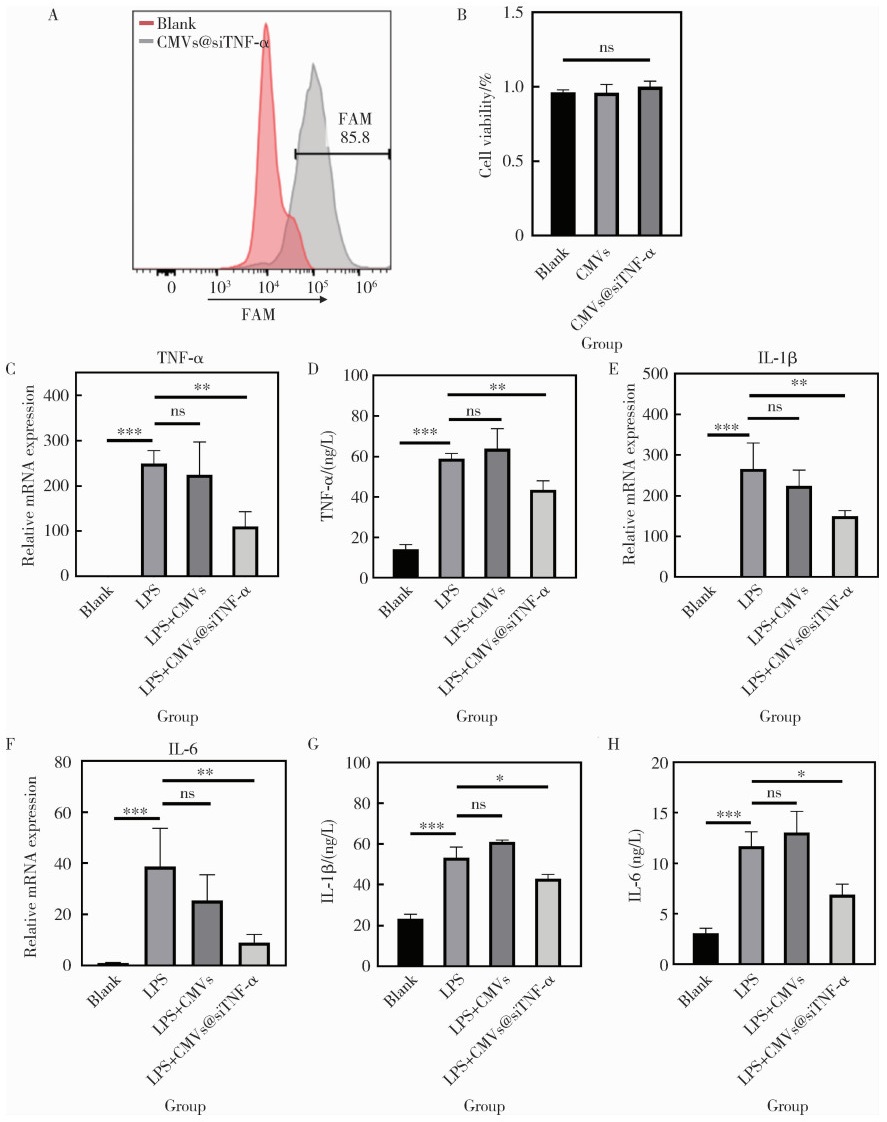
Anti-inflammatory effects of cell membrane vesicle-mediated delivery of small interfering RNA targeting tumor necrosis factor-α on dental pulp stem cells

Ruofan GAO, Tianyu MA, Runkai WANG, Yuchen YIN, Ruidi LI, Dandan WANG*(), Bin XIA*()   

  1. Department of Pediatric Dentistry, Peking University School and Hospital of Stomatology & National Center of Stomatology & National Clinical Research Center for Oral Diseases & National Engineering Laboratory for Digital and Material Technology of Stomatology, Beijing 100081, China
  • Received:2025-09-19 Online:2026-02-18 Published:2025-11-27
  • Contact: Dandan WANG, Bin XIA
  • Supported by:
    the National Natural Science Foundation of China(82401182); the Natural Science Foundation of Beijing-Haidian Original Innovation Joint Fund(L232110)

RICH HTML


Abstract:

Objective: To evaluate the feasibility of using cell membrane vesicles (CMVs) as a delivery system for small interfering RNA (siRNA) and to assess their performance in a lipopolysaccharide (LPS)-induced inflammatory model of human dental pulp stem cells (DPSCs). Methods: CMVs were generated from cytochalasin B-treated 3T3 cells and characterized for their physicochemical properties, including morphology, size distribution, and zeta potential, using confocal microscopy and dynamic light scattering. To construct CMVs@siTNF-α, saponin-mediated transient permeabilization was used to facilitate siRNA loading, after which encapsulation efficiency was evaluated by flow cytometry and confocal imaging. Intracellular uptake behaviors were examined using flow cytometry in DPSCs. LPS (1 mg/L) was employed to establish a robust in vitro inflammatory microenvironment. DPSCs were subsequently treated with CMVs or CMVs@siTNF-α, and cell viability was assessed via CCK-8 (cell counting kit-8). The expression and secretion of tumor necrosis factor-α (TNF-α), interleukin-1β (IL-1β), and interleukin-6 (IL-6) were analyzed using quantitative real-time polymerase chain reaction (qRT-PCR) and enzyme linked immunosorbent assay (ELISA) to evaluate both gene-silencing efficiency and downstream anti-inflammatory effects. Results: CMVs exhibited uniform spherical morphology with an average diameter of approximately 903 nm and a zeta potential of -9.39 mV, confirming successful vesicle formation. CMVs efficiently encapsulated FAM-labeled siRNA, as indicated by a pronounced fluorescence shift in flow cytometry and clear colocalization signals in confocal imaging. DPSCs cultured with CMVs@FAM-siTNF-α demonstrated increased intracellular fluorescence, reflecting efficient vesicle uptake and cytoplasmic siRNA release. Importantly, both CMVs and CMVs@siTNF-α displayed negligible cytotoxicity. LPS stimulation significantly elevated TNF-α, IL-1β, and IL-6 expression, validating the inflammatory model. CMVs alone did not affect cytokine levels, indicating biological inertness. In contrast, CMVs@siTNF-α significantly suppressed the LPS-induced upregulation of TNF-α at both mRNA and protein levels., demonstrating potent gene-silencing activity. Furthermore, suppression of TNF-α led to downstream attenuation of IL-1β and IL-6, with both transcription and secretion significantly decreased compared with the LPS group. These findings collectively confirmed that CMVs enabled efficient intracellular siRNA transport and effectively mitigate inflammatory responses in DPSCs. Conclusion: CMVs represent a biocompatible and effective siRNA delivery platform capable of achieving robust TNF-α knockdown and ameliorating inflammatory cytokine production in vitro, highlighting their potential for future nucleic acid-based anti-inflammatory therapies.

Key words: Small interfering RNA, Cell membrane vesicles, Dental pulp stem cells, Tumor necrosis factor

CLC Number: 

  • R78

Table 1

Quantitative real-time PCR primer sequence"

Gene Forward primer (5′ to 3′) Reverse primer (5′ to 3′)
TNF-α AGCAAGGACAGCAGAGGA GGGGAGAGAGGGTGGAG
IL-1β TGTGCTGAATGTGGACTCA ACAAAAGGGCTGGGGAT
IL-6 TGTGTGAAAGCAGCAAAGA ACCAGGCAAGTCTCCTCA
GAPDH GCCAACGTGTCAGTGGTG AAGGTGGAGGAGTGGGTGT

Figure 1

Preparation and characterization of CMVs A, morphological changes of 3T3 cells without treatment and after 30 min of cytochalasin B exposure under a light microscope (scale bar=100 μm); B, alterations of cell membrane and cytoskeleton in untreated and CB-treated (30 min) 3T3 cells observed by confocal microscopy (red: DiI, green: phalloidin, blue: DAPI; scale bar = 40 μm); C, confocal microscopy images of CMVs (red: DiI, green: cFDA-SE, scale bar = 20 μm) and magnified view (red: DiI, green: cFDA-SE, scale bar = 2 μm); D, size distribution of CMVs; E, zeta potential distribution of CMVs. DAPI, 4', 6-diamidino-2-phenylindole; cFDA-SE, 5-(and-6)-carboxyfluorescein diacetate succinimidyl ester; DiI, 1, 1'-dioctadecyl-3, 3, 3', 3'-tetramethylindocarbocyanine perchlorate; CMVs, cell membrane vesicles; CB, cytochalasin B."

Figure 2

Loading of siTNF-α into CMVs A, flow cytometry analysis of CMVs with or without loading of FAM-siTNF-α; B, confocal microscopy images of CMVs@FAM-siTNF-α (red, DiI; green, FAM; scale bar=2 μm). DiI, 1, 1′-dioctadecyl-3, 3, 3', 3'-tetramethylindocarbocyanine perchlorate; CMVs, cell membrane vesicles; FAM, fluorescein amidite; siTNF-α, small interfering RNA targeting tumor necrosis factor-alpha."

Figure 3

Intracellular delivery and anti-inflammatory effects of CMVs@siTNF-α A, flow cytometry analysis of DPSCs with or without transfection of CMVs@siTNF-α; B, relative cell viability of DPSCs after treatment with CMVs or CMVs@siTNF-α, measured by CCK-8 assay; C, E, F, mRNA expression levels of TNF-α, IL-1β, and IL-6 in DPSCs under different treatments (blank, LPS, LPS+CMVs, or LPS+CMVs@siTNF-α), determined by qRT-PCR; D, G, H, protein secretion levels of TNF-α, IL-1β, and IL-6, assessed by ELISA. *P < 0.05; * *P < 0.01; * * *P0.001; ns, no significance. CMVs, cell membrane vesicles; siTNF-α, small interfering RNA targeting tumor necrosis factor-alpha; FAM, fluorescein amidite; LPS, lipopolysaccharide; TNF-α, tumor necrosis factor-alpha; IL-1β, interleukin-1 beta; IL-6, interleukin-6; DPSCs, dental pulp stem cells; qRT-PCR, quantitative real-time polymerase chain reaction; ELISA, enzyme-linked immunosorbent assay."

1
Jadhav V, Vaishnaw A, Fitzgerald K, et al. RNA interference in the era of nucleic acid therapeutics[J]. Nat Biotechnol, 2024, 42(3): 394- 405.

doi: 10.1038/s41587-023-02105-y
2
Alshaer W, Zureigat H, Al Karaki A, et al. siRNA: Mechanism of action, challenges, and therapeutic approaches[J]. Eur J Pharmacol, 2021, 905, 174178.

doi: 10.1016/j.ejphar.2021.174178
3
Moazzam M, Zhang M, Hussain A, et al. The landscape of nanoparticle-based siRNA delivery and therapeutic development[J]. Mol Ther, 2024, 32(2): 284- 312.

doi: 10.1016/j.ymthe.2024.01.005
4
Kulkarni JA, Witzigmann D, Chen S, et al. Lipid nanoparticle technology for clinical translation of siRNA therapeutics[J]. Acc Chem Res, 2019, 52(9): 2435- 2444.

doi: 10.1021/acs.accounts.9b00368
5
Tang Q, Khvorova A. RNAi-based drug design: Considerations and future directions[J]. Nat Rev Drug Discov, 2024, 23(5): 341- 364.

doi: 10.1038/s41573-024-00912-9
6
Biscans A, Coles A, Haraszti R, et al. Diverse lipid conjugates for functional extra-hepatic siRNA delivery in vivo[J]. Nucleic Acids Res, 2019, 47(3): 1082- 1096.

doi: 10.1093/nar/gky1239
7
Tanowitz M, Hettrick L, Revenko A, et al. Asialoglycoprotein receptor 1 mediates productive uptake of N-acetylgalactosamine-conjugated and unconjugated phosphorothioate antisense oligonucleotides into liver hepatocytes[J]. Nucleic Acids Res, 2017, 45(21): 12388- 12400.

doi: 10.1093/nar/gkx960
8
Schlegel MK, Janas MM, Jiang Y, et al. From bench to bedside: Improving the clinical safety of GalNAc-siRNA conjugates using seed-pairing destabilization[J]. Nucleic Acids Res, 2022, 50(12): 6656- 6670.

doi: 10.1093/nar/gkac539
9
Liu YJ, Wang C. A review of the regulatory mechanisms of extracellular vesicles-mediated intercellular communication[J]. Cell Commun Signal, 2023, 21(1): 77.

doi: 10.1186/s12964-023-01103-6
10
Schulz-Siegmund M, Aigner A. Nucleic acid delivery with extracellular vesicles[J]. Adv Drug Deliv Rev, 2021, 173, 89- 111.

doi: 10.1016/j.addr.2021.03.005
11
Wang D, Jiang S, Zhang F, et al. Cell membrane vesicles with enriched CXCR4 display enhances their targeted delivery as drug carriers to inflammatory sites[J]. Adv Sci, 2021, 8(23): 2101562.

doi: 10.1002/advs.202101562
12
Sama S, Jerz G, Schmieder P, et al. Sapofectosid-ensuring non-toxic and effective DNA and RNA delivery[J]. Int J Pharm, 2017, 534(1/2): 195- 205.
13
Sama S, Woith E, Walther W, et al. Targeted suicide gene transfections reveal promising results in nu/nu mice with aggressive neuroblastoma[J]. J Control Release, 2018, 275, 208- 216.

doi: 10.1016/j.jconrel.2018.02.031
14
Oshchepkova A, Neumestova A, Matveeva V, et al. Cytochalasin-B-inducible nanovesicle mimics of natural extracellular vesicles that are capable of nucleic acid transfer[J]. Micromachines, 2019, 10(11): 750.

doi: 10.3390/mi10110750
15
Ni Z, Cai L, Tsai IC, et al. NAT10 regulates LPS-induced inflammation via stabilization of N4-acetylated PTX3 mRNA in human dental pulp stem cells[J]. Int J Mol Sci, 2025, 26(9): 4325.

doi: 10.3390/ijms26094325
16
Yu S, Liu XM, Liu Y, et al. Inflammatory microenvironment of moderate pulpitis enhances the osteo-/odontogenic potential of dental pulp stem cells by autophagy[J]. Int Endodontic J, 2024, 57(10): 1465- 1477.

doi: 10.1111/iej.14108
17
Lan C, Chen S, Jiang S, et al. Different expression patterns of inflammatory cytokines induced by lipopolysaccharides from Escherichia coli or Porphyromonas gingivalis in human dental pulp stem cells[J]. BMC Oral Health, 2022, 22(1): 121.

doi: 10.1186/s12903-022-02161-x
18
Chang J, Zhang C, Tani-Ishii N, et al. NF-κB activation in human dental pulp stem cells by TNF and LPS[J]. J Dent Res, 2005, 84(11): 994- 998.

doi: 10.1177/154405910508401105
19
Christ R, Siemes D, Zhao S, et al. Inhibition of tumour necrosis factor alpha by Etanercept attenuates Shiga toxin-induced brain pathology[J]. J Neuroinflammation, 2025, 22(1): 33.

doi: 10.1186/s12974-025-03356-z
20
Chen P, Wu L, Zhang S, et al. Combining TNF-α silencing with Wnt3a overexpression: A promising gene therapy for particle-induced periprosthetic osteolysis[J]. Front Cell Dev Biol, 2025, 13, 1511577.

doi: 10.3389/fcell.2025.1511577
21
Pick H, Schmid EL, Tairi AP, et al. Investigating cellular signaling reactions in single attoliter vesicles[J]. J Am Chem Soc, 2005, 127(9): 2908- 2912.

doi: 10.1021/ja044605x
22
Brown CR, Gupta S, Qin J, et al. Investigating the pharmacodynamic durability of GalNAc-siRNA conjugates[J]. Nucleic Acids Res, 2020, 48(21): 11827- 11844.

doi: 10.1093/nar/gkaa670
23
Hosseini-Kharat M, Bremmell KE, Prestidge CA. Why do lipid nanoparticles target the liver? Understanding of biodistribution and liver-specific tropism[J]. Mol Ther Meth Clin Dev, 2025, 33(1): 101436.

doi: 10.1016/j.omtm.2025.101436
24
Peng LH, Zhang YH, Han LJ, et al. Cell membrane capsules for encapsulation of chemotherapeutic and cancer cell targeting in vivo[J]. ACS Appl Mater Interfaces, 2015, 7(33): 18628- 18637.

doi: 10.1021/acsami.5b05065
25
Salloum G, Bresnick AR, Backer JM. Macropinocytosis: Mechanisms and regulation[J]. Biochem J, 2023, 480(5): 335- 362.

doi: 10.1042/BCJ20210584
26
Wang D, Guo Y, Heng BC, et al. Cell membrane vesicles derived from hBMSCs and hUVECs enhance bone regeneration[J]. Bone Res, 2024, 12, 23.

doi: 10.1038/s41413-024-00325-9
27
Yuan Z, Kolluri KK, Gowers KHC, et al. TRAIL delivery by MSC-derived extracellular vesicles is an effective anticancer therapy[J]. J Extracellular Vesicle, 2017, 6, 1265291.

doi: 10.1080/20013078.2017.1265291
28
Smyth T, Petrova K, Payton NM, et al. Surface functionalization of exosomes using click chemistry[J]. Bioconjugate Chem, 2014, 25(10): 1777- 1784.

doi: 10.1021/bc500291r
29
Nawal RR, Yadav S, Duncan HF, et al. Discriminatory performance of the pulpal inflammatory biomarkers; Interleukin-8 and TNF-α in patients with symptoms indicative of reversible and irreversible pulpitis: A diagnostic accuracy study[J]. Int Endodontic J, 2024, 57(9): 1200- 1211.

doi: 10.1111/iej.14078
30
Wang RP, Huang J, Chan KWY, et al. IL-1β and TNF-α play an important role in modulating the risk of periodontitis and Alzheimer's disease[J]. J Neuroinflammation, 2023, 20(1): 71.

doi: 10.1186/s12974-023-02747-4
31
Qin Y, Cai ML, Jin HZ, et al. Age-associated B cells contribute to the pathogenesis of rheumatoid arthritis by inducing activation of fibroblast-like synoviocytes via TNF-α-mediated ERK1/2 and JAK-STAT1 pathways[J]. Ann Rheum Dis, 2022, 81(11): 1504- 1514.

doi: 10.1136/ard-2022-222605
32
Neurath MF. Strategies for targeting cytokines in inflammatory bowel disease[J]. Nat Rev Immunol, 2024, 24(8): 559- 576.

doi: 10.1038/s41577-024-01008-6
33
Chugh V, Vijaya Krishna K, Pandit A. Cell membrane-coated mimics: A methodological approach for fabrication, characterization for therapeutic applications, and challenges for clinical translation[J]. ACS Nano, 2021, 15(11): 17080- 17123.

doi: 10.1021/acsnano.1c03800
[1] Mengdi LI, Lei LEI, Zhongning LIU, Jian LI, Ting JIANG. Gene silencing of Nemo-like kinase promotes neuralized tissue engineered bone regeneration [J]. Journal of Peking University (Health Sciences), 2025, 57(2): 227-236.
[2] Yu-yang YE,Lin YUE,Xiao-ying ZOU,Xiao-yan WANG. Characteristics and microRNA expression profile of exosomes derived from odontogenic dental pulp stem cells [J]. Journal of Peking University (Health Sciences), 2023, 55(4): 689-696.
[3] LOU Xue,LIAO Li,LI Xing-jun,WANG Nan,LIU Shuang,CUI Ruo-mei,XU Jian. Methylation status and expression of TWEAK gene promoter region in peripheral blood of patients with rheumatoid arthritis [J]. Journal of Peking University (Health Sciences), 2021, 53(6): 1020-1025.
[4] Yong-wei HU,Rui LIU,Li LUO. Chronic multifocal osteomyelitis: A case report and literature review [J]. Journal of Peking University (Health Sciences), 2020, 52(6): 1140-1145.
[5] Xiao-min GAO,Xiao-ying ZOU,Lin YUE. Mediated pathways of exosomes uptake by stem cells of apical papilla [J]. Journal of Peking University(Health Sciences), 2020, 52(1): 43-50.
[6] Jing XIE,Yu-ming ZHAO,Nan-quan RAO,Xiao-tong WANG,Teng-jiao-zi FANG,Xiao-xia LI,Yue ZHAI,Jing-zhi LI,Li-hong GE,Yuan-yuan WANG. Comparative study of differentiation potential of mesenchymal stem cells derived from orofacial system into vascular endothelial cells [J]. Journal of Peking University(Health Sciences), 2019, 51(5): 900-906.
[7] JIA Wei-qian, ZHAO Yu-ming, GE Li-hong. Recombinant human transforming growth factor β1 promotes dental pulp stem cells proliferation and mineralization [J]. Journal of Peking University(Health Sciences), 2017, 49(4): 680-681.
Viewed
Full text


Abstract

Cited

  Shared   
  Discussed   
No Suggested Reading articles found!