Journal of Peking University (Health Sciences) ›› 2026, Vol. 58 ›› Issue (1): 145-152. doi: 10.19723/j.issn.1671-167X.2026.01.019

Previous Articles     Next Articles

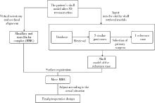
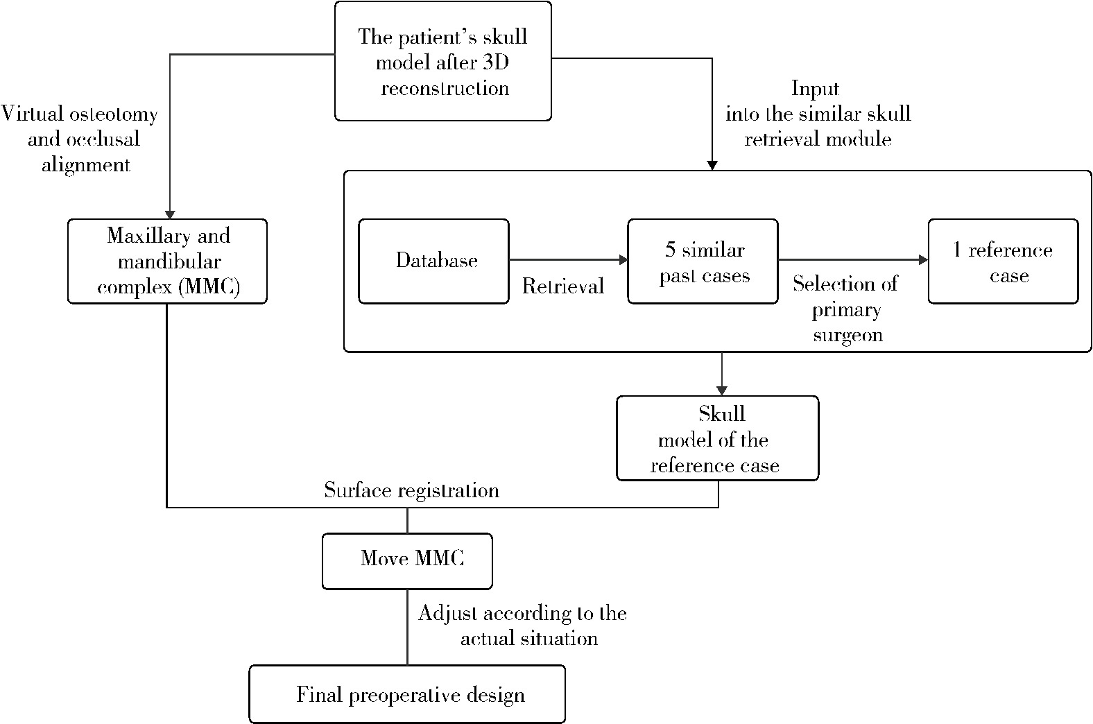
Feasibility study of a surgical planning protocol for orthognathic surgery utilizing similarity retrieval from database: A randomized controlled trial

Lu YU, Ling WU, Xiaojing LIU*(), Zili LI*()   

  1. Department of Oral and Maxillofacial Surgery, Peking University School and Hospital of Stomatology & National Center for Stomatology & National Clinical Research Center for Oral Diseases & National Engineering Research Center of Oral Biomaterials and Digital Medical Devices, Beijing 100081, China
  • Received:2025-10-13 Online:2026-02-18 Published:2026-01-05
  • Contact: Xiaojing LIU, Zili LI
  • Supported by:
    Capital's Funds for Health Improvement and Research(CFH2022-2-4104); Beijing Natural Science Foundation(F2024202104); Beijing Natural Science Foundation(L242111)

RICH HTML


Abstract:

Objective: To establish a surgical planning workflow for orthognathic surgery based on similarity retrieval from a historical patient database and to evaluate its non-inferiority with the expert's surgical plan through a randomized controlled trial. Methods: A prospective randomized controlled trial was conducted involving 60 patients (19 males, 41 females; aged 18-35 years) scheduled for orthognathic surgery in the Department of Oral and Maxillofacial Surgery at Peking University School of Stomatology between June 2023 and June 2024. Participants were randomly assigned to a test group (n=30) or a control group (n=30). In the test group, surgical plans were generated using a database-driven similarity retrieval process while in the control group plans were developed by the expert based on clinical expe-rience. All surgeries were performed by the same expert. Outcome measures assessed at 6 months post-operatively included both subjective and objective indicators. Subjective evaluations comprised patient and surgeon visual analogue scale (VAS) scores, and FACE questionnaire (FACE-Q) scores, with surgeon assessments conducted by five independent senior surgeons. Objective measures included cephalometric angles [sella-nasion-A point (SNA), sella-nasion-B point (SNB), A point-nasion-B point (ANB)] and root mean square error (RMSE) of facial symmetry regions. Results: Postoperative subjective assessments demonstrated significant improvements from baseline in both groups (all P < 0.05). Specifically, the VAS scores increased by 30.10±19.67 in the test group versus 25.43±24.48 in the control group as rated by the patients, and by 28.19±10.21 versus 26.71±7.90 as evaluated by the surgeons. Similarly, the FACE-Q scores showed marked enhancements, with patient-reported scores increasing by 33.41±17.75 in the test group and 32.97±17.65 in the control group, and surgeon-assessed scores improving by 37.75±11.60 versus 38.63±10.23, respectively. However, the magnitude of improvement in all these subjective measures did not differ significantly between the test and control groups (all P>0.05 for intergroup comparisons of the change scores). Analysis of postoperative objective measurements revealed that cephalometric values were within the normal range for both groups: SNA angle was 84.06°±3.73° in the test group compared with 85.23°±3.71° in the control group; SNB angle was 81.78°±3.63° versus 83.51°±3.66°; and ANB angle was 2.28°±1.09° versus 1.72°± 1.25°. No statistically significant differences were observed between the two groups for these cephalometric parameters (all P>0.05). Furthermore, three-dimensional facial symmetry, quantified by the average RMSE value, exhibited significant improvement postoperatively compared with preoperative levels [Test group: from (10.39±2.83) mm to (8.35±2.72) mm; Control group: from (8.55±4.95) mm to (7.59±3.56) mm; P < 0.05 for within-group comparisons]. The postoperative average RMSE values between the test and control groups were not statistically different (P>0.05). Conclusion: Surgical planning based on similarity retrieval from a historical database demonstrated non-inferiority when compared with the conventional expert-driven approach, as evidenced by the absence of statistically significant diffe-rences in both subjective and objective postoperative outcome measures.

Key words: Dentofacial deformities, Orthognathic surgery, Computer-aided design, Database platform, Similar cases

CLC Number: 

  • R782.2

Figure 1

Process of orthognathic surgery design based on previous cases"

Figure 2

Patient versus the best reference case and final design A, new patient and their skull model S0; B, similarity case and their skull model; C, postoperative design reference case Sref."

Figure 3

Orthognathic surgery design based on previous cases A, reference case Sref selection; B, overlap and registrate Sref to new patient S0; C, surgical plan definition."

Figure 4

Preoperative (A) and 6-month postoperative (B) cephalometry indications"

Figure 5

Evaluation of soft tissue (A) and hard tissue (B) symmetry"

Table 1

Baseline and observation indicators of test group and control group"

Items Test group (n=29) Control group (n=30) t P value
Male 11 (37.93) 8 (26.67) -0.917 0.363
Age/years 24.41±4.39 24.07±3.67 -0.330 0.742
VAS (Patient) 59.52±22.15 56.27±19.63 -0.597 0.553
FACE-Q (Patient) 62.90±14.63 58.93±11.33 -1.116 0.249
VAS (Surgeon) 59.68±10.60 60.05±7.89 0.153 0.879
FACE-Q (Surgeon) 53.74±7.77 52.64±8.94 -0.506 0.615
Angle SNA/(°) 80.26±3.70 81.95±3.66 1.763 0.083
Angle SNB/(°) 84.90±3.94 86.67±3.67 1.791 0.079
Angle ANB/(°) -4.63±3.31 -4.72±2.46 -0.119 0.906
Average RMSE/mm 10.39±5.63 8.55±4.95 -1.138 0.186

Table 2

Preoperative and 6-month postoperative VAS and FACE-Q scores"

Items Test group (n=29) Control group (n=30)
Preoperative 6-month postoperative t P Preoperative 6-month postoperative t P
VAS (Patient) 59.52±22.15 89.62±8.68 -8.240 < 0.001 56.27±19.63 81.70±15.21 -5.691 < 0.001
FACE-Q (Patient) 62.90±14.63 96.31±12.92 -10.139 < 0.001 58.93±11.33 91.90±15.11 -10.232 < 0.001
VAS (Surgeon) 59.68±10.60 87.86±5.02 -14.859 < 0.001 60.05±7.89 86.75±4.29 -18.509 < 0.001
FACE-Q (Surgeon) 53.74±7.77 92.00±9.85 -18.619 < 0.001 52.64±8.94 91.27±8.00 -20.674 < 0.001

Table 3

Improvement in VAS and FACE-Q scores"

Items Test group (n=29) Control group (n=30) t P value
ΔVAS (Patient) 30.10±19.67 25.43±24.48 -0.806 0.424
ΔFACE-Q (Patient) 33.41±17.75 32.97±17.65 -0.097 0.923
ΔVAS (Surgeon) 28.19±10.21 26.71±7.90 -0.623 0.535
ΔFACE-Q (Surgeon) 37.75±11.60 38.63±10.23 0.062 0.951

Table 4

Pre- and 6-months postoperative cephalometric values of test group and control group"

Items Test group (n=29) Control group (n=30) t P value
Preoperative
  Angle SNA/(°) 80.26±3.70 81.95±3.66 1.763 0.083
  Angle SNB/(°) 84.90±3.94 86.67±3.67 1.791 0.079
  Angle ANB/(°) -4.63±3.31 -4.72±2.46 -0.119 0.906
6-month postoperative
  Angle SNA/(°) 84.06±3.73 85.23±3.71 1.207 0.233
  Angle SNB/(°) 81.78±3.63 83.51±3.66 1.830 0.073
  Angle ANB/(°) 2.28±1.09 1.72±1.25 -1.852 0.069

Table 5

Pre- and 6-months postoperative average RMSE values (mm) of test group and control group"

Items Test group (n=29) Control group (n=30) t P value
Preoperative 10.39±5.63 8.55±4.95 -1.138 0.186
6-month postoperative 8.35±2.72 7.59±3.56 -0.925 0.359
1
Shetty SK , Kasrija R . Analog to digital diagnosis and planning in orthognathic surgery: A narrative review[J]. Cureus, 2025, 17 (3): e80858.
2
Miloro M , Han MD , Kwon TG , et al. Predicting the future focus of orthognathic surgery: Outcome-driven planning and treatment with function, esthetics, and occlusion as key indicators[J]. J Oral Maxillofac Surg, 2024, 82 (10): 1329- 1335.

doi: 10.1016/j.joms.2024.06.168
3
Rupperti S , Winterhalder P , Rudzki I , et al. Changes in the facial soft-tissue profile after mandibular orthognathic surgery[J]. Clin Oral Investig, 2019, 23 (4): 1771- 1776.

doi: 10.1007/s00784-018-2609-5
4
Wermker K , Kleinheinz J , Jung S , et al. Soft tissue response and facial symmetry after orthognathic surgery[J]. J Craniomaxillofac Surg, 2014, 42 (6): e339- e345.

doi: 10.1016/j.jcms.2014.01.032
5
Rustemeyer J , Martin A . Soft tissue response in orthognathic surgery patients treated by bimaxillary osteotomy: Cephalometry compared with 2-D photogrammetry[J]. Oral Maxillofac Surg, 2013, 17 (1): 33- 41.

doi: 10.1007/s10006-012-0330-0
6
Yamazaki K , Terada K , Nakamura J , et al. Three-dimensional analysis comparing changes in soft tissue with those in hard tissue following orthognathic surgery in patients showing mandibular prognathism with facial asymmetry[J]. Jpn J Jaw Deform, 2005, 15 (2): 87- 94.

doi: 10.5927/jjjd1991.15.87
7
Hu J , Wang D , Luo S , et al. Differences in soft tissue profile changes following mandibular setback in Chinese men and women[J]. J Oral Maxillofac Surg, 1999, 57 (10): 1182- 1186.

doi: 10.1016/S0278-2391(99)90481-0
8
Berends B , Bielevelt F , Baan F , et al. Soft-tissue prediction based on 3D photographs for virtual surgery planning of orthognathic surgery[J]. Comput Biol Med, 2025, 194, 110529.

doi: 10.1016/j.compbiomed.2025.110529
9
Philip MR , AlFotawi R . The accuracy of soft tissue movement using virtual planning for non-syndromic facial asymmetry cases: A systematic review[J]. Oral Maxillofac Surg, 2023, 27 (2): 187- 200.
10
Rupperti S , Winterhalder P , Krennmair S , et al. Changes in the facial soft tissue profile after maxillary orthognathic surgery[J]. J Orofac Orthop, 2022, 83 (3): 215- 220.

doi: 10.1007/s00056-021-00294-2
11
Huang L , Li Z , Yan J , et al. Evaluation of facial soft tissue thickness in asymmetric mandibular deformities after orthognathic surgery[J]. Maxillofac Plast Reconstr Surg, 2021, 43 (1): 37.

doi: 10.1186/s40902-021-00323-5
12
Czako L , Sufliarsky B , Simko K , et al. Exploring the practical applications of artificial intelligence, deep learning, and machine learning in maxillofacial surgery: A comprehensive analysis of published works[J]. Bioengineering, 2024, 11 (7): 679.

doi: 10.3390/bioengineering11070679
13
Deng HH , Liu Q , Chen A , et al. Clinical feasibility of deep learning-based automatic head CBCT image segmentation and landmark detection in computer-aided surgical simulation for orthognathic surgery[J]. Int J Oral Maxillofac Surg, 2023, 52 (7): 793- 800.

doi: 10.1016/j.ijom.2022.10.010
14
Eissa M . The potential of artificial intelligence for personalized surgical planning: A letter to the editor[J]. J Surg Res Rev, 2024, 1 (1): 1.
15
Oeding JF , Yang L , Sanchez-Sotelo J , et al. A practical guide to the development and deployment of deep learning models for the orthopaedic surgeon: Part Ⅲ, focus on registry creation, diagnosis, and data privacy[J]. Knee Surg Phys Traumatol Arthrosc, 2024, 32 (3): 518- 528.

doi: 10.1002/ksa.12085
16
Deng HH, Wang L, Ren Y, et al. Patient-specific reference model for planning orthognathic surgery[M]//Machine learning in dentistry. Cham, Switzerland: Springer International Publishing, 2021: 105-114.
17
吴灵, 方嘉琨, 刘筱菁, 等. 基于牙颌面畸形患者三维颅面特征相似性度量模型的建立及评估[J]. 北京大学学报(医学版), 2025, 57 (1): 128- 135.

doi: 10.19723/j.issn.1671-167X.2025.01.019
18
Araujo GTT , Peralta-Mamani M , Silva AFMD , et al. Influence of cone beam computed tomography versus panoramic radiography on the surgical technique of third molar removal: A systematic review[J]. Int J Oral Maxillofac Surg, 2019, 48 (10): 1340- 1347.

doi: 10.1016/j.ijom.2019.04.003
19
McGuckin T , Crick K , Myroniuk TW , et al. Understanding challenges of using routinely collected health data to address clinical care gaps: A case study in Alberta, Canada[J]. BMJ Open Qual, 2022, 11 (1): e001491.

doi: 10.1136/bmjoq-2021-001491
20
Luo Y. Study on personalized medical decision support system based on big data[C]//Proceedings of the 2024 4th International Conference on Bioinformatics and Intelligent Computing. Beijing, China: ACM, 2024: 293-296.
[1] Lijuan MA, Yonghui TENG, Yong WANG, Yijiao ZHAO, Xinyue ZHANG, Qingzhao QIN, Dong YIN. Three-dimensional finite element analysis of digital wire loop space maintainers for missing deciduous teeth [J]. Journal of Peking University (Health Sciences), 2025, 57(2): 376-383.
[2] Ling WU, Jiakun FANG, Xiaojing LIU, Zili LI, Yang LI, Xiaoxia WANG. Establishment and evaluation of a similarity measurement model for orthognathic patients based on the 3D craniofacial features [J]. Journal of Peking University (Health Sciences), 2025, 57(1): 128-135.
[3] Xinyu XU,Ling WU,Fengqi SONG,Zili LI,Yi ZHANG,Xiaojing LIU. Mandibular condyle localization in orthognathic surgery based on mandibular movement trajectory and its preliminary accuracy verification [J]. Journal of Peking University (Health Sciences), 2024, 56(1): 57-65.
[4] Sui LI,Wenjie MA,Shimin WANG,Qian DING,Yao SUN,Lei ZHANG. Trueness of different digital design methods for incisal guidance of maxillary anterior implant-supported single crowns [J]. Journal of Peking University (Health Sciences), 2024, 56(1): 81-87.
[5] FENG Sha-wei,GUO Hui,WANG Yong,ZHAO Yi-jiao,LIU He. Initial establishment of digital reference standardized crown models of the primary teeth [J]. Journal of Peking University (Health Sciences), 2022, 54(2): 327-334.
[6] LI Yi,WONG Lai U,LIU Xiao-qiang,ZHOU Ti,LYU Ji-zhe,TAN Jian-guo. Marginal features of CAD/CAM laminate veneers with different materials and thicknesses [J]. Journal of Peking University (Health Sciences), 2022, 54(1): 140-145.
[7] QIU Shu-ting,ZHU Yu-jia,WANG Shi-min,WANG Fei-long,YE Hong-qiang,ZHAO Yi-jiao,LIU Yun-song,WANG Yong,ZHOU Yong-sheng. Preliminary clinical application verification of complete digital workflow of design lips symmetry reference plane based on posed smile [J]. Journal of Peking University (Health Sciences), 2022, 54(1): 193-199.
[8] XU Xiao-xiang,CAO Ye,ZHAO Yi-jiao,JIA Lu,XIE Qiu-fei. In vitro evaluation of the application of digital individual tooth tray in the impression making of mandibular full-arch crown abutments [J]. Journal of Peking University (Health Sciences), 2021, 53(1): 54-61.
[9] YUE Zhao-guo,ZHANG Hai-dong,YANG Jing-wen,HOU Jian-xia. Comparison of residual cement between CAD/CAM customized abutments and stock abutments via digital measurement in vitro [J]. Journal of Peking University (Health Sciences), 2021, 53(1): 69-75.
[10] LI Zheng,LIU Yu-shu,WANG Shi-min,ZHANG Rui,JIA Lu,YE Hong-qiang,HU Wen-jie,ZHAO Wen-yan,LIU Yun-song,ZHOU Yong-sheng. Application of biocopy function of temporary crown occlusal morphology in patients with severe attrition [J]. Journal of Peking University (Health Sciences), 2021, 53(1): 62-68.
[11] FANG Shuo-bo,YANG Guang-ju,KANG Yan-feng,SUN Yu-chun,XIE Qiu-fei. Method and accuracy of determining the jaw position of repositioning splint with the aid of digital technique [J]. Journal of Peking University (Health Sciences), 2021, 53(1): 76-82.
[12] Lang YOU,Ke-hui DENG,Wei-wei LI,Yi-jiao ZHAO,Yu-chun SUN,Yong-sheng ZHOU. Visual sensitivity threshold of lateral view of nasolabial Angle changes in edentulous jaw patients [J]. Journal of Peking University(Health Sciences), 2020, 52(1): 107-112.
[13] Lei HOU,Guo-hua YE,Xiao-jing LIU,Zi-li LI. Evaluation of mandibular stability and condylar volume after orthognathic surgery in patients with severe temporomandibular joint osteoarthrosis [J]. Journal of Peking University(Health Sciences), 2020, 52(1): 113-118.
[14] Xian-tao SUN,Wei HE,Xiao-jing LIU,Zi-li LI,Xing WANG. Feasibility of Delaire cephalometric analysis to predict the ideal sagittal position of the maxilla and chin for surgery-first patients in orthognathic surgery [J]. Journal of Peking University(Health Sciences), 2020, 52(1): 90-96.
[15] Shuo CHEN,Yang HE,Jin-gang AN,Yi ZHANG. Application of computer-aided virtual mandibular position in the simultaneous treatment of children with temporomandibular joint ankylosis and jaw deformity [J]. Journal of Peking University(Health Sciences), 2019, 51(5): 954-958.
Viewed
Full text


Abstract

Cited

  Shared   
  Discussed   
No Suggested Reading articles found!