Journal of Peking University (Health Sciences) ›› 2023, Vol. 55 ›› Issue (1): 44-51. doi: 10.19723/j.issn.1671-167X.2023.01.007
Previous Articles Next Articles
Effect of pH on the chelation between strontium ions and decellularized small intestinal submucosal sponge scaffolds
Yu-ke LI1,Mei WANG2,Lin TANG1,Yu-hua LIU1,*( ),Xiao-ying CHEN1
- 1. Department of Prosthodontics, Peking University School and Hospital of Stomatology & National Center of Stomatology & National Clinical Research Center for Oral Diseases & National Engineering Research Center of Oral Biomaterials and Digital Medical Devices & Beijing Key Laboratory of Digital Stomatology, Beijing 100081, China
2. Department of Stomatology, Beijing Chao-Yang Hospital, Capital Medical University, Beijing 100020, China
CLC Number:
- R318.08
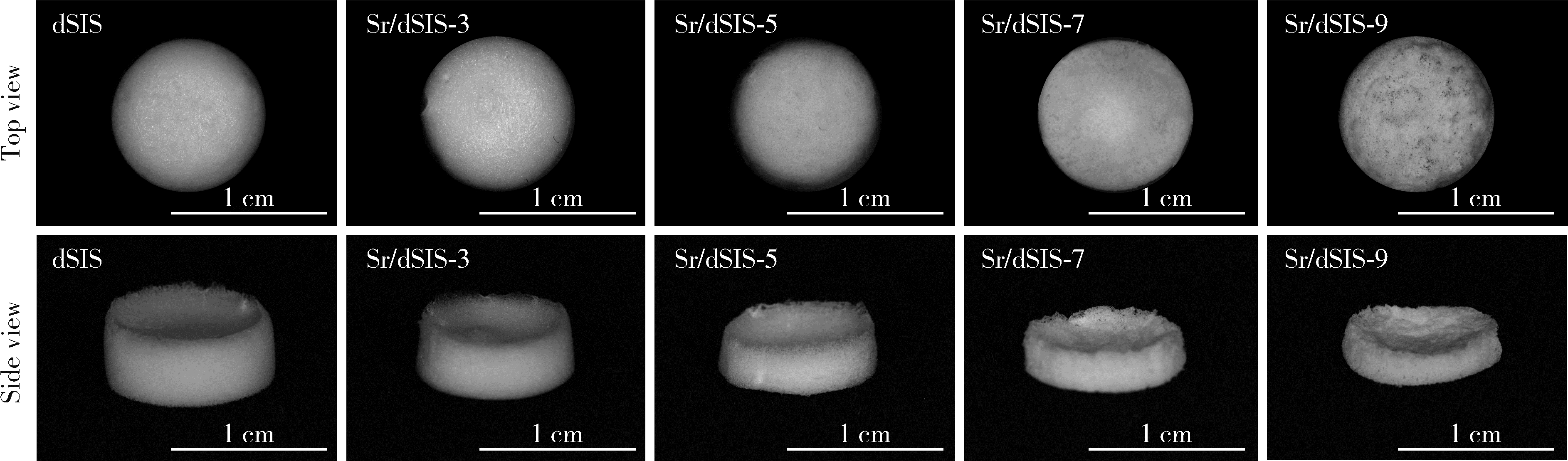
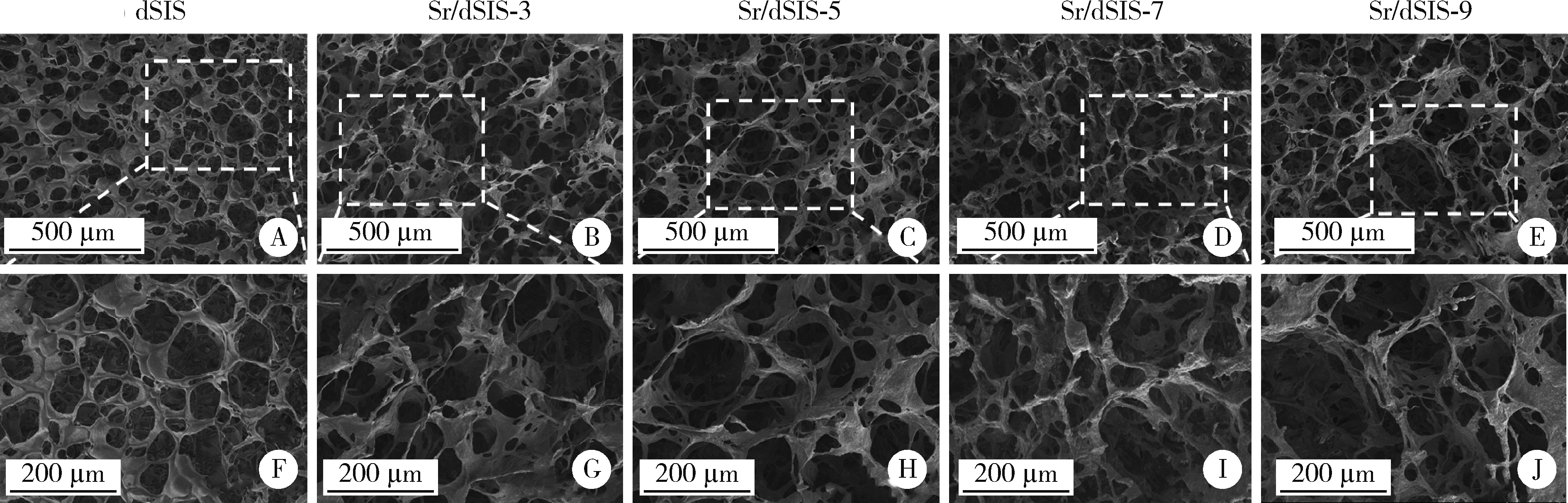

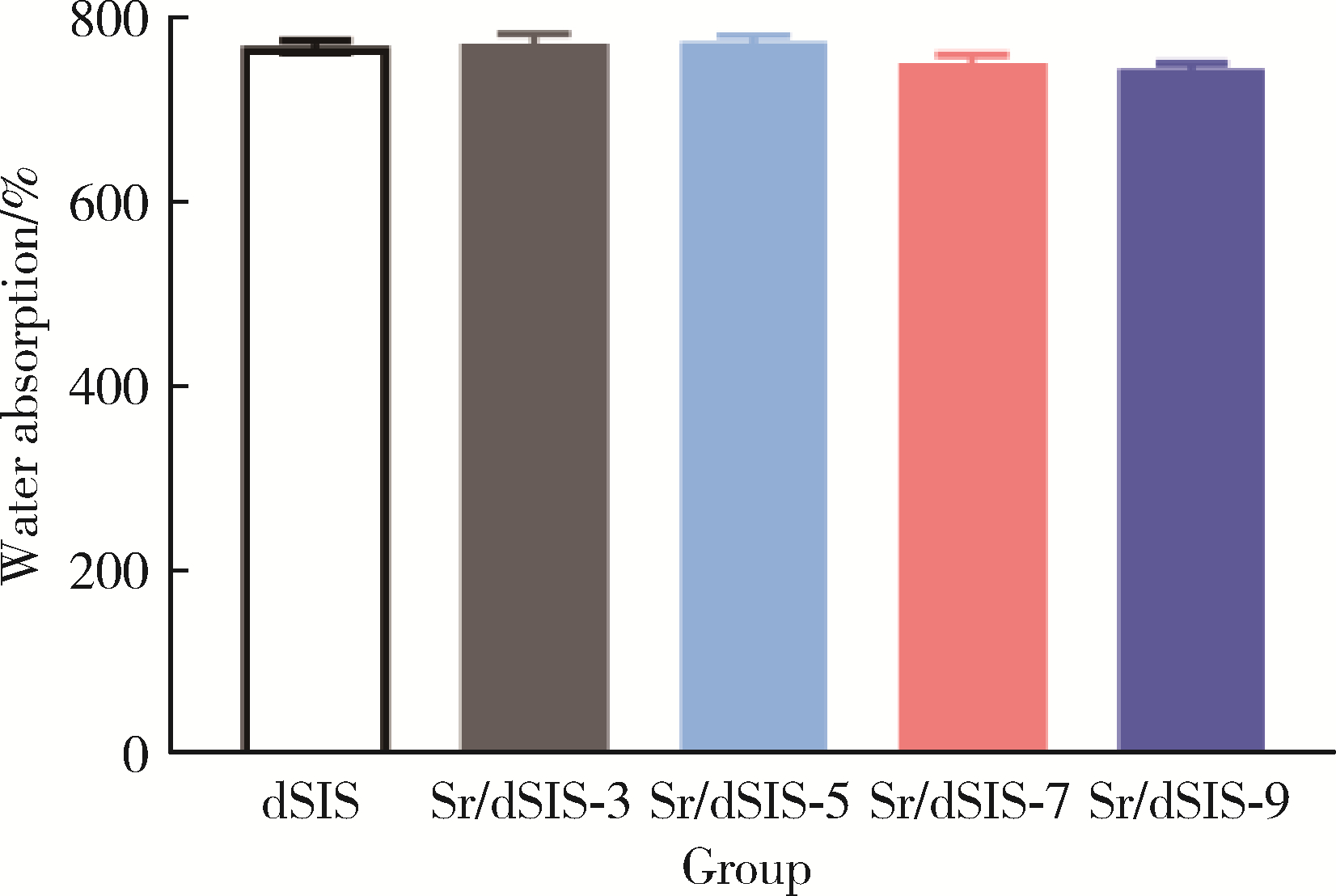
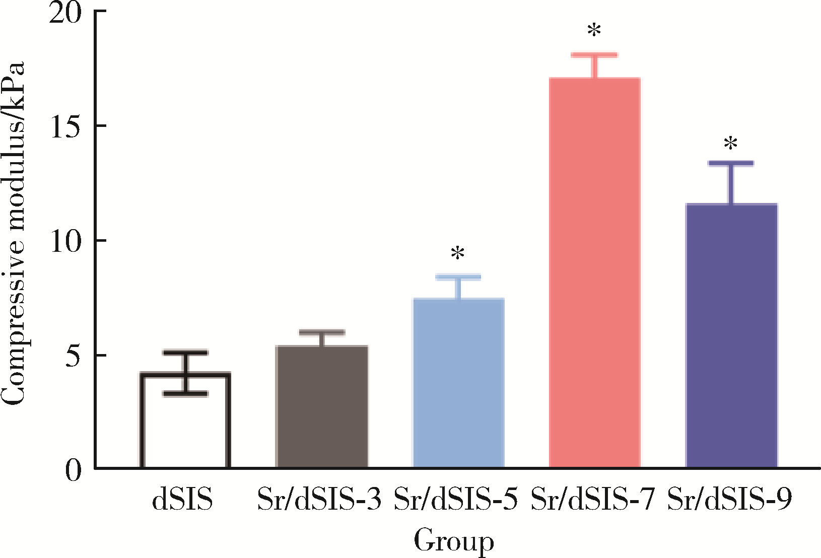
| 1 |
Andrée B , Bär A , Haverich A , et al. Small intestinal submucosa segments as matrix for tissue engineering: review[J]. Tissue Eng Part B Rev, 2013, 19 (4): 279- 291.
doi: 10.1089/ten.teb.2012.0583 |
| 2 | Tian Q , Fan Y , Hao L , et al. A comprehensive review of calcium and ferrous ions chelating peptides: Preparation, structure and transport pathways[J]. Crit Rev Food Sci Nutr, 2021, 61 (11): 1- 13. |
| 3 |
O'Neill E , Awale G , Daneshmandi L , et al. The roles of ions on bone regeneration[J]. Drug Discov Today, 2018, 23 (4): 879- 890.
doi: 10.1016/j.drudis.2018.01.049 |
| 4 | Wu W , He L , Liang Y , et al. Preparation process optimization of pig bone collagen peptide-calcium chelate using response surface methodology and its structural characterization and stability analysis[J]. Food Chem, 2019, 284 (30): 80- 89. |
| 5 |
蔡冰娜, 陈忻, 潘剑宇, 等. 响应面法优化鳕鱼皮胶原蛋白肽螯合铁工艺[J]. 食品科学, 2012, 33 (2): 48- 52.
doi: 10.3969/j.issn.1671-1513.2012.02.010 |
| 6 |
Bi J , Wang X , Zhou Y , et al. Preparation and characterization for peptide-chelated calcium of deer bone[J]. Food Sci Technol Res, 2018, 24 (4): 717- 728.
doi: 10.3136/fstr.24.717 |
| 7 |
韩克光, 甄守艳, 范华, 等. 钙螯合羊骨胶原多肽的制备及表征分析[J]. 农业工程学报, 2015, 31 (21): 301- 307.
doi: 10.11975/j.issn.1002-6819.2015.21.040 |
| 8 | Zhang H , Zhao L , Shen Q , et al. Preparation of cattle bone collagen peptides-calcium chelate and its structural characterization and stability[J]. LWT-Food Sci Technol, 2021, 144 (12): 111264. |
| 9 | 陆剑锋, 孟昌伟, 李进, 等. 斑点叉尾鱼骨胶原多肽螯合钙的制备及其特征[J]. 水产学报, 2012, 36 (2): 314- 320. |
| 10 |
Crapo PM , Gilbert TW , Badylak SF . An overview of tissue and whole organ decellularization processes[J]. Biomaterials, 2011, 32 (12): 3233- 3243.
doi: 10.1016/j.biomaterials.2011.01.057 |
| 11 |
Li B , Wang M , Liu Y , et al. Independent effects of structural optimization and resveratrol functionalization on extracellular matrix scaffolds for bone regeneration[J]. Colloids Surf B Biointerfaces, 2022, 212, 112370.
doi: 10.1016/j.colsurfb.2022.112370 |
| 12 |
Reing JE , Brown BN , Daly KA , et al. The effects of processing methods upon mechanical and biologic properties of porcine dermal extracellular matrix scaffolds[J]. Biomaterials, 2010, 31 (33): 8626- 8633.
doi: 10.1016/j.biomaterials.2010.07.083 |
| 13 | Ji Y , Zhou J , Sun T , et al. Diverse preparation methods for small intestinal submucosa (SIS): Decellularization, components, and structure[J]. J Biomed Mater Res A, 2019, 107 (3): 689- 697. |
| 14 |
Cowles EA , Brailey LL , Gronowicz GA . Integrin-mediated signaling regulates AP-1 transcription factors and proliferation in osteoblasts[J]. J Biomed Mater Res, 2000, 52 (4): 725- 737.
doi: 10.1002/1097-4636(20001215)52:4<725::AID-JBM18>3.0.CO;2-O |
| 15 |
Yi S , Ding F , Gong L , et al. Extracellular matrix scaffolds for tissue engineering and regenerative medicine[J]. Curr Stem Cell Res Ther, 2017, 12 (3): 233- 246.
doi: 10.2174/1574888X11666160905092513 |
| 16 | Liao J , Xu B , Zhang R , et al. Applications of decellularized materials in tissue engineering: Advantages, drawbacks and current improvements, and future perspectives[J]. J Mater Chem B, 2020, 8 (44): 10023- 10049. |
| 17 | Gorschewsky O , Puetz A , Riechert K , et al. Quantitative analysis of biochemical characteristics of bone-patellar tendon-bone allografts[J]. Biomed Mater Eng, 2005, 15 (6): 403- 411. |
| 18 | Bharadwaz A , Jayasuriya AC . Recent trends in the application of widely used natural and synthetic polymer nanocomposites in bone tissue regeneration[J]. Mater Sci Eng C Mater Biol Appl, 2020, 110, 110698. |
| 19 | Chandika P , Ko SC , Oh GW , et al. Fish collagen/alginate/chitooligosaccharides integrated scaffold for skin tissue regeneration application[J]. Int J Biol Macromol, 2015, 81 (8): 504- 513. |
| 20 | Castilla Bolaños MA , Buttigieg J , Briceño Triana JC . Development and characterization of a novel porous small intestine submucosa-hydroxyapatite scaffold for bone regeneration[J]. Mater Sci Eng C Mater Biol Appl, 2017, 72, 519- 525. |
| 21 | Liu J , Zeng H , Xiao P , et al. Sustained release of magnesium ions mediated by a dynamic mechanical hydrogel to enhance BMSC proliferation and differentiation[J]. ACS Omega, 2020, 5 (38): 24477- 24486. |
| [1] | Xiaoqiang BAI, Zhiruo YUAN, Yongsheng ZHOU, Longwei LV. Dynamic stretching promotes osteogenic differentiation of human bone marrow mesenchymal stem cells in three-dimensional culture [J]. Journal of Peking University (Health Sciences), 2026, 58(3): 641-649. |
| [2] | Zheng LI, Longwei LV, Xiao ZHANG, Dandan XIA, Ping ZHANG, Yunsong LIU, Yongsheng ZHOU. Advances in oral and craniofacial bone regeneration modulated by stem cells and biomaterials [J]. Journal of Peking University (Health Sciences), 2026, 58(2): 272-277. |
| [3] | Han ZHANG, Fujia YANG, Ruili YANG. Progress in regulating stem cell functions for repair and regeneration of craniomaxillofacial tissues [J]. Journal of Peking University (Health Sciences), 2026, 58(2): 285-289. |
| [4] | Jiayu LIU, Ning ZHU, Yuchen CHANG, Xianming GAO, Yu ZHANG. Accuracy of dynamic navigation-assisted trephine method for bone harvesting [J]. Journal of Peking University (Health Sciences), 2026, 58(2): 365-371. |
| [5] | Liting ZENG, Kaiyuan CHENG, Zhongning LIU, Jian LI, Jingwen YANG, Ting JIANG. miR-488-5p promotes osteogenic and neurogenic differentiation of rat bone marrow mesenchymal stem cells and enhances neuralized bone regeneration [J]. Journal of Peking University (Health Sciences), 2026, 58(1): 10-21. |
| [6] | Siqiao ZHANG, Jian LIU, Tao XU, Wenjie HU, Haoyun ZHANG, Yiping WEI. Clinical comparison of xenograft versus synthetic bone graft materials in micro crestal flap-alveolar ridge preservation following extraction of molars [J]. Journal of Peking University (Health Sciences), 2026, 58(1): 74-83. |
| [7] | Yulan WANG, Hao ZENG, Yufeng ZHANG. Current situation and exploration of clinical transformation of plasmatrix in oral implantology [J]. Journal of Peking University (Health Sciences), 2025, 57(5): 836-840. |
| [8] | Wei LIU, Wen GUO, Zhe GUO, Chunyan LI, Yunlong LI, Siqi LIU, Liang ZHANG, Hui SONG. Risk factors associated with non-radiographic bone erosion in patients with gout [J]. Journal of Peking University (Health Sciences), 2025, 57(4): 735-739. |
| [9] | Mengdi LI, Lei LEI, Zhongning LIU, Jian LI, Ting JIANG. Gene silencing of Nemo-like kinase promotes neuralized tissue engineered bone regeneration [J]. Journal of Peking University (Health Sciences), 2025, 57(2): 227-236. |
| [10] | Shuyuan MIN, Yun TIAN. Biocompatibility of 3D printed biodegradable WE43 magnesium alloy scaffolds and treatment of bone defects [J]. Journal of Peking University (Health Sciences), 2025, 57(2): 309-316. |
| [11] | Zhenlong LIU, Zhenchen HOU, Xiaoqing HU, Shuang REN, Qinwei GUO, Yan XU, Xi GONG, Yingfang AO. Arthroscopic tissue engineering scaffold repair for cartilage injuries [J]. Journal of Peking University (Health Sciences), 2025, 57(2): 384-387. |
| [12] | Junnan NIE, Jiayun DONG, Ruifang LU. Analysis of soft tissue healing after keratinized tissue augmentation in reconstructed jaws [J]. Journal of Peking University (Health Sciences), 2025, 57(1): 57-64. |
| [13] | Ting SHUAI, Yanyan GUO, Chunping LIN, Xiaomei HOU, Chanyuan JIN. Knockdown of NPTX1 promotes osteogenic differentiation of human bone marrow mesenchymal stem cells [J]. Journal of Peking University (Health Sciences), 2025, 57(1): 7-12. |
| [14] | Yibo HU, Weijia LYU, Wei XIA, Yihong LIU. Hydrodynamic finite element analysis of biological scaffolds with different pore sizes for cell growth and osteogenic differentiation [J]. Journal of Peking University (Health Sciences), 2025, 57(1): 97-105. |
| [15] | Xiaoying CHEN,Yi ZHANG,Yuke LI,Lin TANG,Yuhua LIU. Effects of different polymers on biomimetic mineralization of small intestine submucosal scaffolds [J]. Journal of Peking University (Health Sciences), 2024, 56(1): 17-24. |
|
||