Journal of Peking University (Health Sciences) ›› 2024, Vol. 56 ›› Issue (3): 505-511. doi: 10.19723/j.issn.1671-167X.2024.03.018

Previous Articles     Next Articles

Tofacitinib inhibits the transformation of lung fibroblasts into myofibroblasts through JAK/STAT3 pathway

Shan HE1,2,Xin CHEN1,2,Qi CHENG1,Lingjiang ZHU1,Peiyu ZHANG1,Shuting TONG1,Jing XUE1,Yan DU1,*()   

  1. 1. Department of Rheumatology, the Second Affiliated Hospital of Zhejiang University School of Medicine, Hangzhou, 310009, China
    2. Department of Rheumatology, the Affiliated Jinhua Hospital of Zhejiang University School of Medicine, Jinhua, 321000, Zhejiang, China
  • Received:2022-08-31 Online:2024-06-18 Published:2024-06-12
  • Contact: Yan DU E-mail:duyan2014@zju.edu.cn
  • Supported by:
    the National Natural Science Foundation of China(82271817);the Natural Science Foundation of Zhejiang Province(LY22H100004)

RICH HTML


Abstract:

Objective: To investigate the effect of tofacitinib, a pan-Janus kinase (JAK) inhibitor, on transforming growth factor-beta 1 (TGF-β1)-induced fibroblast to myofibroblast transition (FMT) and to explore its mechanism. To provide a theoretical basis for the clinical treatment of connective tissue disease-related interstitial lung disease (CTD-ILD). Methods: (1) Human fetal lung fibroblast 1 (HFL-1) were cultured in vitro, and 6 groups were established: DMSO blank control group, TGF-β1 induction group, and TGF-β1 with different concentrations of tofacitinib (0.5, 1.0, 2.0, 5.0 μmol/L) drug intervention experimental groups. CCK-8 was used to measure the cell viability, and wound-healing assay was performed to measure cell migration ability. After 48 h of combined treatment, quantitative real-time PCR (RT-PCR) and Western blotting were used to detect the gene and protein expression levels of α-smooth muscle actin (α-SMA), fibronectin (FN), and collagen type Ⅰ (COL1). (2) RT-PCR and enzyme-linked immunosorbnent assay (ELISA) were used to detect the interleukin-6 (IL-6) gene and protein expression changes, respectively. (3) DMSO carrier controls, 1.0 μmol/L and 5.0 μmol/L tofacitinib were added to the cell culture media of different groups for pre-incubation for 30 min, and then TGF-β1 was added to treat for 1 h, 6 h and 24 h. The phosphorylation levels of Smad2/3 and signal transducer and activator of transcription 3 (STAT3) protein were detected by Western blotting. Results: (1) Tofacitinib inhibited the viability and migration ability of HFL-1 cells after TGF-β1 induction. (2) The expression of α-SMA, COL1A1 and FN1 genes of HFL-1 in the TGF-β1-induced groups was significantly up-regulated compared with the blank control group (P < 0.05). Compared with the TGF-β1 induction group, α-SMA expression in the 5.0 μmol/L tofacitinib intervention group was significantly inhi-bited (P < 0.05). Compared with the TGF-β1-induced group, FN1 gene was significantly inhibited in each intervention group at a concentration of 0.5-5.0 μmol/L (P < 0.05). Compared with the TGF-β1-induced group, the COL1A1 gene expression in each intervention group did not change significantly. (3) Western blotting results showed that the protein levels of α-SMA and FN1 in the TGF-β1-induced group were significantly higher than those in the control group (P < 0.05), and there was no significant difference in the expression of COL1A1. Compared with the TGF-β1-induced group, the α-SMA protein level in the intervention groups with different concentrations decreased. And the differences between the TGF-β1-induced group and 2.0 μmol/L or 5.0 μmol/L intervention groups were statistically significant (P < 0.05). Compared with the TGF-β1-induced group, the FN1 protein levels in the intervention groups with different concentrations showed a downward trend, but the difference was not statistically significant. There was no difference in COL1A1 protein expression between the intervention groups compared with the TGF-β1-induced group. (4) After TGF-β1 acted on HFL-1 cells for 48 h, the gene expression of the IL-6 was up-regulated and IL-6 in culture supernatant was increased, the intervention with tofacitinib partly inhibited the TGF-β1-induced IL-6 gene expression and IL-6 in culture supernatant. TGF-β1 induced the increase of Smad2/3 protein phosphorylation in HFL-1 cells for 1 h and 6 h, STAT3 protein phosphorylation increased at 1 h, 6 h and 24 h, the pre-intervention with tofacitinib inhibited the TGF-β1-induced Smad2/3 phosphorylation at 6 h and inhibited TGF-β1-induced STAT3 phosphorylation at 1 h, 6 h and 24 h. Conclusion: Tofacitinib can inhibit the transformation of HFL-1 cells into myofibroblasts induced by TGF-β1, and the mechanism may be through inhibiting the classic Smad2/3 pathway as well as the phosphorylation of STAT3 induced by TGF-β1, thereby protecting the disease progression of pulmonary fibrosis.

Key words: Tofacitinib, Pulmonary fibrosis, Fibroblasts, Myofibroblasts, STAT3 transcription factor

CLC Number: 

  • R563.13

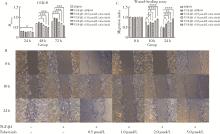
Figure 1

Inhibitory effect of tofacitinib on HFL-1 cells' viability and migration ability * P<0.05, ** P<0.01, *** P<0.001, **** P<0.000 1. A, cell viability in cultured HFL-1 cells maintained in normal growth medium treated with vehicle (DMSO) and treated with tofacitinib at a concentration of 0.5-5.0 μmol/L in combination with stimulation with TGF-β1 for 24 h, 48 h, or 72 h; B and C, migration ability of HFL-1 cells maintained in normal growth medium treated with vehicle (DMSO) and treated with tofacitinib at a concentration of 0.5-5.0 μmol/L in combination with stimulation with TGF-β1 for 10 h or 24 h. TGF-β1, transforming growth factor-beta 1; HFL-1, human fetal lung fibroblast 1."

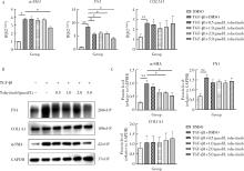
Figure 2

Effects of tofacitinib on the expression of α-SMA, FN1 and COL1A1 in HFL-1 cells induced by TGF-β1 * P<0.05, ** P<0.01. A, quantitative real-time PCR of α-SMA, FN1 and COL1A1 in cultured HFL-1 cells maintained in normal growth medium treated with vehicle (DMSO) and treated with tofacitinib at a concentration of 0.5-5.0 μmol/L in combination with stimulation with TGF-β1 for 48 h; the final results were obtained from 4 independent in vitro experiments (mean±SE). B and C, Western blotting and related densitometric analysis of α-SMA, FN1 and COL1A1 in cultured HFL-1 cells maintained in normal growth medium treated with vehicle (DMSO) and treated with tofacitinib at a concentration of 0.5-5.0 μmol/L in combination with stimulation with TGF-β1 for 48 h; the final results were obtained from 4-6 independent in vitro experiments (mean±SEM). α-SMA, α-smooth muscle actin; FN1, fibronectin 1; COL1A1, type Ⅰ collagen a1; TGF-β1, transforming growth factor-beta 1; HFL-1, human fetal lung fibroblast 1; RQ, relative quantity; SE, standard error; SEM, standard error of the mean."

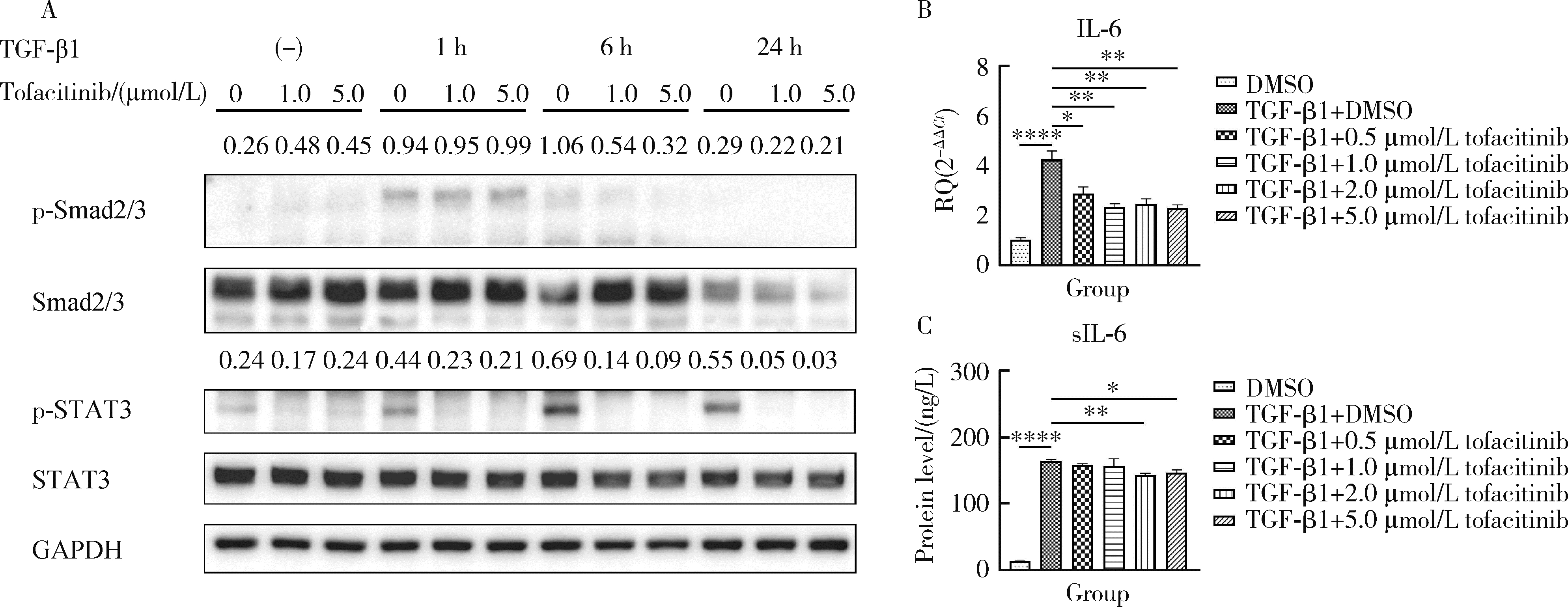
Figure 3

Effects of tofacitinib on related pathways of HFL-1 cells in FMT * P<0.05, ** P<0.01, **** P<0.000 1. A, relative protein abundance of p-Smad2/3 and p-STAT3; B and C, relative RNA transcription level of IL-6 in HFL-1 cell (B) and sIL-6 protein level in the cell culture supernatant (C). The final results were obtained from 3-4 independent in vitro experiments (mean±SEM). Smad2/3, small mothers against decapentaplegic 2 and 3; p-Smad2/3, phosphorylation-Smad2/3; STAT3, signal transducer and activator of transcription 3; p-STAT3, phosphorylation-STAT3; TGF-β1, transforming growth factor-beta 1; HFL-1, human fetal lung fibroblast 1; IL-6, interleukin 6; sIL-6, soluble interleukin 6; FMT, fibroblast to myofibroblast transition; RQ, relative quantity; SEM, standard error of the mean."

1 Shao T , Shi X , Yang S , et al. Interstitial lung disease in connective tissue disease: A common lesion with heterogeneous mechanisms and treatment considerations[J]. Front Immunol, 2021, 12, 684699.
doi: 10.3389/fimmu.2021.684699
2 Cutolo M , Ruaro B , Montagna P , et al. Effects of selexipag and its active metabolite in contrasting the profibrotic myofibroblast activity in cultured scleroderma skin fibroblasts[J]. Arthritis Res Ther, 2018, 20 (1): 77.
doi: 10.1186/s13075-018-1577-0
3 Truchetet ME , Brembilla NC , Chizzolini C . Current concepts on the pathogenesis of systemic sclerosis[J]. Clin Rev Allergy Immunol, 2023, 64 (3): 262- 283.
4 Finnson KW , Almadani Y , Philip A . Non-canonical (non-SMAD2/3) TGF-β signaling in fibrosis: Mechanisms and targets[J]. Semin Cell Dev Biol, 2020, 101, 115- 122.
doi: 10.1016/j.semcdb.2019.11.013
5 You H , Xu D , Zhao J , et al. JAK inhibitors: Prospects in connective tissue diseases[J]. Clin Rev Allergy Immunol, 2020, 59 (3): 334- 351.
doi: 10.1007/s12016-020-08786-6
6 Wang W , Bhattacharyya S , Marangoni RG , et al. The JAK/STAT pathway is activated in systemic sclerosis and is effectively targeted by tofacitinib[J]. J Scleroderma Relat Disord, 2020, 5 (1): 40- 50.
doi: 10.1177/2397198319865367
7 Yoo H , Hino T , Hwang J , et al. Connective tissue disease-related interstitial lung disease (CTD-ILD) and interstitial lung abnorma-lity (ILA): Evolving concept of CT findings, pathology and management[J]. Eur J Radiol Open, 2022, 9, 100419.
doi: 10.1016/j.ejro.2022.100419
8 Montero P , Milara J , Roger I , et al. Role of JAK/STAT in interstitial lung diseases, molecular and cellular mechanisms[J]. Int J Mol Sci, 2021, 22 (12): 6211.
doi: 10.3390/ijms22126211
9 Lescoat A , Lelong M , Jeljeli M , et al. Combined anti-fibrotic and anti-inflammatory properties of JAK-inhibitors on macrophages in vitro and in vivo: Perspectives for scleroderma-associated interstitial lung disease[J]. Biochem Pharmacol, 2020, 178, 114103.
doi: 10.1016/j.bcp.2020.114103
10 Kagan P , Sultan M , Tachlytski I , et al. Both MAPK and STAT3 signal transduction pathways are necessary for IL-6-dependent hepatic stellate cells activation[J]. PLoS One, 2017, 12 (5): e0176173.
doi: 10.1371/journal.pone.0176173
11 Chen W , Li Y , Hsu CT , et al. Connective tissue growth factor in hepatocytes is elevated by carbon tetrachloride via STAT3 activation[J]. Mol Med Rep, 2020, 21 (3): 1390- 1398.
12 Dees C , Tomcik M , Palumbo-Zerr K , et al. JAK-2 as a novel mediator of the profibrotic effects of transforming growth factor β in systemic sclerosis[J]. Arthritis Rheum, 2012, 64 (9): 3006- 3015.
doi: 10.1002/art.34500
13 Chakraborty D , Šumová B , Mallano T , et al. Activation of STAT3 integrates common profibrotic pathways to promote fibroblast activation and tissue fibrosis[J]. Nat Commun, 2017, 8 (1): 1130.
doi: 10.1038/s41467-017-01236-6
14 Li HG , You PT , Xia Y , et al. Yu Gan Long ameliorates hepatic fibrosis by inhibiting PI3K/AKT, Ras/ERK and JAK1/STAT3 signaling pathways in CCl4-induced liver fibrosis rats[J]. Curr Med Sci, 2020, 40 (3): 539- 547.
doi: 10.1007/s11596-020-2211-3
15 Yan J , Zhang Z , Yang J , et al. JAK3/STAT6 stimulates bone marrow-derived fibroblast activation in renal fibrosis[J]. J Am Soc Nephrol, 2015, 26 (12): 3060- 3071.
doi: 10.1681/ASN.2014070717
16 Pedroza M , Le TT , Lewis K , et al. STAT-3 contributes to pulmonary fibrosis through epithelial injury and fibroblast-myofibroblast differentiation[J]. FASEB J, 2016, 30 (1): 129- 140.
doi: 10.1096/fj.15-273953
17 Levine RL , Pardanani A , Tefferi A , et al. Role of JAK2 in the pathogenesis and therapy of myeloproliferative disorders[J]. Nat Rev Cancer, 2007, 7 (9): 673- 683.
doi: 10.1038/nrc2210
[1] Xue ZOU,Xiao-juan BAI,Li-qing ZHANG. Effectiveness of tofacitinib combined with iguratimod in the treatment of difficult-to-treat moderate-to-severe rheumatoid arthritis [J]. Journal of Peking University (Health Sciences), 2023, 55(6): 1013-1021.
[2] GAO Hong-yu,MENG Huan-xin,HOU Jian-xia,HUANG Bao-xin,LI Wei. Expression and distribution of calprotectin in healthy and inflamed periodontal tissues [J]. Journal of Peking University (Health Sciences), 2021, 53(4): 744-749.
[3] Bing-qing SHI,Xiao-jing YUAN,Yu-ming ZHAO. Effects of mineral trioxide aggregate and ethanolic extracts of Shandong propolis on the biological properties of human dental pulp fibroblasts [J]. Journal of Peking University(Health Sciences), 2019, 51(6): 1108-1114.
[4] Miao ZHENG,Ling-lu ZHAN,Zhi-qiang LIU,He-ping LI,Jian-guo TAN. Effect of different plasma treated zirconia on the adhensive behaviour of human gingival fibroblasts [J]. Journal of Peking University(Health Sciences), 2019, 51(2): 315-320.
[5] JIA Shuang-shuang, LI Wei-yang, LIU Xin, LI Li-ying. Transforming growth factor-β1 induces differentiation of bone marrowderived mesenchymal stem cells into myofibroblasts via production of reactive oxygen species [J]. Journal of Peking University(Health Sciences), 2015, 47(5): 737-742.
Viewed
Full text


Abstract

Cited

  Shared   
  Discussed   
No Suggested Reading articles found!