Journal of Peking University (Health Sciences) ›› 2026, Vol. 58 ›› Issue (2): 365-371. doi: 10.19723/j.issn.1671-167X.2026.02.022

Previous Articles     Next Articles

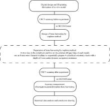
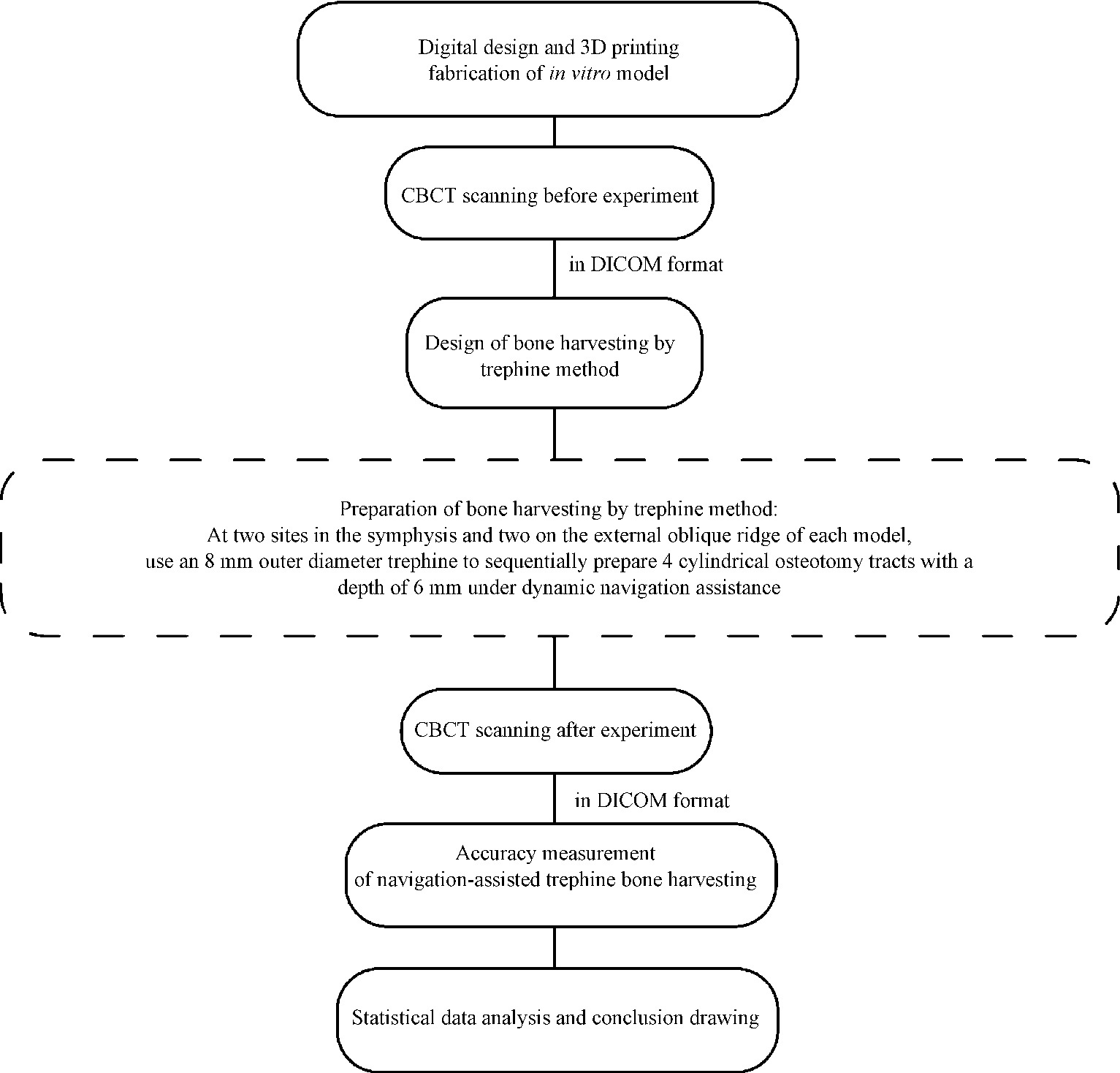
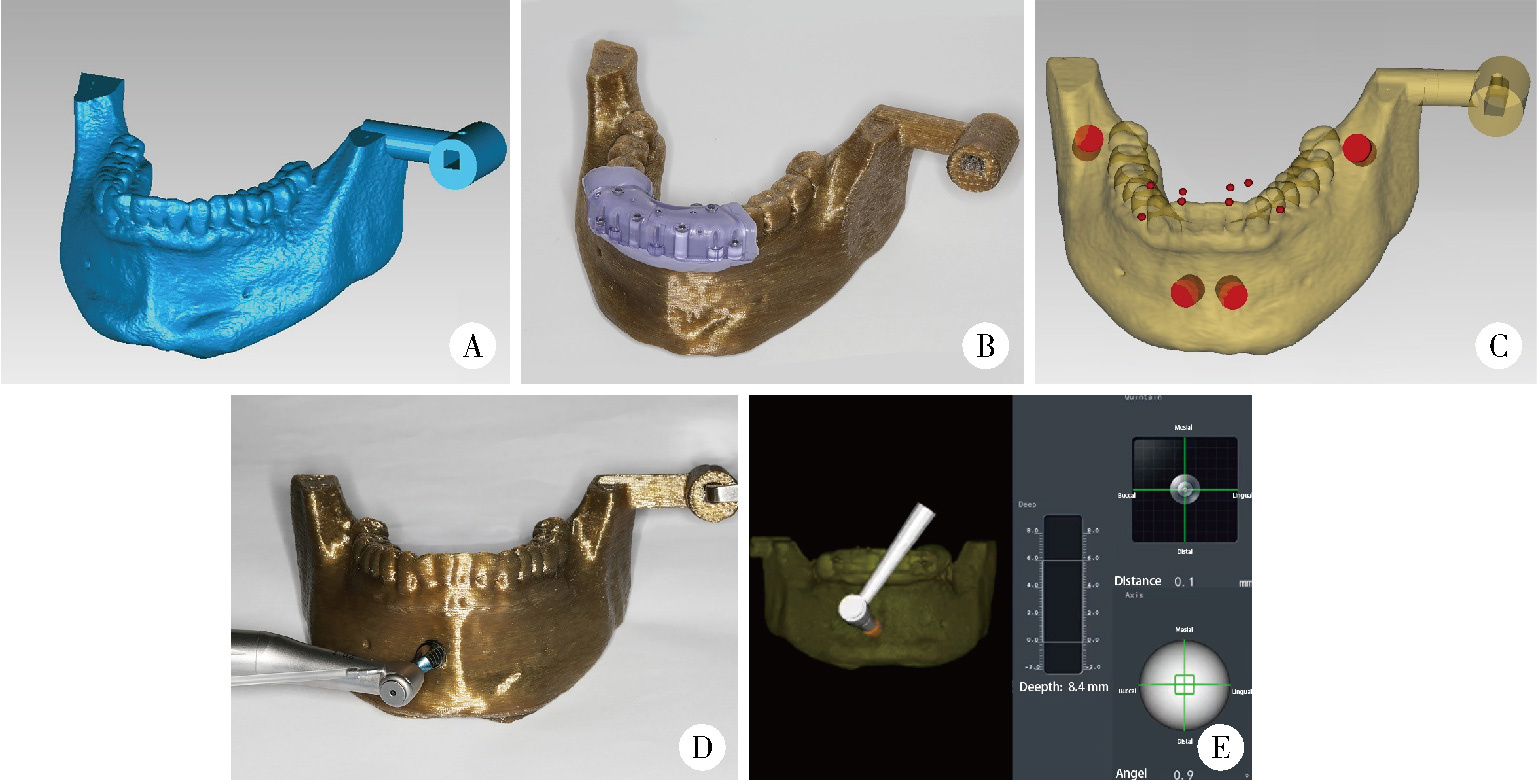
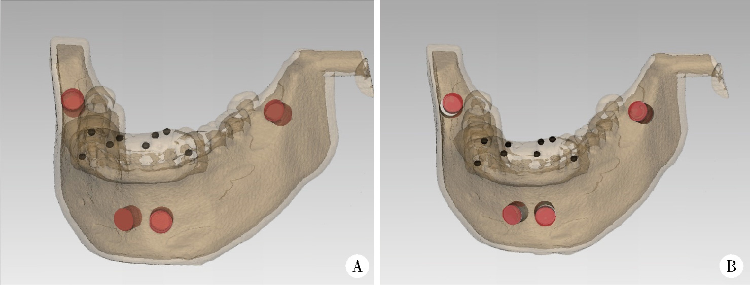
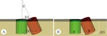
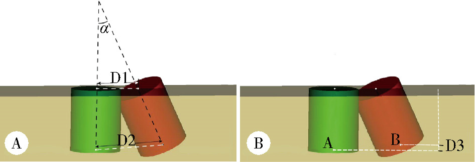
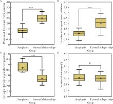
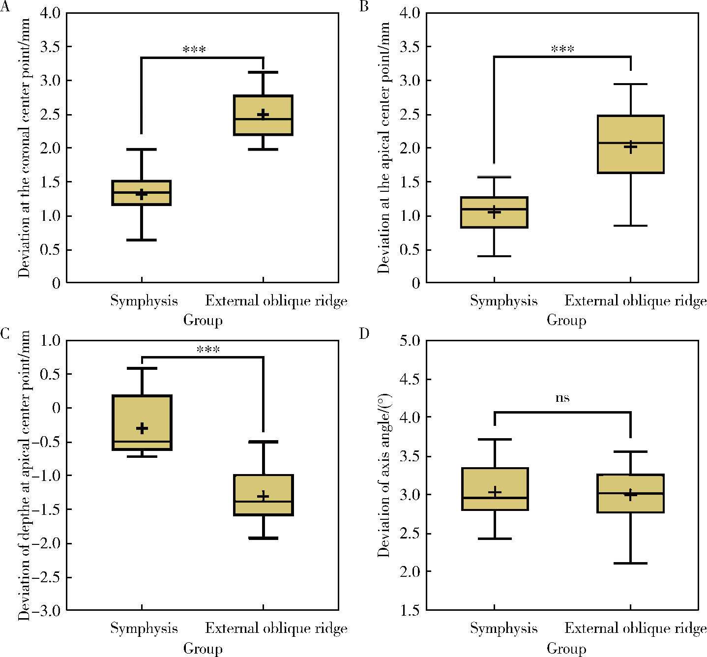
Accuracy of dynamic navigation-assisted trephine method for bone harvesting

Jiayu LIU, Ning ZHU, Yuchen CHANG, Xianming GAO, Yu ZHANG*()   

  1. Department of Oral Implantology, Peking University School and Hospital of Stomatology & National Center for Stomatology & National Clinical Research Center for Oral Diseases & National Engineering Research Center of Oral Biomaterials and Digital Medical Devices & Beijing Key Laboratory of Digital Stomatology, Beijing 100081, China
  • Received:2024-02-05 Online:2026-04-18 Published:2026-02-25
  • Contact: Yu ZHANG
  • Supported by:
    the Program for New Clinical Techniques and Therapies of Peking University School and Hospital of Stomatology(PKUSSNCT-22A01)

RICH HTML


Abstract:

Objective: To evaluate the positional accuracy of dynamic navigation-assisted trephine bone harvesting in the symphysis and external oblique ridge. Methods: Ten standardized mandibular models were 3D-printed using polyetheretherketone (PEEK), mimicking natural mandibular mechanical properties. Pre-operative cone beam CT (CBCT) scans (70 kV, 70 mA, 0.25 mm×0.25 mm ×0.25 mm voxel) were acquired, and data were imported into dynamic navigation software (Dcarer, China). Two donor sites were designed in both the symphysis (≥15 mm from anterior teeth) and external oblique ridge (≥6 mm from molars), with 8 mm-diameter, 6 mm-deep cylindrical osteotomy tracts planned for each site.After calibrating the navigation system with 20 mm and 50 mm spherical burs, an 8 mm outer-diameter trephine prepared 40 tracts under real-time guidance. Post-operative CBCT scans were taken, and Mimics 20.0 software fitted actual tracts to standard cylinders. Superimposing actual and designed tracts via metal registration markers, we measured coronal/apical center point deviation, depth deviation, and axis angle deviation in order to compare site-specific accuracy. Results: Deviations of the dynamic navigation-assisted trephine method for bone harvesting was (1.91±0.69) mm at the coronal center point, (1.54±0.66) mm at the apical center point, (-0.83±0.77) mm at the depth of the apical center point and 3.02°±0.38° at the axis angle. The four deviations in symphysis and external oblique ridge were (1.32±0.36) mm and (2.50±0.35) mm at the coronal center point (P < 0.01), (1.06± 0.31) mm and (2.02±0.56) mm at the apical center point (P < 0.01), (-0.30±0.52) mm and (-1.38±0.57) mm at the depth of apical center point (P < 0.01), 3.03°± 0.38° and 3.00°± 0.39° at axis angle (P=0.80). Conclusion: Within the limitations of this study, dynamic navigation-assisted trephine harvesting shows good accuracy. The symphysis exhibits higher accuracy than the external oblique ridge, possibly due to surface morphology and operability differences. These findings support its clinical potential, but future clinical studies are needed to validate results.

Key words: Dental implant, Surgical navigation systems, Bone transplantation

CLC Number: 

  • R783

Figure 1

Workflow chart of in vitro study on accuracy of dynamic navigation-assisted trephine bone harvesting CBCT, cone beam computed tomography; DICOM, digital imaging and communications in medicine."

Figure 2

Digital plan and conduction of the navigation-assisted trephine method for bone harvesting A, digitally designed model of the in vitro study; B, registration device of dynamic navigation system was positioned on the lower anterior teeth; C, design digital trephine osteotomy tracts (red); D, use trephines to complete the osteotomy tracts under the guidance of dynamic navigation; E, real-time positioning and angular deviations of the trephine are displayed in the dynamic navigation system."

Figure 3

Statistical measurement for dynamic navigation-assisted trephine bone harvesting accuracy A, actual osteotomy tracts were adapted in the post-lab CBCT (red); B, superimpose the actual osteotomy tracts (red) to the planned ones (white) based on the mental fiducial markers on the registration device (black)."

Figure 4

Experimental accuracy evaluation index A, coronal center point deviation D1, apical center point deviation D2 and angulation deviation α; B, deviations in depths of apical centre points D3."

Table 1

Descriptive values of deviations in dynamic navigation-assisted trephine methods for bone harvesting"

Deviations n ${\bar x}$±s Minimum Maximum
Coronal center point/mm 40 1.91±0.69 0.64 3.12
Apical center point/mm 40 1.54±0.66 0.41 2.94
Depth of apical center point/mm 40 -0.84±0.77 -2.22 0.63
Axis angle/(°) 40 3.02±0.38 2.11 3.72

Table 2

Descriptive values of deviations in dynamic navigation-assisted trephine methods for bone harvesting in the symphysis and external oblique line area"

Deviations Donor sites n ${\bar x}$±s Minimum Maximum t P
Coronal center point/mm Symphysis 20 1.32±0.36 0.64 1.98 -10.42 <0.01
External oblique line 20 2.50±0.35 1.98 3.12
Apical center point/mm Symphysis 20 1.06±0.31 0.41 1.57 -6.70 <0.01
External oblique line 20 2.02±0.56 0.86 2.94
Depth of apical center point/mm Symphysis 20 -0.30±0.52 -1.13 0.63 4.81 <0.01
External oblique line 20 -1.38±0.57 -2.22 -0.55
Axis angle/(°) Symphysis 20 3.03±0.38 2.43 3.72 0.26 0.80
External oblique line 20 3.00±0.39 2.11 3.56

Figure 5

Analysis of differences in bone harvesting accuracy using dynamic navigation-assisted trephine in extraoral oblique line and symphysis A, deviations in coronal center points; B, deviations in apical center points; C, deviations in depth of apical center points; D, deviations in the axial angles. ns, correlation is not significant at the 0.05 level (2-tailed); * * * correlation is significant at the 0.001 level (2-tailed)."

1
Lichte P , Pape HC , Pufe T , et al. Scaffolds for bone healing: Concepts, materials and evidence[J]. Injury, 2011, 42 (6): 569- 573.

doi: 10.1016/j.injury.2011.03.033
2
Omara M , Abdelwahed N , Ahmed M , et al. Simultaneous implant placement with ridge augmentation using an autogenous bone ring transplant[J]. Int J Oral Maxillofac Surg, 2016, 45 (4): 535- 544.

doi: 10.1016/j.ijom.2015.11.001
3
Khoury F , Hanser T . Mandibular bone block harvesting from the retromolar region: A 10-year prospective clinical study[J]. Int J Oral Maxillofac Implants, 2015, 30 (3): 688- 697.

doi: 10.11607/jomi.4117
4
Senos R , Hankenson KD . Calvaria critical-size defects in rats using piezoelectric equipment: A comparison with the classic trephine[J]. Injury, 2020, 51 (7): 1509- 1514.

doi: 10.1016/j.injury.2020.04.041
5
Abdulrazaq SS , Issa SA , Abdulrazzak NJ . Evaluation of the trephine method in harvesting bone graft from the anterior iliac crest for oral and maxillofacial reconstructive surgery[J]. J Craniofacial Surg, 2015, 26 (8): e744- e746.

doi: 10.1097/SCS.0000000000002177
6
Chandra R , Suvvari N , Reddy A . Trephine core procedure versus bone-added osteotome sinus floor elevation in the augmentation of the sinus floor: A comparative clinical and radiographic study[J]. Int J Oral Maxillofac Implants, 2018, 33 (2): 425- 432.

doi: 10.11607/jomi.5998
7
Takamoto M , Takechi M , Ohta K , et al. Risk of bacterial contamination of bone harvesting devices used for autogenous bone graft in implant surgery[J]. Head Face Med, 2013, 9 (1): 3.

doi: 10.1186/1746-160X-9-3
8
Maridati P , Dellavia C , Pellegrini G , et al. Histologic and radiographic comparison of bone scraper and trephine bur for autologous bone harvesting in maxillary sinus augmentation[J]. Int J Oral Maxillofac Implants, 2015, 30 (5): 1128.
9
Miron RJ , Gruber R , Hedbom E , et al. Bigger size and defatting of bone chips will increase cup stability[J]. Clin Implant Dent Rel Res, 2013, 15 (4): 481- 489.

doi: 10.1111/j.1708-8208.2012.00440.x
10
Urban IA , Monje A , Lozada JL , et al. Long-term evaluation of peri-implant bone level after reconstruction of severely atrophic edentulous maxilla via vertical and horizontal guided bone regeneration in combination with sinus augmentation: A case series with 1 to 15 years of loading[J]. Clin Implant Dent Rel Res, 2017, 19 (1): 46- 55.

doi: 10.1111/cid.12431
11
Streckbein P , Kähling C , Wilbrand JF , et al. Horizontal alveolar ridge augmentation using autologous press fit bone cylinders and micro-lag-screw fixation: Technical note and initial experience[J]. J Cranio Maxillofac Surg, 2014, 42 (5): 387- 391.

doi: 10.1016/j.jcms.2014.01.011
12
Giesenhagen B . Klinische fünfjahresergebnisse nach einzeitiger augmentation mit autologen knochenringen und ankylos-implantaten im atrophierten unterkiefer[J]. Zeitschrift Für Zahnärztliche Implantologie, 2015, 31 (1): 52- 62.
13
Streckbein P , Meier M , Kähling C , et al. Donor-site morbidity after retromolar bone harvesting using a standardised press fit cylinder protocol[J]. Materials, 2019, 12 (22): 3802.

doi: 10.3390/ma12223802
14
Yeung R . Simultaneous placement of implant and bone graft in the anterior maxilla: A case report[J]. Int J Oral Maxillofac Implants, 2004, 19 (6): 892- 895.
15
Lin WS , Yang CC , Polido WD , et al. CAD-CAM cobalt-chromium surgical template for static computer-aided implant surgery: A dental technique[J]. J Prosthet Dent, 2020, 123 (1): 42- 44.

doi: 10.1016/j.prosdent.2019.04.014
16
Zhu N , Liu J , Ma T , et al. A fully digital workflow for prosthetically driven alveolar augmentation with intraoral bone block and implant rehabilitation in an atrophic anterior maxilla[J]. J Prosthet Dent, 2023, 130 (5): 668- 673.

doi: 10.1016/j.prosdent.2021.11.034
17
Block MS , Emery RW . Static or dynamic navigation for implant placement-choosing the method of guidance[J]. J Oral Maxillofac Surg, 2016, 74 (2): 269- 277.

doi: 10.1016/j.joms.2015.09.022
18
Vasak C , Watzak G , Gahleitner A , et al. Computed tomography-based evaluation of template (NobelGuideTM)-guided implant positions: A prospective radiological study[J]. Clin Oral Implants Res, 2011, 22 (10): 1157- 1163.

doi: 10.1111/j.1600-0501.2010.02070.x
19
Panchal N , Mahmood L , Retana A , et al. Dynamic navigation for dental implant surgery[J]. Oral Maxillofac Surg Clin N Am, 2019, 31 (4): 539- 547.

doi: 10.1016/j.coms.2019.08.001
20
刘思民, 赵一姣, 王晓燕, 等. 动态导航下不同深度环钻定位精确度的体外评价[J]. 北京大学学报(医学版), 2022, 54 (2): 146- 152.

doi: 10.19723/j.issn.1671-167X.2022.01.023
21
Wu D , Zhou L , Yang J , et al. Accuracy of dynamic navigation compared to static surgical guide for dental implant placement[J]. Int J Implant Dent, 2020, 6 (1): 78.

doi: 10.1186/s40729-020-00272-0
22
Cecchetti F , Di Girolamo M , Ippolito DG , et al. Computer-guided implant surgery: Analysis of dynamic navigation systems and digital accuracy[J]. J Bio Regulators Homeostatic Agents, 2020, 34 (3 Suppl 1): 9- 17.
23
Ma L , Jiang W , Zhang B , et al. Augmented reality surgical navigation with accurate CBCT-patient registration for dental implant placement[J]. Med Biol Eng Comput, 2019, 57 (1): 47- 57.

doi: 10.1007/s11517-018-1861-9
24
满毅, 周楠, 杨醒眉. 动态实时导航在口腔种植领域中的临床应用及新进展[J]. 口腔疾病防治, 2020, 28 (6): 341- 348.
25
Tao B , Shen Y , Sun Y , et al. Comparative accuracy of cone-beam CT and conventional multislice computed tomography for real-time navigation in zygomatic implant surgery[J]. Clin Implant Dent Rel Res, 2020, 22 (6): 747- 755.

doi: 10.1111/cid.12958
26
陶宝鑫, 蓝耕良, 黄伟, 等. 动态导航技术辅助无牙颌种植精度分析[J]. 上海交通大学学报(医学版), 2022, 42 (9): 1353- 1360.
27
王磊, 伍颖颖, 满毅. 动态导航技术中植入位点骨质量对种植精度的影响[J]. 口腔颌面外科杂志, 2022, 32 (3): 174- 181.
28
Wei SM , Zhu Y , Wei JX , et al. Accuracy of dynamic navigation in implant surgery: A systematic review and meta-analysis[J]. Clin Oral Implants Res, 2021, 32 (4): 383- 393.

doi: 10.1111/clr.13719
29
Aydemir CA , Arısan V . Accuracy of dental implant placement via dynamic navigation or the freehand method: A split-mouth randomized controlled clinical trial[J]. Clin Oral Implants Res, 2020, 31 (3): 255- 263.

doi: 10.1111/clr.13563
30
Mediavilla Guzmán A , Riad Deglow E , Zubizarreta-Macho á , et al. Accuracy of computer-aided dynamic navigation compared to computer-aided static navigation for dental implant placement: An in vitro study[J]. J Clin Med, 2019, 8 (12): 2123.

doi: 10.3390/jcm8122123
31
林后学, 郭松松, 何佳宜, 等. 下颌骨颏部骨皮质增龄性变化的锥形束CT研究[J]. 中国CT和MRI杂志, 2016, 14 (9): 15- 18.
[1] Yongtao YANG, Yuwen TIAN, Shenyao SHAN, Wenbo LI, Xiangyi SHANG, Yizhen WANG, Shuwei GUO, Zixiang GAO, Aonan WEN, Yijiao ZHAO, Yong WANG. A multi-view stereo vision methodology for digital soft-tissue impressions in fixed implant rehabilitation of edentulous patients [J]. Journal of Peking University (Health Sciences), 2026, 58(1): 126-132.
[2] Yulan WANG, Hao ZENG, Yufeng ZHANG. Current situation and exploration of clinical transformation of plasmatrix in oral implantology [J]. Journal of Peking University (Health Sciences), 2025, 57(5): 836-840.
[3] Ziyang YU, Houzuo GUO, Xi JIANG, Weihua HAN, Ye LIN. Imaging study of osteogenesis in maxillary sinus segment of zygomatic implants [J]. Journal of Peking University (Health Sciences), 2025, 57(5): 967-974.
[4] Fangru LIN, Zhihui TANG. Correlation analysis of peri-implant health after single-tooth dental implant [J]. Journal of Peking University (Health Sciences), 2025, 57(2): 347-353.
[5] Yutong SHI, Yiping WEI, Wenjie HU, Tao XU, Haoyun ZHANG. Evaluation of micro crestal flap-alveolar ridge preservation following extraction of mandibular molars with severe periodontitis [J]. Journal of Peking University (Health Sciences), 2025, 57(1): 33-41.
[6] Junnan NIE, Jiayun DONG, Ruifang LU. Analysis of soft tissue healing after keratinized tissue augmentation in reconstructed jaws [J]. Journal of Peking University (Health Sciences), 2025, 57(1): 57-64.
[7] Juan WANG, Lixin QIU, Huajie YU. Influence of emergence profile designs on the peri-implant tissue in the mandibular molar: A randomized controlled trial [J]. Journal of Peking University (Health Sciences), 2025, 57(1): 65-72.
[8] Hong LI, Feifei MA, Jinlong WENG, Yang DU, Binzhang WU, Feng SUN. Accuracy of dynamic navigation system for immediate dental implant placement [J]. Journal of Peking University (Health Sciences), 2025, 57(1): 85-90.
[9] Han ZHANG,Yixuan QIN,Diyuan WEI,Jie HAN. A preliminary study on compliance of supportive treatment of patients with periodontitis after implant restoration therapy [J]. Journal of Peking University (Health Sciences), 2024, 56(1): 39-44.
[10] Congwei WANG,Min GAO,Yao YU,Wenbo ZHANG,Xin PENG. Clinical analysis of denture rehabilitation after mandibular fibula free-flap reconstruction [J]. Journal of Peking University (Health Sciences), 2024, 56(1): 66-73.
[11] Sui LI,Wenjie MA,Shimin WANG,Qian DING,Yao SUN,Lei ZHANG. Trueness of different digital design methods for incisal guidance of maxillary anterior implant-supported single crowns [J]. Journal of Peking University (Health Sciences), 2024, 56(1): 81-87.
[12] Xiaoqiang LIU,Yin ZHOU. Risk factors of perioperative hypertension in dental implant surgeries with bone augmentation [J]. Journal of Peking University (Health Sciences), 2024, 56(1): 93-98.
[13] Da-wei WANG,Hua-dong WANG,Li LI,Xin YIN,Wei HUANG,Ji-dong GUO,Ya-feng YANG,Yi-hao LIU,Yang ZHENG. Efficacy analysis of autologous facet joint bone block in lumbar interbody fusion of osteoporosis patients [J]. Journal of Peking University (Health Sciences), 2023, 55(5): 899-909.
[14] Qian DING,Wen-jin LI,Feng-bo SUN,Jing-hua GU,Yuan-hua LIN,Lei ZHANG. Effects of surface treatment on the phase and fracture strength of yttria- and magnesia-stabilized zirconia implants [J]. Journal of Peking University (Health Sciences), 2023, 55(4): 721-728.
[15] Meng-en OU,Yun DING,Wei-feng TANG,Yong-sheng ZHOU. Three-dimensional finite element analysis of cement flow in abutment margin-crown platform switching [J]. Journal of Peking University (Health Sciences), 2023, 55(3): 548-552.
Viewed
Full text


Abstract

Cited

  Shared   
  Discussed   
No Suggested Reading articles found!