Journal of Peking University (Health Sciences) ›› 2025, Vol. 57 ›› Issue (5): 967-974. doi: 10.19723/j.issn.1671-167X.2025.05.023

Previous Articles     Next Articles

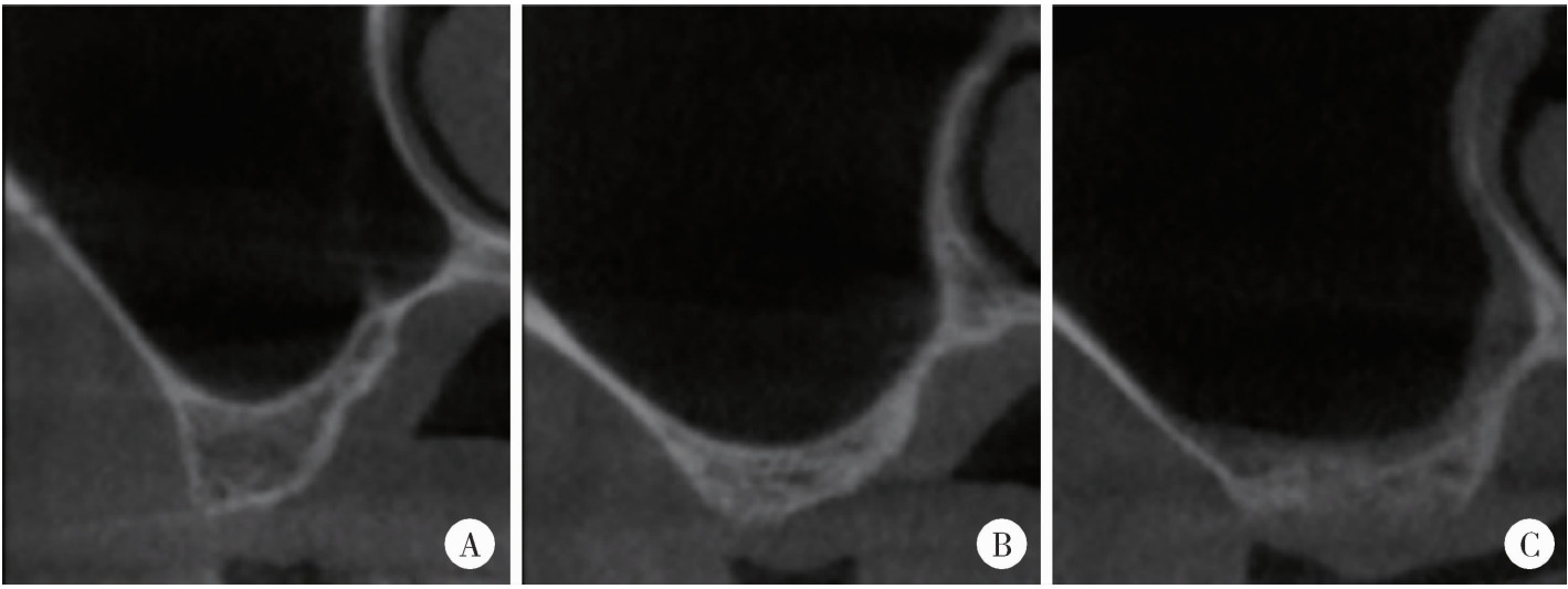
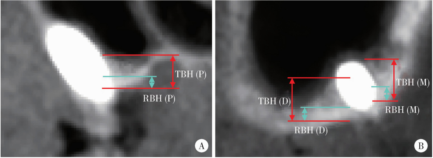
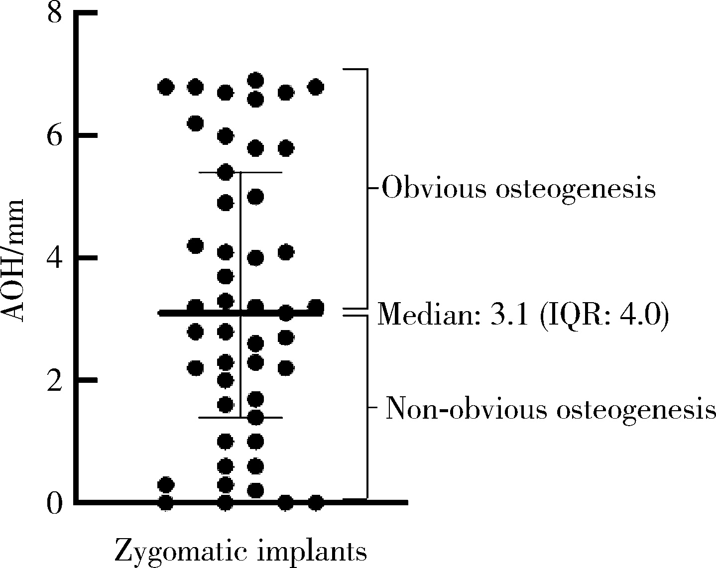
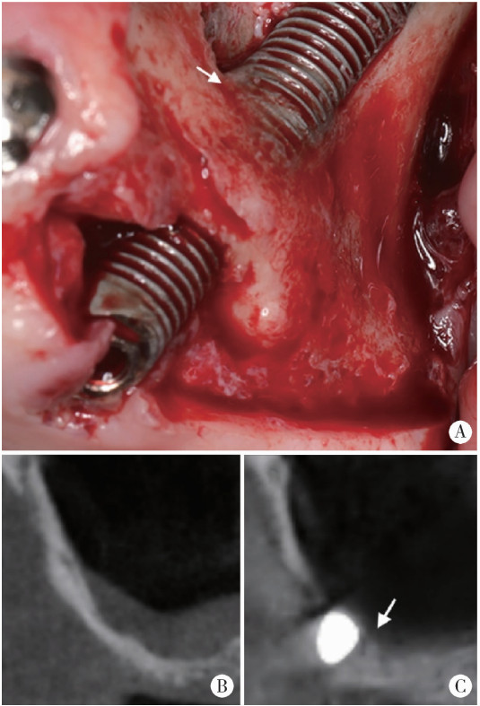
Imaging study of osteogenesis in maxillary sinus segment of zygomatic implants

Ziyang YU1, Houzuo GUO1, Xi JIANG1, Weihua HAN2, Ye LIN1,*()   

  1. 1. Department of Implantology, Peking University School and Hospital of Stomatology & National Center for Stomatology & National Clinical Research Center for Oral Diseases & National Engineering Research Center of Oral Biomaterials and Digi-tal Medical Devices & Beijing Key Laboratory of Digital Stomatology, Beijing 100081, China
    2. Department of Stomatology, Peking Union Medical College Hospital, Peking Union Medical College & Chinese Academy of Medical Science, Beijing 100730, China
  • Received:2023-02-23 Online:2025-10-18 Published:2025-09-05
  • Contact: Ye LIN
  • Supported by:
    the National Central Healthcare Research Project(2022—2027); the National Central Healthcare Research Project(2022ZD18)

RICH HTML


Abstract: Objective: To assess the osteogenesis height in maxillary sinus segment one year after zygomatic implantation by imaging methods, and evaluate the influence of patient factors, maxillary sinus anatomical factors and surgical factors on postoperative osteogenesis height. Methods: This study is a retrospective study, including patients who underwent zygomatic implantation and whose zygomatic implants passed through the maxillary sinus at the Department of Implantology, Peking University School and Hospital of Stomatology from July 2017 to January 2022. Preoperative and postoperative cone beam CT (CBCT)was taken to measure and calculate the average osteogenesis height (AOH) in maxillary sinus segment of the zygomatic implants, then the residual bone height, the width and morphology of the maxillary sinus floor in the buccal and palatal directions were measured. Besides, the integrity of Schneiderian membrane during implant surgery, and the general information of the patients and zygomatic implants were recorded. By comparing anatomical situations and surgical characteristics, the differences of AOH under different conditions were analyzed. Then AOH was divided into two groups (obvious osteogenesis group and non-obvious osteogenesis group) using the median as the threshold, and the influencing factors of osteogenesis were evaluated using mixed effect generalized linear model univariable and multivariable analysis. Results: A total of 47 zygomatic implants were implanted in 24 patients. During the average follow-up period of 12.1 months, there was no implant failure, and the implant survival rate was 100%. Postoperative CBCT showed that 43 zygomatic implants had osteogenic images in the maxillary sinus segment, most of which originated from the floor of the maxillary sinus, and the median AOH was 3.1 mm [interquartile range (IQR): 4.0 mm]. In terms of maxillary sinus width, there were 31 cases (66.0%) of wide type and 16 cases (34.0%) of narrow type. In the aspect of buccal and palatal morphology, 17 cases were taper (36.2%), 20 cases were round (42.6%), and 10 cases were flat (21.3%). The median of residual bone height was 2.8 mm (IQR: 2.2 mm) before operation. Univa-riate analysis of mixed effect generalized linear model showed that postoperative obvious osteogenic rate was related to the residual bone height (OR=2.09, P=0.006). Multivariate analysis showed that the resi-dual bone height (OR=2.55, P=0.022) and the shape of a taper maxillary sinus (OR=11.44, P=0.040) had a significant impact on the postoperative obvious osteogenic rate. Conclusion: The maxillary sinus floor showed osteogenic images 1 year after the zygomatic implantation surgery. Larger residual bone height and the shape of a taper maxillary sinus may be favorable factors for osteogenesis.

Key words: Dental implants, Zygoma, Maxillary sinus, Osteogenesis

CLC Number: 

  • R782.12

Figure 1

Division of edentulous maxilla by Bedrossian"

Figure 2

Measurement of RBH and PBD IM, implantation point of zygomatic implant; PBD, distance between the palatal and buccal wall; RBH, residual bone height."

Figure 3

Morphology of maxillary sinus in the buccal and palatal directions A, taper; B, round; C, flat."

Figure 4

Measurement of osteogenic height in maxillary sinus floor TBH, total bone height; RBH, residual bone height; P, palatal; M, mesial; D, distal."

Table 1

General information of patients (n=24)"

Items Data
Gender, n(%)
  Male 16 (66.7)
  Female 8 (33.3)
Age/years, n(%)
  30- 1 (4.2)
  40- 8 (33.3)
  50- 8 (33.3)
  60-69 7 (29.2)
General condition, n(%)
  Hypertension 6 (25.0)
  Diabetes 2 (8.3)
  Cardiovascular disease 1 (4.2)
Smoking history, n(%) 3 (12.5)

Table 2

General information of zygomatic implants (n=47)"

Items Data
Length of zygomatic implants/mm, ${\bar x}$±s 43.78±4.91
Site, n(%)
  Left 24 (51.1)
  Right 23 (48.9)
Survival, n(%)
  No 0 (0.0)
  Yes 47 (100.0)
Schneiderian membrane, n(%)
  Perforated 10 (21.3)
  Non-perforated 37 (78.7)

Table 3

Residual bone height of zygomatic implants"

Items n RBH/mm, median (IQR) P
Gender 0.025
  Male 31 2.40 (1.70)
  Female 16 3.90 (3.93)
Site 0.873
  Left 24 2.60 (2.43)
  Right 23 2.80 (2.20)
Total 47 2.80 (2.20)

Figure 5

Postoperative 1-year AOH of zygomatic implants AOH, average osteogenesis height; IQR, interquartile range."

Table 4

Univariate analysis of osteogenesis of maxillary sinus floor"

Examined variable n Obvious osteogenic rate/% β OR (95%CI) P
Gender
  Male 31 62.5 Reference
  Female 16 41.9 -0.842 0.43 (0.09, 1.52) 0.197
Age 47 48.9 0.038 1.04 (0.96, 1.14) 0.320
Smoker
  No 41 51.2 Reference
  Yes 6 33.3 -0.752 0.47 (0.05, 3.09) 0.426
Diabetes
  No 43 48.8 Reference
  Yes 4 50.0 -0.002 1.00 (0.05, 9.37) 0.998
RBH 47 48.9 0.738 2.09 (1.32, 4.57) 0.006
PBD
  Wide 31 48.4 Reference
  Narrow 16 50.0 0.094 1.10 (0.31, 5.44) 0.886
Morphology
  Round 20 35.0 Reference
  Taper 17 70.6 1.706 5.51 (0.84, 36.0) 0.075
  Flat 10 40.0 0.310 1.36 (0.23, 8.16) 0.734
Perforation
  No 28 57.1 Reference
  Yes 19 36.8 -0.827 0.44 (0.13, 1.48) 0.175
Site
  Left 24 50.0 Reference
  Right 23 47.8 -0.091 0.91 (0.28, 2.95) 0.878

Table 5

Multivariate analysis of osteogenesis of maxillary sinus floor"

Examined variables β OR (95%CI) P VIF
Morphology
  Round Reference
  Taper 0.937 11.44 (1.11, 117.4) 0.040 1.128
RBH 2.437 2.55 (1.14, 5.69) 0.022 1.272

Figure 6

Osteogenesis of the maxillary sinus floor during surgical exploration in a patient with peri-zygomatic infection A, surgical exploration, arrow indicating new bone formation; B, pre-operative cone beam CT (CBCT); C, CBCT at 12 months after surgery, arrow indicating new bone formation."

1
Tavelli C, Tedesco A. Survival and complication rate of zygomatic implants: A systematic review[J/OL]. J Oral Implantol (2022-12-06) [2023-01-24]. doi: 10.1563/aaid-joi-D-22-00008.
2
Brånemark PI , Svensson B , van Steenberghe D . Ten-year survival rates of fixed prostheses on four or six implants ad modum Brånemark in full edentulism[J]. Clin Oral Implants Res, 1995, 6 (4): 227- 231.

doi: 10.1034/j.1600-0501.1995.060405.x
3
Higuchi KW . The zygomaticus fixture: An alternative approach for implant anchorage in the posterior maxilla[J]. Ann R Australas Coll Dent Surg, 2000, 15, 28- 33.
4
Goker F , Grecchi F , Grecchi E , et al. Clinical outcomes of fully and partially threaded zygomatic implants in a cohort of patients with minimum 7.5-year follow-up[J]. Eur Rev Med Pharmacol Sci, 2022, 26 (Suppl 3): 35- 44.
5
Vrielinck L , Moreno-Rabie C , Coucke W , et al. Retrospective cohort assessment of survival and complications of zygomatic implants in atrophic maxillae[J]. Clin Oral Implants Res, 2023, 34 (2): 148- 156.

doi: 10.1111/clr.14027
6
Petrungaro PS , Gonzales S , Villegas C , et al. A retrospective study of a multi-center case series of 452 zygomatic implants placed over 5 years for treatment of severe maxillary atrophy[J]. Compend Contin Educ Dent, 2020, 41 (4): 232- 241.
7
周国辉. 穿颧骨植体在无牙上颌的应用[J]. 中国口腔种植学杂志, 2009, 14 (2): 28- 29.
8
Freedman M , Ring M , Stassen LA . Effect of alveolar bone support on zygomatic implants: A finite element analysis study[J]. Int J Oral Maxillofac Surg, 2013, 42 (5): 671- 676.

doi: 10.1016/j.ijom.2012.12.006
9
Freedman M , Ring M , Stassen LA . Effect of alveolar bone support on zygomatic implants in an extra-sinus position—a finite element analysis study[J]. Int J Oral Maxillofac Surg, 2015, 44 (6): 785- 790.

doi: 10.1016/j.ijom.2015.01.009
10
林柏均, 吕鸣樾, 袁泉. 影响经牙槽嵴顶上颌窦底提升术成骨效果的解剖因素分析[J]. 口腔医学, 2022, 42 (3): 193- 199.
11
Aparicio C . A proposed classification for zygomatic implant patient based on the zygoma anatomy guided approach (ZAGA): A cross-sectional survey[J]. Eur J Oral Implantol, 2011, 4 (3): 269- 275.
12
Bedrossian E , Sullivan RM , Fortin Y , et al. Fixed-prosthetic implant restoration of the edentulous maxilla: A systematic pretreatment evaluation method[J]. J Oral Maxillofac Surg, 2008, 66 (1): 112- 122.

doi: 10.1016/j.joms.2007.06.687
13
Aparicio C , López-Piriz R , Albrektsson T . ORIS criteria of success for the zygoma-related rehabilitation: The (revisited) zygoma success code[J]. Int J Oral Maxillofac Implants, 2020, 35 (2): 366- 378.

doi: 10.11607/jomi.7488
14
Avila G , Wang HL , Galindo-Moreno P , et al. The influence of the bucco-palatal distance on sinus augmentation outcomes[J]. J Periodontol, 2010, 81 (7): 1041- 1050.

doi: 10.1902/jop.2010.090686
15
Soardi CM , Spinato S , Zaffe D , et al. Atrophic maxillary floor augmentation by mineralized human bone allograft in sinuses of different size: An histologic and histomorphometric analysis[J]. Clin Oral Implants Res, 2011, 22 (5): 560- 566.

doi: 10.1111/j.1600-0501.2010.02034.x
16
Spinato S , Bernardello F , Galindo-Moreno P , et al. Maxillary sinus augmentation by crestal access: A retrospective study on cavity size and outcome correlation[J]. Clin Oral Implants Res, 2015, 26 (12): 1375- 1382.

doi: 10.1111/clr.12477
17
Niu L , Wang J , Yu H , et al. New classification of maxillary sinus contours and its relation to sinus floor elevation surgery[J]. Clin Implant Dent Relat Res, 2018, 20 (4): 493- 500.

doi: 10.1111/cid.12606
18
Stacchi C , Rapani A , Lombardi T , et al. Does new bone formation vary in different sites within the same maxillary sinus after lateral augmentation? A prospective histomorphometric study[J]. Clin Oral Implants Res, 2022, 33 (3): 322- 332.

doi: 10.1111/clr.13891
19
Lie SN , Claessen RA , Leung CW , et al. Non-grafted versus grafted sinus lift procedures for implantation in the atrophic maxilla: A systematic review and meta-analysis of randomized controlled trials[J]. Int J Oral Maxillofac Surg, 2022, 51 (1): 122- 132.

doi: 10.1016/j.ijom.2021.03.016
20
Rues S , Schmitter M , Kappel S , et al. Effect of bone quality and quantity on the primary stability of dental implants in a simulated bicortical placement[J]. Clin Oral Investig, 2021, 25 (3): 1265- 1272.

doi: 10.1007/s00784-020-03432-z
21
Danesh-Sani SA , Engebretson SP , Janal MN . Histomorphometric results of different grafting materials and effect of healing time on bone maturation after sinus floor augmentation: A systematic review and meta-analysis[J]. J Periodontal Res, 2017, 52 (3): 301- 312.

doi: 10.1111/jre.12402
22
Seike T , Hashimoto I , Matsumoto K , et al. Early postoperative evaluation of secondary bone grafting into the alveolar cleft and its effects on subsequent orthodontic treatment[J]. J Med Invest, 2012, 59 (1/2): 152- 165.
23
Dragonas P , Katsaros T , Schiavo J , et al. Osteogenic capacity of the sinus membrane following maxillary sinus augmentation procedures: A systematic review[J]. Int J Oral Implantol (Berl), 2020, 13 (3): 213- 232.
24
Jungner M , Cricchio G , Salata LA , et al. On the early mechanisms of bone formation after maxillary sinus membrane elevation: An experimental histological and immunohistochemical study[J]. Clin Implant Dent Relat Res, 2015, 17 (6): 1092- 1102.

doi: 10.1111/cid.12218
25
Hernández-Alfaro F , Torradeflot MM , Marti C . Prevalence and management of Schneiderian membrane perforations during sinus-lift procedures[J]. Clin Oral Implants Res, 2008, 19 (1): 91- 98.

doi: 10.1111/j.1600-0501.2007.01372.x
26
林野. 上颌窦植骨与种植[M]. 北京: 北京大学医学出版社, 2020: 205.
27
Lombardi T , Stacchi C , Berton F , et al. Influence of maxillary sinus width on new bone formation after transcrestal sinus floor elevation: A proof-of-concept prospective cohort study[J]. Implant Dent, 2017, 26 (2): 209- 216.

doi: 10.1097/ID.0000000000000554
28
Zheng X , Teng M , Zhou F , et al. Influence of maxillary sinus width on transcrestal sinus augmentation outcomes: Radiographic evaluation based on cone beam CT[J]. Clin Implant Dent Relat Res, 2016, 18 (2): 292- 300.

doi: 10.1111/cid.12298
29
郑小菲, 莫安春, 朱娟芳, 等. 上颌窦解剖因素对经牙槽嵴顶上颌窦底提升术成骨效果的影响[J]. 华西口腔医学杂志, 2020, 38 (6): 652- 656.
[1] Yulan WANG, Hao ZENG, Yufeng ZHANG. Current situation and exploration of clinical transformation of plasmatrix in oral implantology [J]. Journal of Peking University (Health Sciences), 2025, 57(5): 836-840.
[2] Fangru LIN, Zhihui TANG. Correlation analysis of peri-implant health after single-tooth dental implant [J]. Journal of Peking University (Health Sciences), 2025, 57(2): 347-353.
[3] Juan WANG, Lixin QIU, Huajie YU. Influence of emergence profile designs on the peri-implant tissue in the mandibular molar: A randomized controlled trial [J]. Journal of Peking University (Health Sciences), 2025, 57(1): 65-72.
[4] Congwei WANG,Min GAO,Yao YU,Wenbo ZHANG,Xin PENG. Clinical analysis of denture rehabilitation after mandibular fibula free-flap reconstruction [J]. Journal of Peking University (Health Sciences), 2024, 56(1): 66-73.
[5] Qian DING,Wen-jin LI,Feng-bo SUN,Jing-hua GU,Yuan-hua LIN,Lei ZHANG. Effects of surface treatment on the phase and fracture strength of yttria- and magnesia-stabilized zirconia implants [J]. Journal of Peking University (Health Sciences), 2023, 55(4): 721-728.
[6] Fei SUN,Jian LIU,Si-qi LI,Yi-ping WEI,Wen-jie HU,Cui WANG. Profiles and differences of submucosal microbial in peri-implantitis and health implants: A cross-sectional study [J]. Journal of Peking University (Health Sciences), 2023, 55(1): 30-37.
[7] LAN Lin,HE Yang,AN Jin-gang,ZHANG Yi. Relationship between prognosis and different surgical treatments of zygomatic defects: A retrospective study [J]. Journal of Peking University (Health Sciences), 2022, 54(2): 356-362.
[8] LI Yi,YU Hua-jie,QIU Li-xin. Clinical classification and treatment decision of implant fracture [J]. Journal of Peking University (Health Sciences), 2022, 54(1): 126-133.
[9] DU Wen-yu,YANG Jing-wen,JIANG Ting. Early constant observation of the effect of deferoxamine mesylate on improvement of vascularized bone regeneration in SD rat skull critical size defect model [J]. Journal of Peking University (Health Sciences), 2021, 53(6): 1171-1177.
[10] WANG Jing-qi,WANG Xiao. In vivo study of strontium-doped calcium phosphate cement for biological properties [J]. Journal of Peking University (Health Sciences), 2021, 53(2): 378-383.
[11] ZHANG Sheng-nan,AN Na,OUYANG Xiang-ying,LIU Ying-jun,WANG Xue-kui. Role of growth arrest-specific protein 6 in migration and osteogenic differentiation of human periodontal ligament cells [J]. Journal of Peking University (Health Sciences), 2021, 53(1): 9-15.
[12] LI Peng,PIAO Mu-zi,HU Hong-cheng,WANG Yong,ZHAO Yi-jiao,SHEN Xiao-jing. Radiography study on osteotome sinus floor elevation with placed implant simultaneously with no graft augmentation [J]. Journal of Peking University (Health Sciences), 2021, 53(1): 95-101.
[13] Mei WANG, Bo-wen LI, Si-wen WANG, Yu-hua LIU. Preparation and osteogenic effect study of small intestinal submucosa sponge [J]. Journal of Peking University (Health Sciences), 2020, 52(5): 952-958.
[14] Zhong ZHANG,Huan-xin MENG,Jie HAN,Li ZHANG,Dong SHI. Effect of vertical soft tissue thickness on clinical manifestation of peri-implant tissue in patients with periodontitis [J]. Journal of Peking University (Health Sciences), 2020, 52(2): 332-338.
[15] Chun-ping LIN,Song-he LU,Jun-xin ZHU,Hong-cheng HU,Zhao-guo YUE,Zhi-hui TANG. Influence of thread shapes of custom-made root-analogue implants on stress distribution of peri-implant bone: A three-dimensional finite element analysis [J]. Journal of Peking University(Health Sciences), 2019, 51(6): 1130-1137.
Viewed
Full text


Abstract

Cited

  Shared   
  Discussed   
No Suggested Reading articles found!