Journal of Peking University (Health Sciences) ›› 2026, Vol. 58 ›› Issue (1): 126-132. doi: 10.19723/j.issn.1671-167X.2026.01.016

Previous Articles     Next Articles

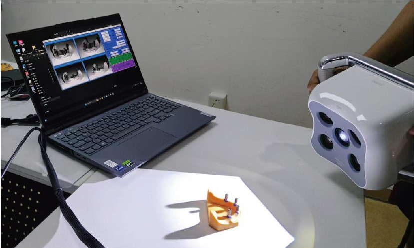
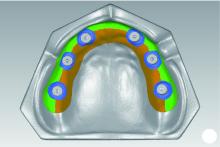
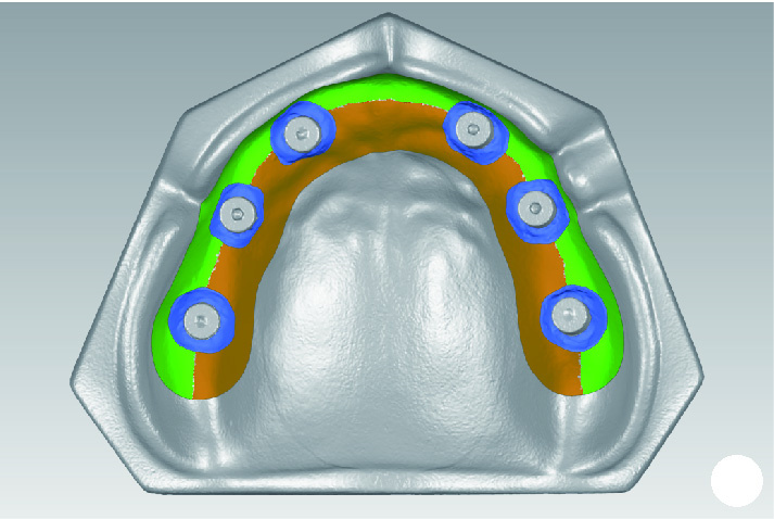
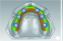
A multi-view stereo vision methodology for digital soft-tissue impressions in fixed implant rehabilitation of edentulous patients

Yongtao YANG1,2, Yuwen TIAN2, Shenyao SHAN2, Wenbo LI2, Xiangyi SHANG2, Yizhen WANG2, Shuwei GUO1, Zixiang GAO1, Aonan WEN1, Yijiao ZHAO1,2,*(), Yong WANG1,2,*()   

  1. 1. Center of Digital Dentistry, Department of Prosthodontics, Peking University School and Hospital of Stomatology & National Center for Stomatology & National Clinical Research Center for Oral Diseases & National Engineering Research Center of Oral Biomaterials and Digital Medical Devices & Beijing Key Laboratory of Digital Stomatology & NHC Key Laboratory of Digital Stomatology, Beijing 100081, China
    2. Institute of Medical Technology, Peking University Health Science Center, Beijing 100191, China
  • Received:2025-10-13 Online:2026-02-18 Published:2025-12-10
  • Contact: Yijiao ZHAO, Yong WANG
  • Supported by:
    the National Natural Science Foundation of China(82271039); the National Key Research and Development Program of China(2022YFC2405401); the Beijing Natural Science Foundation(L232100); the Beijing Natural Science Foundation(L242132)

RICH HTML


Abstract:

Objective: To explore the methodology and feasibility of reconstructing soft tissue morphology for fixed implant rehabilitation in edentulous patients using multi-view stereo vision technology, and to conduct a preliminary evaluation of the method's in vitro accuracy. Methods: A pair of edentulous resin models were designed and printed, with 6 implant analogs placed in the maxilla and 4 in the mandible. The experimental group (n=10) utilized a self-developed photogrammetric quad-camera system and the automated reconstruction software RealityScan 2.0.1. Self-developed scan bodies were attached to the analogs, and the handheld camera system was used to capture images of the models in vitro. The images were imported into the software to reconstruct the 3D models, and the data were exported as ".stl" files. The control group (n=10) used an intraoral scanner. Scan caps were attached to the analogs, and the models were scanned to generate ".stl" data. Reference data were obtained by scanning the maxillary and mandibular resin models once each with a desktop scanner (EX-PRO). All data were imported into Geomagic Wrap 2021. The root mean square (RMS) was calculated by comparing the 3D morphology of the experimental and control group data against the reference data to represent the magnitude of the 3D morphological deviation and evaluate accuracy. The evaluation was conducted in 4 specific regions: the alveolar ridge, peri-implant soft tissue, buccal, and lingual areas. Results: In the maxilla, the RMS of the experimental group was significantly higher than the control group in the alveolar ridge [(124.89±21.30) μm vs. (53.90±8.93) μm, P < 0.001], peri-implant soft tissue [(157.74±19.13) μm vs. (67.03±3.94) μm, P < 0.001], and lingual areas [(146.01±33.87) μm vs. (46.20±11.19) μm, P < 0.001]. The RMS in the buccal area was lower for the experimental group than the control group [(50.56±8.34) μm vs. (53.83±12.66) μm], but the difference was not statistically significant (P=0.571). In the mandible, the RMS of the experimental group was significantly higher than the control group in the alveolar ridge [(254.04±88.42) μm vs. (58.28±38.96) μm, P < 0.001], peri-implant soft tissue [(165.18±21.30) μm vs. (70.48±28.20) μm, P < 0.001], and lingual areas [(421.75±59.51) μm vs. (54.59±36.77) μm, P < 0.001]. When comparing the buccal and lingual sides, the lingual RMS was significantly higher than the buccal RMS for the experimental group in both the maxilla (P < 0.001) and mandible (P < 0.001). For the control group, the maxillary lingual RMS was significantly lower than the buccal RMS (P < 0.05), while the mandibular lingual RMS was higher than the buccal, but the difference was not statistically significant (P=0.378). Conclusion: The self-developed quad-camera system, combined with multi-view stereo vision reconstruction software, can successfully record the 3D morphology of soft tissue. This study provides a research foundation for the development of extraoral photogrammetric devices capable of simultaneously determining the spatial positions of multiple implant units and acquiring soft tissue morphology.

Key words: Multi-view stereo, Jaw, edentulous, Digital impression, Dental implantation

CLC Number: 

  • R783.6

Figure 1

Edentulous maxillary implant model"

Figure 2

Edentulous mandibular implant model"

Figure 3

Handheld scanning procedure with the self-developed camera system"

Figure 4

The boundary scope of the respective regions of the maxilla"

Figure 5

The boundary scope of the respective regions of the mandible"

Figure 6

Color map of maxillary 3D morphological deviation analysis A, alveolar ridge; B, peri-implant soft tissue; C, buccal area; D, lingual area."

Figure 7

Color map of mandibular 3D morphological deviation analysis A, alveolar ridge; B, peri-implant soft tissue; C, buccal area; D, lingual area."

Table 1

Comparison of morphological trueness between experimental group and control group in the maxillary and mandibular"

Items Alveolar ridge Peri-implant soft tissue Buccal area Lingual area
Maxillary
  Experimental group/μm, ${\bar x}$±s 124.89±21.30 157.74±19.13 50.56±8.34 146.01±33.87
  Control group/μm, ${\bar x}$±s 53.90±8.93 67.03±3.94 53.83±12.66 46.20±11.19
  t 9.949 15.643 -0.587 8.984
  P < 0.001 < 0.001 0.571 < 0.001
Mandibular
  Experimental group/μm, ${\bar x}$±s 254.04±88.42 165.18±21.30 57.32±8.26 421.75±59.51
  Control group/μm, ${\bar x}$±s 58.28±38.96 70.48±28.20 49.71±49.21 54.59±36.77
  t 7.031 11.170 0.448 17.466
  P < 0.001 < 0.001 0.665 < 0.001

Table 2

Comparison of morphological trueness between the buccal and lingual areas"

Items Experimental group in mandibular Control group in mandibular Experimental group in maxillary Control group in maxillary
Buccal area/μm, ${\bar x}$±s 50.56±8.34 53.83±12.66 57.32±8.26 49.71±49.21
Lingual area/μm, ${\bar x}$±s 146.01±33.87 46.20±11.19 421.75±59.51 54.59±36.77
t -9.476 2.876 -18.262 -0.928
P < 0.001 < 0.05 < 0.001 0.378
1
周永胜. 口腔修复学[M]. 3版 北京: 北京大学医学出版社, 2020.
2
Rutkūnas V, Gedrimiene A, Mischitz I, et al. EPA consensus project paper: Accuracy of photogrammetry devices, intraoral scanners, and conventional techniques for the full-arch implant impressions: A systematic review [J/OL]. Eur J Prosthodont Restor Dent, 2023 [2023-6-13]. https://pubmed.ncbi.nlm.nih.gov/37314199/.
3
Abdelrehim A , Etajuri EA , Sulaiman E , et al. Magnitude of misfit threshold in implant-supported restorations: A systematic review[J]. J Prosthet Dent, 2024, 132 (3): 528- 535.

doi: 10.1016/j.prosdent.2022.09.010
4
Hussein MO . Photogrammetry technology in implant dentistry: A systematic review[J]. J Prosthet Dent, 2023, 130 (3): 318- 326.

doi: 10.1016/j.prosdent.2021.09.015
5
杨咏涛, 温奥楠, 商相宜, 等. 基于摄影测量技术的无牙颌种植口外扫描系统的研发和精度评价[J]. 中华口腔医学杂志, 2025, 60 (8): 863- 870.
6
Brakoč J , Todorović A , Mangano FG , et al. Accuracy of intraoral photogrammetry versus direct digital implant impressions in the fully edentulous lower jaw: An in vitro study[J]. J Dent, 2025, 156, 105654.

doi: 10.1016/j.jdent.2025.105654
7
张力, 刘玉轩, 孙洋杰, 等. 数字航空摄影三维重建理论与技术发展综述[J]. 测绘学报, 2022, 51 (7): 1437- 1457.
8
龚健雅, 季顺平. 从摄影测量到计算机视觉[J]. 武汉大学学报(信息科学版), 2017, 42 (11): 1518-1522, 1615.
9
Revilla-León M, Cascos R, Barmak AB, et al. Registration accuracy of soft tissue information scan captured using an intraoral scanner and implant position scan recorded using extraoral and intraoral photogrammetry systems [J/OL]. J Prosthet Dent, 2025: S0022-S3913(25)00392-0 [2025-05-30]. https://pubmed.ncbi.nlm.nih.gov/40450447/.
10
Revilla-León M , Cascos R , Barmak AB , et al. Alignment accuracy of soft tissue and implant position scans captured by using four intraoral scanners and an intraoral photogrammetry system with and without coded healing abutments[J]. J Prosthet Dent, 2025, 134 (4): 1259. e1- 1259. e10.

doi: 10.1016/j.prosdent.2025.06.015
11
Ke Y , Zhang Y , Tian S , et al. Accuracy of digital implant impressions using a novel structured light scanning system assisted by a planar mirror in the edentulous maxilla: An in vitro study[J]. Clin Oral Implants Res, 2024, 35 (8): 876- 887.

doi: 10.1111/clr.14208
12
Mourouzis P . Critical methodological factors influencing the accuracy of intraoral scanners in digital dentistry research[J]. Comput Biol Med, 2025, 187, 109780.

doi: 10.1016/j.compbiomed.2025.109780
13
Limones A , Cascos R , Molinero-Mourelle P , et al. Impact of the superimposition methods on accuracy analyses in complete-arch digital implant investigation[J]. J Dent, 2024, 147, 105081.

doi: 10.1016/j.jdent.2024.105081
14
樊铭瑞, 申冰可, 牛文龙, 等. 基于深度学习的多视图立体视觉综述[J]. 软件学报, 2025, 36 (4): 1692- 1714.
15
Li J, Chen Z, Liu F, et al. Obtaining full-arch implant scan with smartphone video and deep learning: An in vitro investigation on trueness and precision [J/OL]. J Prosthodont, 2025 [2025-03-08]. https://pubmed.ncbi.nlm.nih.gov/40055947/.
16
Kernen-Gintaute A , Akulauskas M , Kernen F , et al. Accuracy of digital and conventional implant impressions in edentulous jaws: A clinical comparative study[J]. J Dent, 2025, 153, 105559.

doi: 10.1016/j.jdent.2025.105559
[1] Hong LI, Feifei MA, Jinlong WENG, Yang DU, Binzhang WU, Feng SUN. Accuracy of dynamic navigation system for immediate dental implant placement [J]. Journal of Peking University (Health Sciences), 2025, 57(1): 85-90.
[2] Xiaoqiang LIU,Yin ZHOU. Risk factors of perioperative hypertension in dental implant surgeries with bone augmentation [J]. Journal of Peking University (Health Sciences), 2024, 56(1): 93-98.
[3] Meng-en OU,Yun DING,Wei-feng TANG,Yong-sheng ZHOU. Three-dimensional finite element analysis of cement flow in abutment margin-crown platform switching [J]. Journal of Peking University (Health Sciences), 2023, 55(3): 548-552.
[4] WANG Juan,YU Hua-jie,SUN Jing-de,QIU Li-xin. Application evaluation of prefabricated rigid connecting bar in implants immediate impression preparation of edentulous jaw [J]. Journal of Peking University (Health Sciences), 2022, 54(1): 187-192.
[5] LIU Xiao-qiang,YANG Yang,ZHOU Jian-feng,LIU Jian-zhang,TAN Jian-guo. Blood pressure and heart rate changes of 640 single dental implant surgeries [J]. Journal of Peking University (Health Sciences), 2021, 53(2): 390-395.
[6] Ke-yi HAO,Jia LUO,Ping DI,Hou-zuo GUO,Hui-dan SHEN,Yan-ping LIU,Yu ZHANG,Ye LIN. Validation of the digital integration technology for evaluating the nasolabial morphology variation after the cross-arch fixed restoration of maxillary implant-supported prostheses [J]. Journal of Peking University (Health Sciences), 2020, 52(5): 924-930.
[7] Yue CAO,Jun-kai CHEN,Ke-hui DENG,Yong WANG,Yu-chun SUN,Yi-jiao ZHAO. Accuracy of three intraoral scans for primary impressions of edentulous jaws [J]. Journal of Peking University(Health Sciences), 2020, 52(1): 129-137.
[8] Wei-ting LI,Peng LI,Mu-zi PIAO,Fang ZHANG,Jie DI. Study on bone volume harvested from the implant sites with different methods [J]. Journal of Peking University(Health Sciences), 2020, 52(1): 103-106.
[9] Qiang LUO,Qian DING,Lei ZHANG,Qiu-fei XIE. Quantitative analysis of occlusal changes in posterior partial fixed implant supported prostheses [J]. Journal of Peking University(Health Sciences), 2019, 51(6): 1119-1123.
[10] CHAI Jin-you, LIU Jian-zhang, WANG Bing, QU Jian, SUN Zhen, GAO Wen-hui, GUO Tian-hao, FENG Hai-lan, PAN Shao-xia. Evaluation of the fabrication deviation of a kind of milling digital implant surgical guides#br# [J]. Journal of Peking University(Health Sciences), 2018, 50(5): 892-898.
[11] WU Min-jie, ZOU Li-dong, LIANG Feng. Clinical observation on soft and hard tissue changes of immediate implantation and immediate reconstruction in anterior region after loading 3 years [J]. Journal of Peking University(Health Sciences), 2018, 50(4): 694-699.
[12] ZHANG Hai-dong, ZHANG Li, SHI Dong, HAN Jie, YAN Xia, XIE Ye-si, MENG Huan-xin. Clinical study of locking-taper implants in patients treated for periodontitis [J]. Journal of Peking University(Health Sciences), 2018, 50(2): 300-307.
[13] LI Shi-ying, LI Gang, FENG Hai-lan, PAN Shao-xia. Influence of the interforaminal arch form of edentulous mandibles on design of “All-on-4”: preliminary research based on conebeam computed tomography [J]. Journal of Peking University(Health Sciences), 2017, 49(4): 699-703.
[14] CHEN Quan, ZHANG Xiao, ZHANG Zhi-yong, GAO Wei, LIU Wen-shu, MENG Tian, CHEN Yu-huan, WANG Hui-li. Detection and management of the vascular paths in the lateral sinus wall using cone beam computed tomography [J]. Journal of Peking University(Health Sciences), 2017, 49(3): 540-546.
[15] ZHAO Li-ping, ZHAN Ya-lin, HU Wen-jie, WANG Hao-jie, WEI Yi-ping, ZHEN Min, Xu Tao, LIU Yun-song. Dental implantation and soft tissue augmentation after ridge preservation in a molar site: a case report [J]. Journal of Peking University(Health Sciences), 2016, 48(6): 1090-1094.
Viewed
Full text


Abstract

Cited

  Shared   
  Discussed   
No Suggested Reading articles found!