Journal of Peking University(Health Sciences) ›› 2019, Vol. 51 ›› Issue (4): 753-757. doi: 10.19723/j.issn.1671-167X.2019.04.028
Previous Articles Next Articles
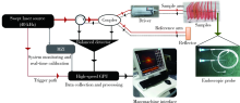
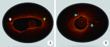
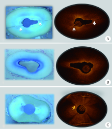
Construction of swept source optical coherence tomography imaging system for root canal endoscopy and application in diagnosis of root fractures
Li-yuan QI1,Chen CHEN2,Lan JIANG2,Jia-nan LI3,△( ),Yu-hong LIANG1,4,△()
- 1. Department of Cariology and Endodontology,Peking University School and Hospital of Stomatology & National Clinical Research Center for Oral Diseases & National Engineering Laboratory for Digital and Material Technology of Stomatology & Beijing Key Laboratory of Digital Stomatology,Beijing 100081,China
2. First Clinical Division,Peking University School and Hospital of Stomatology,Beijing 100034,China
3. State Key Laboratory of Transient Optics and Photonics,Xian Institute of Optics and Precision Mechanics,Chinese Academy of Sciences,Xian,Shanxi 710000,China
4. Department of Stomatology,Peking University International Hospital,Beijing 102206,China
CLC Number:
- R781


| [1] | Fercher AF, Drexler W, Hitzenberger CK , et al. Optical coherence tomography-principles and applications[J]. Rep Prog Phys, 2003,66(2):239. |
| [2] | Oliveira BPD , CÂmara AC, Duarte DA, et al. Detection of apical root cracks using spectral domain and swept-source optical co-herence tomography [J]. Int Endod J, 2017,43(7):1148-1151. |
| [3] | Lavinsky F, Lavinsky D . Novel perspectives on swept-source optical coherence tomography[J]. Int J Retina Vitreous, 2016,2(1):25. |
| [4] | Ha FJ, Giblett JP, Nerlekar N , et al. Optical coherence tomography guided percutaneous coronary intervention[J]. Heart Lung Circ, 2017,26(12):1267-1276. |
| [5] | Colston B, Everett M, Da Silva LB , et al. Imaging of hard-and soft-tissue structure in the oral cavity by optical coherence tomography[J]. Applied Optics, 1998,37(16):3582-3585. |
| [6] | Baumgartner A, Dichtl S, Hitzenberger CK , et al. Polarization-sensitive optical coherence tomography of dental structures[J]. Caries Res, 1999,34(1):59-69. |
| [7] | Shemesh H, van Soest G, Wu MK , et al. Diagnosis of vertical root fractures with optical coherence tomography[J]. J Endod, 2008,34(6):739-742. |
| [8] | Zain E, Zakian CM, Chew HP . Influence of the loci of non-cavitated fissure caries on its detection with optical coherence tomography[J]. J Dent, 2018,71(4):31-37. |
| [9] | Majkut P, Sadr A, Shimada Y , et al. Validation of optical coherence tomography against micro-computed tomography for evaluation of remaining coronal dentin thickness[J]. J Endod, 2015,41(8):1349-1352. |
| [10] | Han SH, Sadr A, Tagami J , et al. Non-destructive evaluation of an internal adaptation of resin composite restoration with swept-source optical coherence tomography and micro-CT[J]. Dent Mater, 2015,32(1):e1-e7 |
| [11] | Yoshioka T, Sakaue H, Ishimura H , et al. Detection of root surface fractures with swept-source optical coherence tomography (SS-OCT)[J]. Photomed Laser Surg, 2013,31(1):23-27. |
| [12] | 陈晨, 章文欣, 戚苈源 , 等. 光学相干断层扫描技术诊断牙根裂的实验研究[J]. 北京大学学报(医学版), 2018,50(3):547-552. |
| [13] | Wang P, Yan X, Liu D , et al. Detection of dental root fractures by using cone-beam computed tomography[J]. Dentomaxillofac Radiol, 2011,40(5):290-298. |
| [14] | Li G . Patient radiation dose and protection from cone-beam computed tomography[J]. Imaging Sci Dent, 2013,43(2):63-69. |
| [15] | Paul RA, Tamse A, Rosenberg E . Cracked and broken teeth definitions, differential diagnosis and treatment[J]. Refuat Hapeh Vehashinayim, 2007,24(2):7-12. |
| [16] | Huang D, Swanson EA, Lin CP , et al. Optical coherence tomography[J]. Science, 1991,254(5035):1178-1181 |
| [17] | Bahcall JK, Barss JT . Fiberoptic endoscope usage for intracanal visualization[J]. J Endod, 2001,27(2):128-129. |
| [18] | Hassan B, Metska ME, Ozok AR , et al. Detection of vertical root fractures in endodontically treated teeth by a cone beam computed tomography scan[J]. J Endod, 2009,35(5):719-722. |
| [19] | Özer SY . Detection of vertical root fractures of different thicknesses in endodontically enlarged teeth by cone beam computed tomography versus digital radiography[J]. J Endod, 2010,36(7):1245-1249. |
| [20] | Patel S, Brady E, Wilson R , et al. The detection of vertical root fractures in root filled teeth with periapical radiographs and CBCT scans[J]. Int Endod J, 2013,46(12):1140-1152. |
| [21] | Chavda R, Mannocci F, Andiappan M , et al. Comparing the in vivo diagnostic accuracy of digital periapical radiography with cone-beam computed tomography for the detection of vertical root fracture[J]. J Endod, 2014,40(10):1524-1529. |
| [22] | Makeeva IM, Byakova SF, Novozhilova NE , et al. Detection of artificially induced vertical root fractures of different widths by cone beam computed tomography in vitro and in vivo[J]. Int Endod J, 2016,49(10):980-989. |
| [1] | Jing YANG, Xiaoyun XU, Danni ZHENG, Xiaotong LING, Liuyang QU, Denggao LIU. Clinical and imaging characteristics and etiology of 544 cases with chronic sialadenitis [J]. Journal of Peking University (Health Sciences), 2026, 58(3): 650-657. |
| [2] | Xinying WANG, Xueyuan CHENG, Mengjun ZHANG, Fei LI, Jinyu DUAN, Jing QIAO. Effect of concentrated growth factors in guided tissue regeneration for the treatment of mandibular molar furcation lesions [J]. Journal of Peking University (Health Sciences), 2026, 58(2): 372-379. |
| [3] | Xin CONG, Jiazeng SU, Liling WU, Chong DING, Wei LI, Guangyan YU. Research progress in diagnosis and treatment of non-tumorous salivary gland diseases [J]. Journal of Peking University (Health Sciences), 2026, 58(1): 1-9. |
| [4] | Yuting YANG, Liuyang QU, Danni ZHENG, Xiaotong LING, Xiaoyun XU, Denggao LIU. Demographic characteristic and clinical features in 1 812 patients with salivary gland stones [J]. Journal of Peking University (Health Sciences), 2026, 58(1): 153-159. |
| [5] | Hailing ZANG, Yuhong LIANG. Endodontic retreatment of a maxillary second molar with chronic apical periodontitis and separated instrument: A case report [J]. Journal of Peking University (Health Sciences), 2026, 58(1): 214-219. |
| [6] | Lianfei PAN, Wenjing LI, Ruiyang WANG, Jian JIAO, Zhanqiang CAO, Li GAO, Dong SHI. Short-term efficacy and influencing factors of systemic antibiotics as an adjunct to mechanical periodontal therapy for stages Ⅲ/Ⅳ periodontitis [J]. Journal of Peking University (Health Sciences), 2026, 58(1): 30-36. |
| [7] | Fei SUN, Cui WANG, Siqi LI, Yiping WEI, Riyue YU, Wenjie HU. Treatment of peri-implant mucositis using an erythritol air-polishing or ultrasonic device: A randomized controlled trial [J]. Journal of Peking University (Health Sciences), 2026, 58(1): 37-42. |
| [8] | Rentao TANG, Liuchang YANG, Jie NIE, Xiaoyan WANG. Microbial communities in extraradicular infections of post-treatment apical periodontitis without or with sinus tracts [J]. Journal of Peking University (Health Sciences), 2026, 58(1): 43-49. |
| [9] | Ziyu HE, Hui ZHANG, Zhibin CHEN, Haixia XING, Jie PAN. Isolation, identification, and metabolic characterization of a Veillonella parvula isolated from supragingival plaque in a patient with rampant caries [J]. Journal of Peking University (Health Sciences), 2026, 58(1): 50-59. |
| [10] | Yanting CHI, Hongjie JIANG, Yan CHEN, Zhixiu XU, Binbin LI. Value of direct immunofluorescence in the diagnosis of oral mucosal pemphigus vulgaris: A retrospective study based on multi-index combined analysis [J]. Journal of Peking University (Health Sciences), 2026, 58(1): 68-73. |
| [11] | Baojin MA, Jianhua LI, Yuanhua SANG, Yang YU, Jichuan QIU, Jinlong SHAO, Kai LI, Shiyue LIU, Mi DU, Lingling SHANG, Shaohua GE. Establishment and application of key technologies for periodontal tissue regeneration based on microenvironment and stem cell regulation [J]. Journal of Peking University (Health Sciences), 2025, 57(5): 841-846. |
| [12] | Pei CAO, Qingxian LUAN. Periodontitis and systemic diseases: Thinking and explorations [J]. Journal of Peking University (Health Sciences), 2025, 57(5): 852-858. |
| [13] | Xiangyu SUN, Chao YUAN, Xinzhu ZHOU, Jing DIAO, Shuguo ZHENG. Application of salivary micro-ecosystem in early prevention and control of oral and systemic diseases [J]. Journal of Peking University (Health Sciences), 2025, 57(5): 859-863. |
| [14] | Zhenying BAO, Yajie WANG. Application of combined detection of inflammatory indexes and cytokines in chronic periodontitis [J]. Journal of Peking University (Health Sciences), 2025, 57(4): 772-778. |
| [15] | 朱慧, 闵赛南, 苏家增, 陈艳, 彭歆, 于尧, 俞光岩. [J]. Journal of Peking University (Health Sciences), 2025, 57(3): 620-625. |
|
||