Journal of Peking University(Health Sciences) ›› 2020, Vol. 52 ›› Issue (1): 107-112. doi: 10.19723/j.issn.1671-167X.2020.01.017
Previous Articles Next Articles
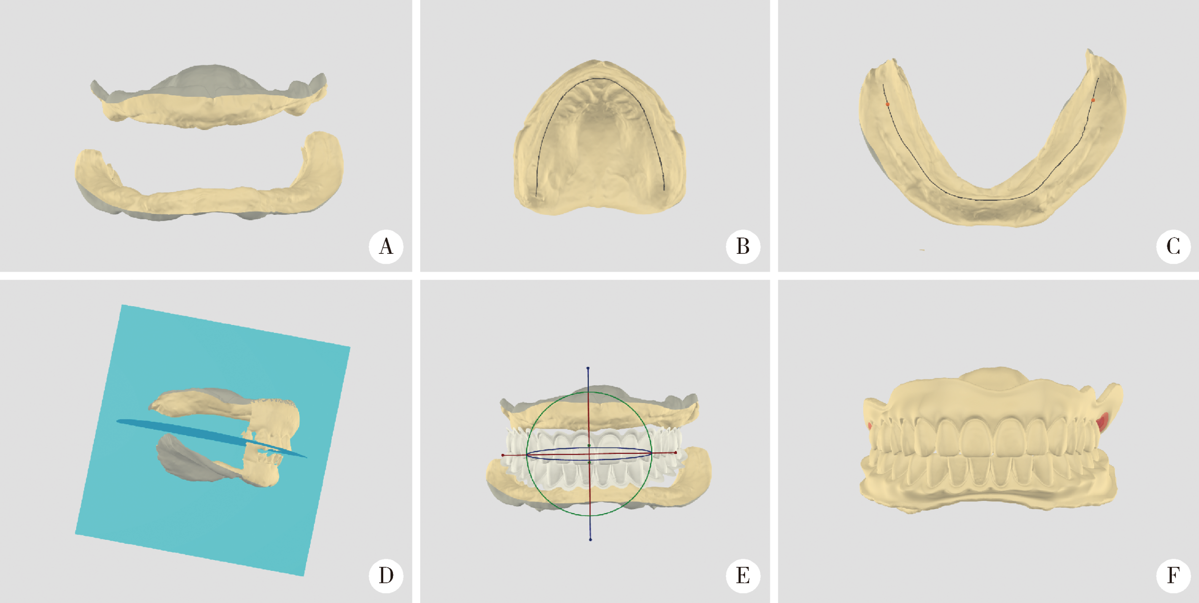
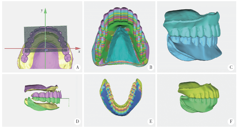
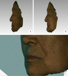
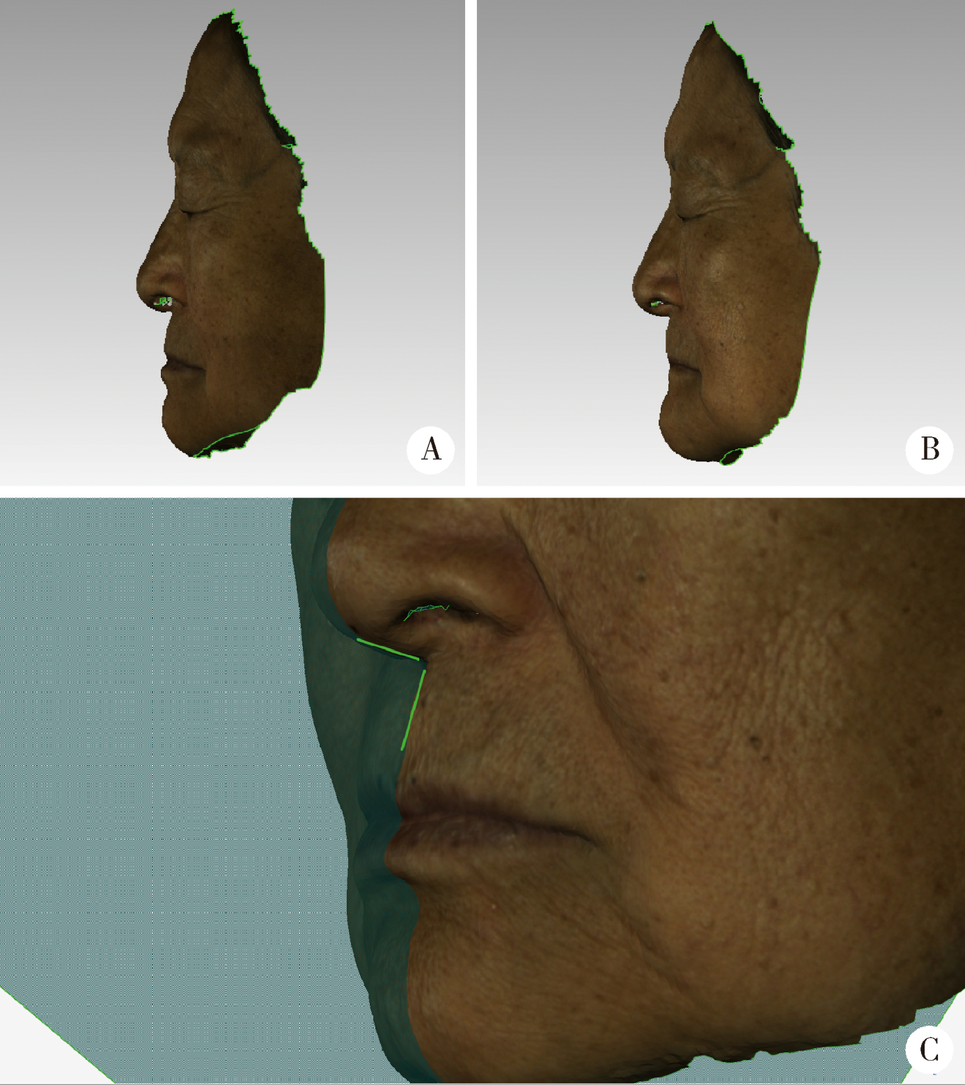
Visual sensitivity threshold of lateral view of nasolabial Angle changes in edentulous jaw patients
Lang YOU1,Ke-hui DENG2,Wei-wei LI2,Yi-jiao ZHAO2,△( ),Yu-chun SUN2,△(),Yong-sheng ZHOU1
- 1. Center of Digital Dentistry, Peking University School and Hospital of Stomatology & Department of Prosthodontics, Beijing 100081, China
2. Center of Digital Dentistry, Peking University School and Hospital of Stomatology & National Clinical Research Center for Oral Diseases & National Engineering Laboratory for Digital and Material Technology of Stomatology & Beijing Key Laboratory of Digital Stomatology, Beijing 100081, China
CLC Number:
- R783


| [1] | Harris R, Nagarkar P, Amirlak B . Varied definitions of nasolabial angle[J]. Plast Reconstr Surg Glob Open, 2016,4(6):e752. |
| [2] | 徐安秀, 邓锋, 王芬芬 , 等. 鼻唇角改变对骨性Ⅰ类软组织侧貌影响的审美评价[J]. 华西口腔医学杂志, 2015,33(5):492-496. |
| [3] | 马玥, 任嫒姝, 付钢 , 等. 鼻唇角、颏唇角改变对骨性Ⅱ类和Ⅲ类患者面容影响的三维美学评价[J]. 实用口腔医学杂志, 2017,33(5):647-652. |
| [4] | Desesa CR, Metzler P, Sawh-Martinez R , et al. Three-dimensional nasolabial morphologic alterations following Le Fort I[J]. Plast Reconstr Surg Glob Open, 2016,4(8):e848. |
| [5] | Aniruddh YV, Ravi K, Edeinton A. Comparative evaluation of soft tissue changes in Class Ⅰ borderline patients treated with extraction and nonextraction modalities[J]. Dental Press J Orthod, 2016,21(4):50-59. |
| [6] | Kamashita Y, Kamada Y, Kawahata N , et al. Influence of lip support on the soft-tissue profile of complete denture wearers[J]. J Oral Rehabil, 2006,33(2):102-109. |
| [7] | Kaipatur NR, Flores-Mir C . Accuracy of computer programs in predicting orthognathic surgery soft tissue response[J]. J Oral Maxillofac Surg, 2009,67(4):751-759. |
| [8] | Raschke GF, Rieger UM, Bader RD , et al. Perioral aging: an anthropometric appraisal[J]. J Craniomaxillofac Surg, 2014,42(5):e312-e317. |
| [9] | Sierpinska T, Golebiewska M, Kuc J , et al. The influence of the occlusal vertical dimension on masticatory muscle activities and hyoid bone position in complete denture wearers[J]. Adv Med Sci, 2009,54(1):104-108. |
| [10] | Krajicek DD . Guides for natural facial appearance as related to complete denture construction[J]. J Prosthet Dent, 1969,21(6):654-662. |
| [11] | Coleman SR, Grover R . The anatomy of the aging face: volume loss and changes in 3-dimensional topography[J]. Aesthet Surg J, 2006,26(Suppl 1):S4-S9. |
| [12] | Owens EG, Goodacre CJ, Loh PL , et al. A multicenter interracial study of facial appearance. Part 1: A comparison of extraoral parameters[J]. Int J Prosthodont, 2002,15(3):273-282. |
| [13] | Yuan F, Cheng C, Dai N , et al. Prediction of aesthetic reconstruction effects in edentulous patients[J]. Sci Rep, 2017,7(1):18077. |
| [14] | Denes BJ, Bolton C, Illsley CS , et al. Notch coordinates periodontal ligament maturation through regulating lamin A[J]. J Dent Res, 2019,98(12):1357-1366. |
| [15] | Katase H, Kanazawa M, Inokoshi M , et al. Face simulation system for complete dentures by applying rapid prototyping[J]. J Prosthet Dent, 2013,109(6):353-360. |
| [16] | Schweiger J, Güth JF, Edelhoff D , et al. Virtual evaluation for cad-cam-fabricated complete dentures[J]. J Prosthet Dent, 2017,117(1):28-33. |
| [17] | Franco SGC, Libdy MR, Normando D . Scan time, reliability and accuracy of craniofacial measurements using a 3D light scanner[J]. J Oral Biol Craniofac Res, 2019,9(4):331-335. |
| [18] | Modabber A, Peters F, Kniha K , et al. Evaluation of the accuracy of a mobile and a stationary system for three-dimensional facial scanning[J]. J Craniomaxillofac Surg, 2016,44(10):1719-1724. |
| [19] | Grant CA, Johnston M, Adam CJ , et al. Accuracy of 3D surface scanners for clinical torso and spinal deformity assessment[J]. Med Eng Phys, 2019,63:63-71. |
| [20] | Li H, Lyu P, Sun Y , et al. A quantitative study of 3D-scanning frequency and Δd of tracking points on the tooth surface[J]. Sci Rep, 2015,5(2):14350. |
| [21] | 萧宁, 王勇, 赵一姣 . 三维颜面部软组织正中矢状面确定方法的研究进展[J]. 中华口腔医学杂志, 2018,53(7):495-499. |
| [22] | Lee JK, Jung PK, Moon CH . Three-dimensional cone beam computed tomographic image reorientation using soft tissues as reference for facial asymmetry diagnosis[J]. Angle Orthod, 2014,84(1):38-47. |
| [23] | Nur RB, Çakan DG, Arun T . Evaluation of facial hard and soft tissue asymmetry using cone-beam computed tomography[J]. Am J Orthod Dentofacial Orthop, 2016,149(2):225-237. |
| [1] | Xiaoqiang BAI, Zhiruo YUAN, Yongsheng ZHOU, Longwei LV. Dynamic stretching promotes osteogenic differentiation of human bone marrow mesenchymal stem cells in three-dimensional culture [J]. Journal of Peking University (Health Sciences), 2026, 58(3): 641-649. |
| [2] | Aonan WEN, Xiaohui ZHANG, Yongtao YANG, Zixiang GAO, Wenbo LI, Shenyao SHAN, Xiangyi SHANG, Yuwen TIAN, Shuwei GUO, Yizhen WANG, Yong WANG, Yijiao ZHAO. Method of constructing 3D facial smile simulation sequence data based on non-rigid registration [J]. Journal of Peking University (Health Sciences), 2026, 58(1): 139-144. |
| [3] | Lu YU, Ling WU, Xiaojing LIU, Zili LI. Feasibility study of a surgical planning protocol for orthognathic surgery utilizing similarity retrieval from database: A randomized controlled trial [J]. Journal of Peking University (Health Sciences), 2026, 58(1): 145-152. |
| [4] | Yuting YANG, Liuyang QU, Danni ZHENG, Xiaotong LING, Xiaoyun XU, Denggao LIU. Demographic characteristic and clinical features in 1 812 patients with salivary gland stones [J]. Journal of Peking University (Health Sciences), 2026, 58(1): 153-159. |
| [5] | Liang SHAO, Wenjie MA, Ying CHEN, Qian DING, Lei ZHANG. Digital measurement and analysis of anatomical characteristics of protrusive and intercuspal position occlusal contacts in maxillary incisors [J]. Journal of Peking University (Health Sciences), 2026, 58(1): 99-106. |
| [6] | Cuiping WANG, Zhe CHEN, Yongjing CHENG. Correlation study of superb microvascular imaging on knee osteoarthritis [J]. Journal of Peking University (Health Sciences), 2025, 57(6): 1096-1100. |
| [7] | Jianxiao ZHAO, Qian DING, Wenjin LI, Quanquan MA, Yixiao LAN, Lei ZHANG, Jianmin HAN. Effect of porous surface structure on fatigue strength of 3D printed zirconia [J]. Journal of Peking University (Health Sciences), 2025, 57(6): 1165-1173. |
| [8] | Yujia XIAO, Bochun MAO, Yanheng ZHOU. Three-dimensional morphological analysis of posed smile [J]. Journal of Peking University (Health Sciences), 2025, 57(5): 989-995. |
| [9] | Zeyuan WANG, Shuanbao YU, Haoke ZHENG, Jin TAO, Yafeng FAN, Xuepei ZHANG. A preoperative prediction model for pelvic lymph node metastasis in prostate cancer: Integrating clinical characteristics and multiparametric MRI [J]. Journal of Peking University (Health Sciences), 2025, 57(4): 684-691. |
| [10] | Jianjun SUN, Qianquan MA, Xiaoliang YIN, Chenlong YANG, Jia ZHANG, Suhua CHEN, Chao WU, Jingcheng XIE, Yunfeng HAN, Guozhong LIN, Yu SI, Jun YANG, Haibo WU, Qiang ZHAO. Significance of precise classification of sacral meningeal cysts by multiple dimensions radiographic reconstruction MRI in guiding operative strategy and rehabilitation [J]. Journal of Peking University (Health Sciences), 2025, 57(2): 303-308. |
| [11] | Shuyuan MIN, Yun TIAN. Biocompatibility of 3D printed biodegradable WE43 magnesium alloy scaffolds and treatment of bone defects [J]. Journal of Peking University (Health Sciences), 2025, 57(2): 309-316. |
| [12] | Shiyu QIU, Yang LIAN, Yifan KANG, Lei ZHANG, Yiwang CAI, Xiaofeng SHAN, Zhigang CAI. Personalized mandibular reconstruction assisted by three-dimensional retrieval model based on fully connected neural network and a database of mandibles [J]. Journal of Peking University (Health Sciences), 2025, 57(2): 360-368. |
| [13] | Lijuan MA, Yonghui TENG, Yong WANG, Yijiao ZHAO, Xinyue ZHANG, Qingzhao QIN, Dong YIN. Three-dimensional finite element analysis of digital wire loop space maintainers for missing deciduous teeth [J]. Journal of Peking University (Health Sciences), 2025, 57(2): 376-383. |
| [14] | Yujia ZHU, Hua SHEN, Aonan WEN, Zixiang GAO, Qingzhao QIN, Shenyao SHAN, Wenbo LI, Xiangling FU, Yijiao ZHAO, Yong WANG. Deep learning algorithms for intelligent construction of a three-dimensional maxillofacial symmetry reference plane [J]. Journal of Peking University (Health Sciences), 2025, 57(1): 113-120. |
| [15] | Wenxin CAI, Qiongying YANG, Dan HAN, Zhe CHEN, Yongjing CHENG. Application and prospects of infrared thermography in rheumatic diseases [J]. Journal of Peking University (Health Sciences), 2024, 56(6): 1132-1136. |
|
||