Journal of Peking University (Health Sciences) ›› 2023, Vol. 55 ›› Issue (1): 124-132. doi: 10.19723/j.issn.1671-167X.2023.01.019

Previous Articles     Next Articles

Changes of gut microflora in newly diagnosed IgA nephropathy patients and its correlation with clinical risk factors

Wen-han BAO,Wen TANG*()   

  1. Department of Nephrology, Peking University Third Hospital, Beijing 100191, China
  • Received:2021-02-05 Online:2023-02-18 Published:2023-01-31
  • Contact: Wen TANG E-mail:tanggwen@126.com
  • Supported by:
    the Clinical Medical Research Special Fund Project of Chinese Medical Association(14050460583);the China International Medical Foundation(Z-2017-24-203)

RICH HTML


Abstract:

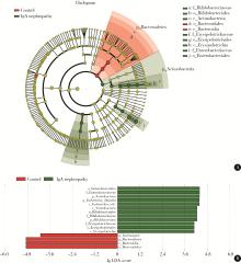
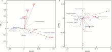
Objective: To investigate the gut microbiota in newly diagnosed IgA nephropathy patients with chronic kidney disease (CKD) stages 1-2 and the association between the gut microbiota and the clinical risk factors of IgA nephropathy. Methods: Fresh fecal samples were collected from nineteen newly diagnosed IgA nephropathy patients with CKD stages 1-2 and fifteen age- and sex-matched healthy controls. Fecal bacterial DNA was extracted and microbiota composition were characterized using 16S ribosomal RNA (16S rRNA) high-throughput sequencing for the V3-V4 region. The Illumina Miseq platform was used to analyze the results of 16S rRNA high-throughput sequencing of fecal flora. At the same time, the clinical risk factors of IgA nephropathy patients were collected to investigate the association between the gut microbiota and the clinical risk factors. Results: (1) At the phylum level, the abundance of Bacteroidetes was significantly reduced (P=0.046), and the abundance of Actinobacteria was significantly increased (P=0.001). At the genus level, the abundance of Escherichia-Shigella, Bifidobacte-rium, Dorea and others were significantly increased (P < 0.05). The abundance of Lachnospira, Coprococcus_2 and Sutterella was significantly reduced (P < 0.05). (2) There was no significant difference in the abundance of gut microbiota between the newly diagnosed IgA nephropathy patients and the healthy control group (P>0.05), but there were differences in the structure of the gut microbiota between the two groups. The results of LEfSe analysis showed that there were 16 differential bacteria in the newly diagnosed IgA nephropathy patients and healthy controls. Among them, the abundance of the newly diagnosed IgA nephropathy patients was increased in Enterobacteriales, Actinobacteria, Escherichia-Shigella, etc. The healthy control group was increased in Bacteroidetes and Lachnospira. (3) The result of redundancy analysis (RDA) showed that Bifidobacterium was positively correlated with serum IgA levels, 24-hour urinary protein levels and the presence of hypertension. Lachnoclostridium was positively correlated with the presence of hypertension. Escherichia-Shigella was positively correlated with urine red blood cells account. Bifidobacterium was positively correlated with the proliferation of capillaries. Faecalibacterium was positively correlated with cell/fibrocytic crescents. Ruminococcus_2 was positively correlated with mesangial cell proliferation, glomerular segmental sclerosis and renal tubular atrophy/interstitial fibrosis. Conclusion: The gut microbiota in the newly diagnosed IgA nephropathy patients with CKD stages 1-2 is different from that of the healthy controls. Most importantly, some gut bacteria are related to the clinical risk factors of IgA nephropathy. Further research is needed to understand the potential role of these bacteria in IgA nephropathy.

Key words: Glomerulonephritis, immunoglobulin A, Gastrointestinal microbiome, Disease attributes, RNA, ribosomal, 16S

CLC Number: 

  • R692.3

Table 1

Basic characteristics of IgA nephropathy patients and health controls"

Items IgA nephropathy (n=19) Health controls (n=15) P
Age/years, $\bar x \pm s$ 33.53±3.96 34.95±8.58 0.529
Male 53% 47% 0.790
Serum creatinine/(μmol/L), $\bar x \pm s$ 84.9±19.3 64.4±6.7 < 0.001
CKD-EPI eGFR/[mL/(min·1.73 m2)], $\bar x \pm s$ 90.7±17.7 126.0±21.0 < 0.001
Pathological features of IgA nephropathy renal biopsy*
  Mesangial hypercellularity (M) M0=0%, M1=94.74%, M2=5.26%, M3=0%
  Endocapillary proliferation (E) E0=26.32%, E1=73.68%
  Segmental sclerosis (S) S0=68.42%, S1=31.58%
  Tubular atrophy/interstitial fibrosis (T) T0=73.68%, T1=26.32%, T2=0%
  Cellular/fibrocytic crescent (C) C0=84.21%, C1=15.79%, C2=0%

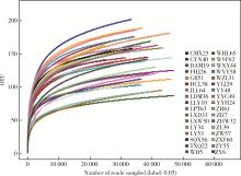
Figure 1

Sample rarefaction curves Alphabetic abbreviations in the figure are the patients' initials. OUT, operational taxonomic units."

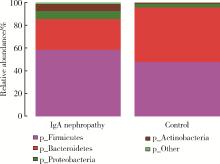
Figure 2

Comparison of gut microflora at phylum level in IgA nephropathy patients and health controls"

Table 2

Change in relative abundance of gut microflora in IgA nephropathy patients and health controls"

Items IgA nephropathy (n=19), M (min, max) Controls (n=15), M (min, max) P
Phylum
  Bacteroidetes 3.13×10⁻¹ (3.86×10⁻², 7.65×10⁻¹) 4.13×10⁻¹ (8.29×10⁻², 8.61×10⁻¹) 0.046
  Actinobacteria 2.47×10⁻² (6.84×10⁻⁴, 1.91×10⁻¹) 2.59×10⁻³ (2.63×10⁻⁴, 7.52×10⁻²) 0.001
  Lentisphaerae 0 (0, 2.93×10⁻⁵) 0 (0, 6.81×10⁻³) 0.011
  Saccharibacteria 5.07×10⁻⁵ (0, 6.40×10⁻⁴) 0 (0, 4.25×10⁻⁵) < 0.001
Genus
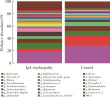
  Escherichia-Shigella 2.88×10⁻³ (7.79×10⁻⁵, 3.88×10⁻¹) 3.79×10⁻⁴ (0, 6.38×10⁻²) 0.025
  Bifidobacterium 1.17×10⁻² (2.409×10⁻⁵, 1.87×10⁻¹) 1.15×10⁻³ (5.42×10⁻⁵, 6.84×10⁻²) 0.036
  Lachnospira 1.43×10⁻³ (0, 1.82×10⁻²) 1.73×10⁻² (0, 1.25×10⁻¹) 0.002
  Dorea 8.84×10⁻³ (7.77×10⁻⁴, 1.03×10⁻¹) 4.67×10⁻³ (1.29×10⁻³, 1.07×10⁻²) 0.025
  Erysipelotrichaceae_UCG-003 7.42×10⁻³ (0, 1.65×10⁻¹) 1.75×10⁻³ (1.78×10⁻⁴, 3.03×10⁻²) 0.013
  Coprococcus_2 0 (0, 8.76×10⁻³) 3.97×10⁻³ (0, 4.59×10⁻²) 0.028
  Sutterella 0 (0, 1.87×10⁻³) 3.73×10⁻³ (0, 4.08×10⁻²) < 0.001
  Tyzzerella_3 0 (0, 2.52×10⁻²) 2.90×10⁻³ (0, 1.61×10⁻²) 0.012
  Thalassospira 0 (0, 1.73×10⁻³) 0 (0, 5.52×10⁻²) 0.015
  Lachnospiraceae_UCG-004 2.78×10⁻⁴ (0, 5.13×10⁻³) 2.50×10⁻³ (2.93×10⁻⁴, 1.27×10⁻²) 0.001
  Eubacterium_ventriosum_group 3.04×10⁻⁴ (0, 1.02×10⁻²) 1.31×10⁻³ (2.13×10⁻⁵, 1.27×10⁻²) 0.042
  Lachnospiraceae_ND3007_group 1.37×10⁻⁴ (0, 1.95×10⁻³) 1.68×10⁻³ (2.44×10⁻⁵, 1.08×10⁻²) 0.002
  Eggerthella 1.11×10⁻³ (0, 1.23×10⁻²) 0 (0, 6.58×10⁻⁵) < 0.001
  Ruminiclostridium_5 8.97×10⁻⁴ (7.78×10⁻⁵, 7.58×10⁻³) 4.14×10⁻⁴ (0, 1.34×10⁻³) 0.030
  Lachnospiraceae_UCG-010 5.06×10⁻⁵ (0, 3.48×10⁻³) 5.72×10⁻⁴ (0, 2.68×10⁻³) 0.049
  Butyricimonas 2.60×10⁻⁵ (0, 1.86×10⁻³) 3.73×10⁻⁴ (0, 8.27×10⁻³) 0.013
  Flavonifractor 2.63×10⁻⁴ (0, 5.19×10⁻³) 2.96×10⁻⁵ (0, 6.30×10⁻⁴) 0.035
  Desulfovibrio 0 (0, 1.82×10⁻³) 0 (0, 1.25×10⁻²) 0.045
  Ruminococcaceae_UCG-003 2.60×10⁻⁴ (0, 7.00×10⁻⁴) 4.44×10⁻⁴ (4.25×10⁻⁵, 3.12×10⁻³) 0.006
  Terrisporobacter 2.53×10⁻⁵ (0, 8.71×10⁻³) 0 (0, 6.58×10⁻⁴) 0.003
  Granulicatella 0.30×10⁻⁵ (0, 6.34×10⁻³) 0 (0, 5.72×10⁻⁵) 0.003
  Erysipelatoclostridium 4.05×10⁻⁵ (0, 3.34×10⁻³) 0 (0, 5.48×10⁻⁵) 0.009
  Ruminiclostridium_9 2.16×10⁻⁵ (0, 1.19×10⁻³) 2.36×10⁻⁴ (0, 1.14×10⁻³) 0.015
  Victivallis 0 (0, 2.93×10⁻⁵) 0 (0, 6.81×10⁻³) 0.011
  Cercis_gigantea 2.53×10⁻⁵ (0, 4.07×10⁻³) 0 (0, 3.33×10⁻⁵) 0.015
  Ezakiella 0 (0, 3.88×10⁻³) 0 (0, 2.74×10⁻⁵) 0.021
  Family_XIII_UCG-001 0 (0, 6.07×10⁻⁴) 6.91×10⁻⁵ (0, 3.52×10⁻⁴) 0.042
  Rothia 0 (0, 2.47×10⁻³) 0 (0, 8.91×10⁻⁵) 0.030
  Gemella 3.00×10⁻⁵ (0, 1.45×10⁻³) 0 (0, 8.26×10⁻⁵) 0.001
  Solobacterium 2.16×10⁻⁵ (0, 7.25×10⁻⁴) 0 (0, 3.33×10⁻⁵) 0.045
  Acinetobacter 0 (0, 2.13×10⁻⁴) 0 (0, 2.74×10⁻⁵) 0.028
  Corynebacterium 0 (0, 2.13×10⁻⁴) 0 (0, 0) 0.005
  Olsenella 2.16×10⁻⁵ (0, 8.09×10⁻⁵) 0 (0, 3.33×10⁻⁵) 0.007
  Atopobium 0 (0, 1.20×10⁻⁴) 0 (0, 0) 0.035
  Leptotrichia 0 (0, 1.20×10⁻⁴) 0 (0, 0) 0.010
  Methylobacterium 0 (0, 8.69×10⁻⁵) 0 (0, 0) 0.019

Figure 3

Comparison of gut microflora at genus level in IgA nephropathy patients and health controls"

Table 3

The α diversity analysis of gut microflora in IgA nephropathy patients and health controls"

Items IgA nephropathy (n=19), $\bar x \pm s$ Controls (n=15), $\bar x \pm s$ P
Shannon index 4.31±0.47 4.05±0.99 0.316
Chao1 index 158±32 157±35 0.942

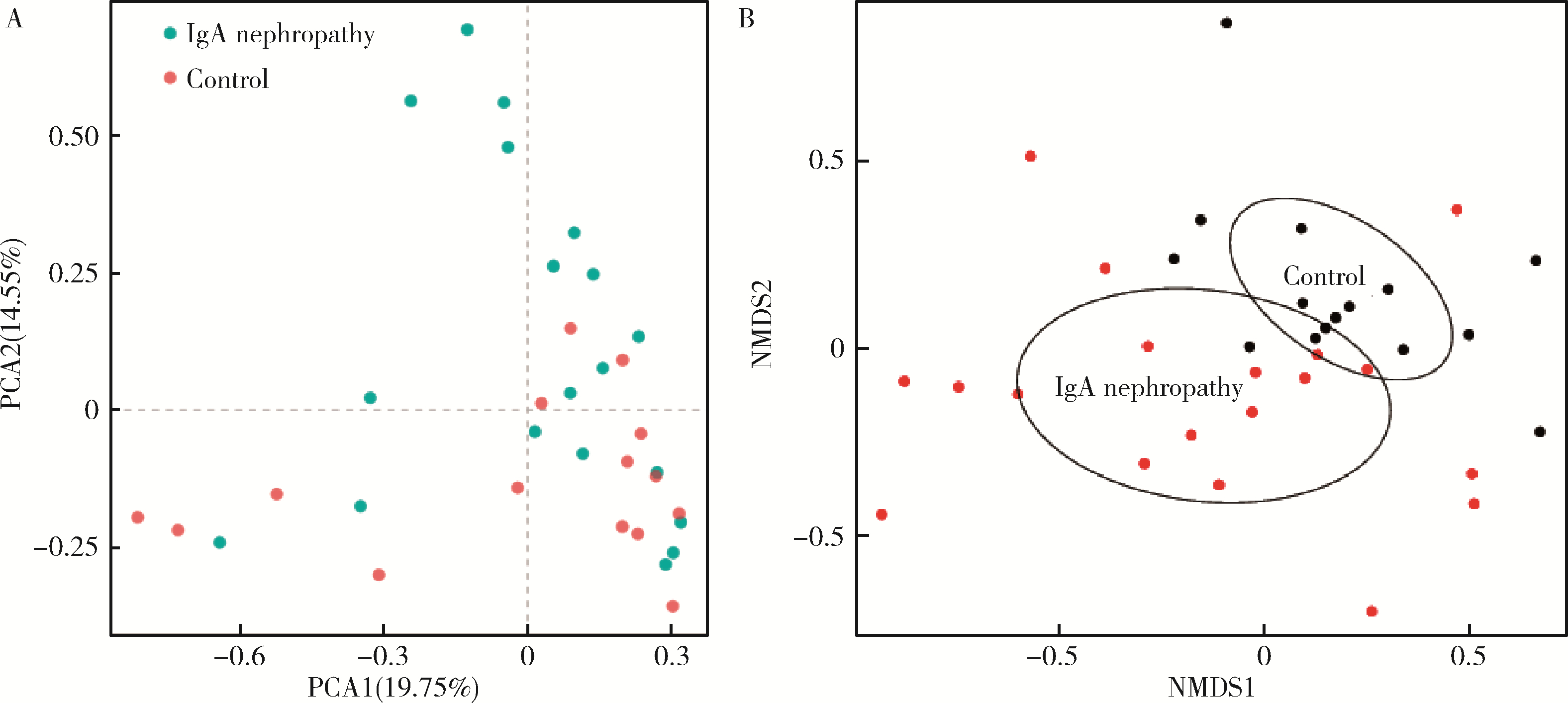
Figure 4

The β diversity analysis in IgA nephropathy patients and health controls A, Principal component analysis in IgA nephropathy patients and health controls; B, Nonmetric muhidimensional scaling in IgA nephropathy patients and health controls. PCA, principal component analysis; NMDS, nonmetric muhidimensional scaling."

Figure 5

LEfSe in IgA nephropathy patients and health controls A, branch evolution diagram of LEfSe in IgA nephropathy patients and health controls; B, histogram of LEfSe in IgA nephropathy patients and health controls. LDA, linear discriminant analysis."

Table 4

The clinical risk factors of IgA nephropathy patients"

Items IgA nephropathy (n=19)
Hypertention (yes/no) 7/12
Systolic pressure/mmHg 133 (121, 189)
Diastolic pressure/mmHg 88.32±11.56
Serum IgA levels/(mg/dL) 4.15 (1.78, 47.60)
Urine red blood cells account (/μL) 175 (12, 1 886)
24-hour urinary protein levels/g 1.86 (0.26, 9.37)

Figure 6

Correlation analysis of gut microflora and clinical risk factors or renal biopsy in IgA nephropathy patients A, correlation analysis of gut microflora and clinical risk factors in IgA nephropathy patients; B, correlation analysis of gut microflora and renal biopsy in IgA nephropathy patients. RDA, also known as multiple direct gradient analysis, is based on linear model analysis, and combines correspondence analysis with multiple regression analysis. Each step of calculation is regression with disease progression-related factors and renal pathological staging, which can be used to detect the relationship between the progression-related factors, renal biopsy and gut microflora. The red rays represent influencing factors, and the length of the rays represents the degree of influence of the influencing factors on the composition of the flora. The direction of the blue vector indicates the direction in which the abundance of the species increases. The angle between the two represents the correlation between influencing factors and species (acute angle represents positive correlation, obtuse angle represents negative correlation). HBP, high blood pressure; SIgA, serum IgA; U-RBC, urine red blood cells account; 24UTP, 24-hour urinary protein. RDA, redundancy analysis. M, mesangial hypercellularity; E, endo-capillary proliferation; S, segmental sclerosis; T, tubular atrophy/interstitial fibrosis; C, cellular/fibrocytic crescent."

1 Schena FP , Nistor I . Epidemiology of IgA nephropathy: A global perspective[J]. Semin Nephrol, 2018, 38 (5): 435- 442.
doi: 10.1016/j.semnephrol.2018.05.013
2 Wang M , Lv J , Zhang X , et al. Secondary IgA nephropathy shares the same immune features with primary IgA nephropathy[J]. Kidney Int Rep, 2020, 5 (2): 165- 172.
doi: 10.1016/j.ekir.2019.10.012
3 Hu X , Du J , Xie Y , et al. Fecal microbiota characteristics of Chinese patients with primary IgA nephropathy: A cross-sectional study[J]. BMC Nephrol, 2020, 21 (1): 97.
doi: 10.1186/s12882-020-01741-9
4 De Angelis M , Montemurno E , Piccolo M , et al. Microbiota and metabolome associated with immunoglobulin A nephropathy (IgAN)[J]. PLoS One, 2014, 9 (6): e99006.
doi: 10.1371/journal.pone.0099006
5 Hobby GP , Karaduta O , Dusio GF , et al. Chronic kidney disease and the gut microbiome[J]. Am J Physiol Renal Physiol, 2019, 316 (6): F1211- F1217.
doi: 10.1152/ajprenal.00298.2018
6 Munyaka PM , Eissa N , Bernstein CN , et al. Antepartum antibio-tic treatment increases offspring susceptibility to experimental colitis: A role of the gut microbiota[J]. PLoS One, 2015, 10 (11): e0142536.
doi: 10.1371/journal.pone.0142536
7 Edgar RC . UPARSE: highly accurate OTU sequences from microbial amplicon reads[J]. Nat Methods, 2013, 10 (10): 996- 998.
doi: 10.1038/nmeth.2604
8 Cole JR , Wang Q , Cardenas E , et al. The ribosomal database project: Improved alignments and new tools for rRNA analysis[J]. Nucleic Acids Res, 2009, 37 (Suppl 1): D141- D145.
9 Segata N , Izard J , Waldron L , et al. Metagenomic biomarker discovery and explanation[J]. Genome Biol, 2011, 12 (6): R60.
doi: 10.1186/gb-2011-12-6-r60
10 Mitchell RJ , Hewison RL , Fielding DA , et al. Decline in atmospheric sulphur deposition and changes in climate are the major drivers of long-term change in grassland plant communities in Scotland[J]. Environ Pollut, 2018, 235, 956- 964.
doi: 10.1016/j.envpol.2017.12.086
11 Li H , Li T , Beasley DE , et al. Diet diversity is associated with beta but not alpha diversity of pika gut microbiota[J]. Front Microbiol, 2016, 7, 1169.
12 Suzuki H , Kiryluk K , Novak J , et al. The pathophysiology of IgA nephropathy[J]. J Am Soc Nephrol, 2011, 22 (10): 1795- 1803.
doi: 10.1681/ASN.2011050464
13 Nakajima A , Vogelzang A , Maruya M , et al. IgA regulates the composition and metabolic function of gut microbiota by promoting symbiosis between bacteria[J]. J Exp Med, 2018, 215 (8): 2019- 2034.
doi: 10.1084/jem.20180427
14 Floege J , Feehally J . The mucosa-kidney axis in IgA nephropathy[J]. Nat Rev Nephrol, 2016, 12 (3): 147- 156.
doi: 10.1038/nrneph.2015.208
15 Olive C , Allen AC , Harper SJ , et al. Expression of the mucosal T cell receptor V region repertoire in patients with IgA nephropathy[J]. Kidney Int, 1997, 52 (4): 1047- 1053.
doi: 10.1038/ki.1997.427
16 McCarthy DD , Kujawa J , Wilson C , et al. Mice overexpressing BAFF develop a commensal flora-dependent, IgA-associated nephropathy[J]. J Clin Invest, 2011, 121 (10): 3991- 4002.
doi: 10.1172/JCI45563
17 Kiryluk K , Li Y , Scolari F , et al. Discovery of new risk loci for IgA nephropathy implicates genes involved in immunity against intestinal pathogens[J]. Nat Genet, 2014, 46 (11): 1187- 1196.
doi: 10.1038/ng.3118
18 Grosserichter-Wagener C , Radjabzadeh D , van der Weide H , et al. Differences in systemic IgA reactivity and circulating Th subsets in healthy volunteers with specific microbiota enterotypes[J]. Front Immunol, 2019, 10, 341.
doi: 10.3389/fimmu.2019.00341
19 Barko PC , McMichael MA , Swanson KS , et al. The gastrointestinal microbiome: A review[J]. J Vet Intern Med, 2018, 32 (1): 9- 25.
doi: 10.1111/jvim.14875
20 Gomaa EZ . Human gut microbiota/microbiome in health and diseases: A review[J]. Antonie Van Leeuwenhoek, 2020, 113 (12): 2019- 2040.
doi: 10.1007/s10482-020-01474-7
21 包文晗, 王悦. 慢性肾脏病患者肠道菌群的变化及影响研究[J]. 中国全科医学, 2018, 21 (24): 2927- 2931.
doi: 10.3969/j.issn.1007-9572.2018.00.168
22 Gibiino G , Lopetuso LR , Scaldaferri F , et al. Exploring Bacteroidetes: Metabolic key points and immunological tricks of our gut commensals[J]. Dig Liver Dis, 2018, 50 (7): 635- 639.
doi: 10.1016/j.dld.2018.03.016
23 Lo Presti A , Zorzi F , Del Chierico F , et al. Fecal and mucosal microbiota profiling in irritable bowel syndrome and inflammatory bowel disease[J]. Front Microbiol, 2019, 10, 1655.
doi: 10.3389/fmicb.2019.01655
24 Amabebe E , Robert FO , Agbalalah T , et al. Microbial dysbiosis-induced obesity: Role of gut microbiota in homoeostasis of energy metabolism[J]. Br J Nutr, 2020, 123 (10): 1127- 1137.
doi: 10.1017/S0007114520000380
25 Coppo R . The gut-kidney axis in IgA nephropathy: Role of microbiota and diet on genetic predisposition[J]. Pediatr Nephrol, 2018, 33 (1): 53- 61.
doi: 10.1007/s00467-017-3652-1
26 Liong MT . Beneficial microorganisms in medical and health applications[M]. Switzerland: Springer International Publishing, 2015.
[1] Jia-he ZHANG,Jia-qi SHI,Zhang-jian CHEN,Guang JIA. Effects of nano titanium dioxide on gut microbiota based on human digestive tract microecology simulation system in vitro [J]. Journal of Peking University (Health Sciences), 2022, 54(3): 468-476.
[2] WANG Zi-jing,LI Zai-ling. Characteristics of gastric microbiota in children with Helicobacter pylori infection family history [J]. Journal of Peking University (Health Sciences), 2021, 53(6): 1115-1121.
[3] Mei-xiang ZHANG,Wen-zhi SHI,Jian-xin LIU,Chun-jian WANG,Yan LI,Wei WANG,Bin JIANG. Clinical characteristics and prognosis of MLL-AF6 positive patients with acute myeloid leukemia [J]. Journal of Peking University (Health Sciences), 2021, 53(5): 915-920.
[4] YAN Yong-Qing, ZHANG Pei-Xun- , WANG Tian-Bing- , CHEN Jian-Hai, JIANG Bao-Guo. Multi-center retrospective analysis of clinical and related sociologic characteristics of postoperative distal radius fracture patients [J]. Journal of Peking University(Health Sciences), 2014, 46(2): 254-257.
Viewed
Full text


Abstract

Cited

  Shared   
  Discussed   
No Suggested Reading articles found!