Journal of Peking University (Health Sciences) ›› 2025, Vol. 57 ›› Issue (4): 684-691. doi: 10.19723/j.issn.1671-167X.2025.04.009

Previous Articles     Next Articles

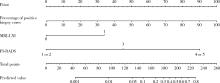
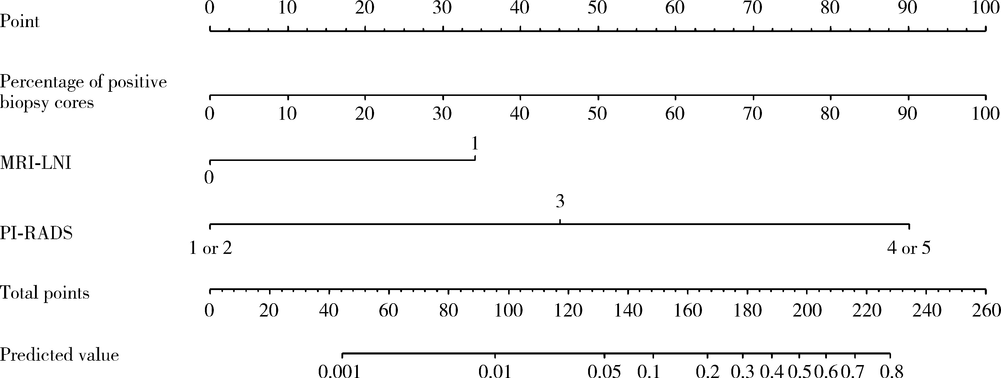
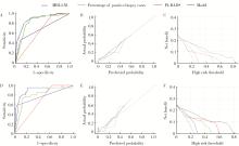
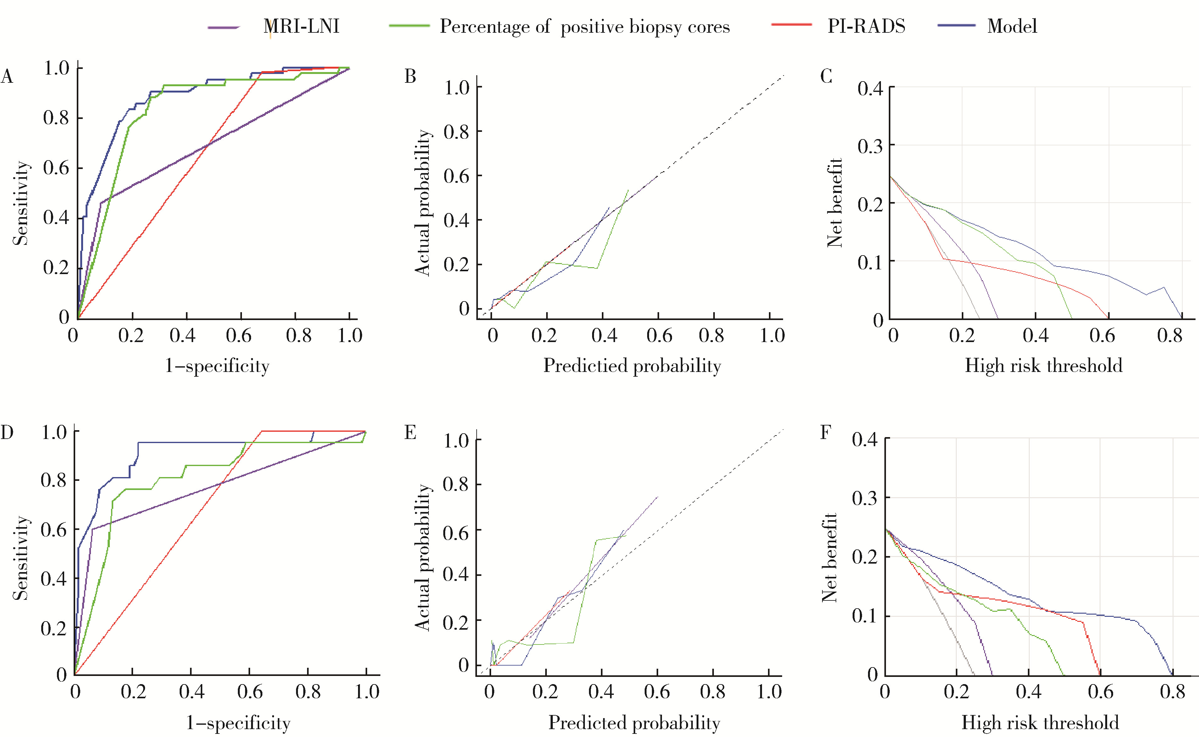
A preoperative prediction model for pelvic lymph node metastasis in prostate cancer: Integrating clinical characteristics and multiparametric MRI

Zeyuan WANG, Shuanbao YU, Haoke ZHENG, Jin TAO, Yafeng FAN, Xuepei ZHANG*()   

  1. Department of Urology, the First Affiliated Hospital of Zhengzhou University, Zhengzhou 450000, China
  • Received:2025-02-27 Online:2025-08-18 Published:2025-08-02
  • Contact: Xuepei ZHANG
  • Supported by:
    the Youth Science Fund of Henan Province(232300420254); the Key Scientific and Technological Projects of Henan Province(242102311074)

RICH HTML


Abstract:

Objective: To analyze the clinical features associated with pelvic lymph node metastasis (PLNM) in prostate cancer and to construct a preoperative prediction model for PLNM, thereby reducing unnecessary extended pelvic lymph node dissection (ePLND). Methods: Based on predefined inclusion and exclusion criteria, 344 patients who underwent radical prostatectomy and ePLND at the First Affiliated Hospital of Zhengzhou University between 2014 and 2024 were retrospectively enrolled, among whom, 77 patients (22.4%) were pathologically confirmed to have lymph node-positive disease. The clinical characteristics, MRI reports, and pathological results were collected. The data were then randomly divi-ded into a training cohort (241 cases, 70%) and a validation cohort (103 cases, 30%). Univariate and multivariate Logistic regression analysis were employed to construct a preoperative prediction model for PLNM. Results: Univariate Logistic regression analysis revealed that total prostate specific antigen (tPSA) (P=0.021), free prostate specific antigen (fPSA) (P=0.002), fPSA to tPSA ratio (fPSA/tPSA) (P=0.011), percentage of positive biopsy cores (P < 0.001), prostate imaging reporting and data system (PI-RADS) score (P=0.004), biopsy Gleason score ≥8 (P=0.005), clinical T stage (P < 0.001), and MRI-indicated lymph node involvement (MRI-LNI) (P < 0.001) were significant predictors of PLNM. Multivariate Logistic regression analysis demonstrated that the percentage of positive biopsy cores (OR=91.24, 95%CI: 13.34-968.68), PI-RADS score (OR=7.64, 95%CI: 1.78-138.06), and MRI-LNI (OR=4.67, 95%CI: 1.74-13.24) were independent risk factors for PLNM. And a novel nomogram for predicting PLNM was developed by integrating all these three variables. Compared with the individual predictors: percentage of positive biopsy cores [area under curve (AUC)=0.806], PI-RADS score (AUC=0.679), and MRI-LNI (AUC=0.768), the multivariate model incorporating all three variables demonstrated significantly superior predictive performance (AUC=0.883). Consistently, calibration curves and decision curve analyses confirmed that the multivariable model had high predictive accuracy and provided significant net clinical benefit relative to single-variable models. And using a cutoff of 6%, the multiparameter model missed only approximately 5.2% of PLNM cases (4/77), while reducing approximately 53% of ePLND procedures (139/267), demonstrating favorable predictive efficacy. Conclusion: Percentage of positive biopsy cores, PI-RADS score and MRI-LNI are independent risk factors for PLNM. The constructed multivariate model significantly improves predictive efficacy, offering a valuable tool to guide clinical decisions on ePLND.

Key words: Prostatic neoplasms, Lymphatic metastasis, Multiparametric magnetic resonance imaging, Biopsy

CLC Number: 

  • R737.25

Table 1

Comparison of clinical characteristics between pelvic lymph node metastasis and non-metastasis groups"

Parameter Total (n=344) Pelvic lymph node metastasis Statistic P
Positive (n=77) Negative (n=267)
Age/years, M (P25, P75) 68 (64, 73) 68 (64, 73) 68 (64, 75) Z=-0.39 0.699
BMI/(kg/m2), M (P25, P75) 24.9 (23.0, 26.7) 25.4 (23.7, 26.6) 24.8 (22.9, 26.8) Z=-0.71 0.475
tPSA/(ng/mL), M (P25, P75) 19.5 (11.6, 50.4) 48.5 (24.2, 100.0) 16.8 (10.1, 34.3) Z=-6.44 < 0.001
fPSA/(ng/mL), M (P25, P75) 2.28 (1.13, 5.57) 5.28 (2.71, 11.90) 1.68 (0.99, 4.02) Z=-6.03 < 0.001
fPSA/tPSA, M (P25, P75) 0.103 (0.074, 0.150) 0.121 (0.082, 0.175) 0.098 (0.071, 0.148) Z=-2.59 0.01
PSAD/(ng/mL2), M (P25, P75) 0.62 (0.33, 1.24) 1.08 (0.61, 2.18) 0.54 (0.28, 0.95) Z=-4.90 < 0.001
Volume/mL, M (P25, P75) 40 (29, 59) 46 (34, 71) 38 (28, 55) Z=-2.30 0.021
Positive biopsy cores/%, M (P25, P75) 0.63 (0.38, 1.00) 1.00 (0.92, 1.00) 0.53 (0.29, 0.85) Z=-7.84 < 0.001
PI-RADS score, n (%) χ2=31.77 < 0.001
  ≤2 25 (7.27) 0 (0) 25 (9.36)
  3 65 (18.90) 1 (1.30) 64 (23.97)
  ≥4 254 (73.84) 76 (98.70) 178 (66.67)
Biopsy Gleason score, n (%) χ2=42.01 < 0.001
  3+3 48 (13.95) 3 (3.90) 45 (16.85)
  3+4 65 (18.90) 7 (9.09) 58 (21.72)
  4+3 74 (21.51) 7 (9.09) 67 (25.09)
  ≥8 157 (45.64) 60 (77.92) 97 (36.33)
Clinical T stage, n (%) χ2=47.16 < 0.001
  ≤T2 249 (72.38) 32 (41.56) 217 (81.27)
  >T2 95 (27.62) 45 (58.44) 50 (18.73)
MRI-LNI, n (%) χ2=75.97 < 0.001
  N0 284 (82.56) 38 (49.35) 246 (92.13)
  N1 60 (17.44) 39 (50.65) 21 (7.87)

Table 2

Comparison of baseline characteristics between training and validation cohort"

Characteristic Training cohort (n=241) Validation cohort (n=103) Statistic P
Age/years, M (P25, P75) 69 (64, 73) 68 (64, 74) Z=-0.05 0.959
BMI/(kg/m2), M (P25, P75) 25.0 (22.8, 26.8) 4.5 (23.5, 25.9) Z=-0.35 0.726
tPSA/(ng/mL), M (P25, P75) 19.1 (11.1, 47.7) 20.2 (13.1, 51.7) Z=-0.88 0.376
fPSA/(ng/mL), M (P25, P75) 2.41 (1.07, 5.39) 2.20 (1.28, 6.12) Z=-0.52 0.606
Positive biopsy cores/%, M (P25, P75) 0.62 (0.39, 1.00) 0.63 (0.38, 0.92) Z=-0.78 0.433
Volume/mL, M (P25, P75) 39.0 (29.0, 55.0) 43.0 (27.5, 61.5) Z=-0.66 0.511
PSAD/(ng/mL2), M (P25, P75) 0.63 (0.32, 1.23) 0.62 (0.33, 1.25) Z=-0.09 0.929
fPSA/tPSA, M (P25, P75) 0.10 (0.08, 0.14) 0.11 (0.07, 0.16) Z=-0.001 0.999
PI-RADS score, n (%) χ2=0.09 0.955
  ≤2 17 (7.05) 8 (7.77)
  3 45 (18.67) 20 (19.42)
  ≥4 179 (74.27) 75 (72.82)
Biopsy Gleason score, n (%) χ2=3.17 0.365
  3+3 29 (12.03) 19 (18.45)
  3+4 49 (20.33) 16 (15.53)
  4+3 51 (21.16) 23 (22.33)
  ≥8 112 (46.47) 45 (43.69)
Clinical T stage, n (%) χ2=2.06 0.152
  ≤T2 169 (70.12) 80 (77.67)
  >T2 72 (29.88) 23 (22.33)
MRI-LNI, n (%) χ2=0.30 0.583
  N0 189 (78.42) 78 (75.73)
  N1 52 (21.58) 25 (24.27)

Table 3

Univariate and multivariate Logistic regression analyses of clinical parameters for predicting PLNM in the training cohort"

Characteristic Univariate analysis Multivariable analysis
OR (95%CI) Z P Coefficient Z OR (95%CI) P
Age 1.006 (0.957, 1.057) 0.231 0.817
BMI 1.093 (0.924, 1.294) 1.039 0.299
tPSA 1.007 (1.001, 1.014) 2.315 0.021
fPSA 1.099 (1.037, 1.166) 3.171 0.002
fPSA/tPSA 591.8 (4.3, 81 857.0) 2.538 0.011
PSAD 1.111 (0.926, 1.333) 1.136 0.256
PV 1.008 (0.998, 1.019) 1.556 0.120
Positive biopsy cores 232.4 (30.3, 1 780.0) 5.245 < 0.001 4.513 4.188 91.24 (13.34, 968.68) < 0.001
PI-RADS score 18.30 (2.56, 130.60) 2.898 0.004 2.034 2.078 7.64 (1.78, 138.06) 0.038
Biopsy Gleason score
  3+3 1.000 (Reference)
  3+4 0.880 (0.138, 5.606) -0.135 0.893
  4+3 1.149 (0.197, 6.692) 0.154 0.877
≥8 8.413 (1.904, 37.180) 2.809 0.005
Clinical T stage 0.150 (0.077, 0.291) -5.594 < 0.001
MRI-LNI 9.268 (4.386, 19.580) 5.834 < 0.001 1.540 2.995 4.67 (1.74, 13.24) 0.003

Figure 1

Nomogram for predicting PLNM in prostate cancer PLNM, pelvic lymph node metastasis; PI-RADS, prostate imaging reporting and data system; MRI-LNI, MRI-indicated lymph node involvement."

Figure 2

ROC curve, calibration plots and DCA of MRI-LNI, PI-RADS, percentage of positive biopsy cores, and the multivariable model for suspected PLNM A, ROC curve in the training cohort; B, calibration plot in the training cohort; C, DCA in the training cohort; D, ROC curve in the validation cohort; E, calibration plot in the validation cohort; F, DCA in the validation cohort. ROC, receiver operating characteristic; DCA, decision curve analysis; PLNM, pelvic lymph node metastasis; PI-RADS, prostate imaging reporting and data system; MRI-LNI, MRI-indicated lymph node involvement."

Table 4

Diagnostic performance of the multivariable model at different cutoff values for PLNM"

Cutoff value Below the cutoff (PLND not recommended), n (%) Above the cutoff (PLND recommended), n (%)
Total Without PLNM With PLNM Total Without PLNM With PLNM
2% 92 (26.7) 90 (97.8) 2 (2.2) 252 (73.3) 177 (70.2) 75 (29.8)
3% 111 (32.3) 108 (97.3) 3 (2.7) 233 (67.7) 159 (68.2) 74 (31.8)
4% 120 (34.9) 116 (96.7) 4 (3.3) 224 (65.1) 151 (67.4) 73 (32.6)
5% 129 (37.5) 125 (96.9) 4 (3.1) 215 (62.5) 142 (66.0) 73 (34.0)
6% 143 (41.6) 139 (97.2) 4 (2.8) 201 (58.4) 128 (63.7) 73 (36.3)
7% 146 (42.4) 141 (96.6) 5 (3.4) 198 (57.6) 126 (63.6) 72 (36.4)
8% 159 (46.2) 152 (95.6) 7 (4.4) 185 (53.7) 115 (62.2) 70 (37.8)
9% 167 (48.5) 159 (95.2) 8 (4.8) 177 (51.5) 108 (61.0) 69 (39.0)
10% 182 (52.9) 174 (95.6) 8 (4.4) 162 (47.1) 93 (57.4) 69 (42.6)
1
Briganti A, Larcher A, Abdollah F, et al. Updated nomogram predicting lymph node invasion in patients with prostate cancer undergoing extended pelvic lymph node dissection: the essential importance of percentage of positive cores[J]. Eur Urol, 2012, 61(3): 480- 487.
2
Sandhu S, Moore CM, Chiong E, et al. Prostate cancer[J]. Lancet, 2021, 398(10305): 1075- 1090.
3
Briganti A, Blute ML, Eastham JH, et al. Pelvic lymph node dissection in prostate cancer[J]. Eur Urol, 2009, 55(6): 1251- 1265.
4
Haiquel L, Cathelineau X, Sanchez-Salas R, et al. Pelvic lymph node dissection in high-risk prostate cancer[J]. Int Braz J Urol, 2022, 48(1): 54- 66.
5
Ploussard G, Briganti A, de la Taille A, et al. Pelvic lymph node dissection during robot-assisted radical prostatectomy: Efficacy, limitations, and complications: A systematic review of the literature[J]. Eur Urol, 2014, 65(1): 7- 16.
6
Dong B, Zhan H, Luan T, et al. The role and controversy of pelvic lymph node dissection in prostate cancer treatment: A focused review[J]. World J Surg Oncol, 2024, 22(1): 68.
7
Fossati N, Willemse PM, Van den Broeck T, et al. The benefits and harms of different extents of lymph node dissection during radical prostatectomy for prostate cancer: A systematic review[J]. Eur Urol, 2017, 72(1): 84- 109.
8
Touijer K, Fuenzalida RP, Rabbani F, et al. Extending the indications and anatomical limits of pelvic lymph node dissection for prostate cancer: Improved staging or increased morbidity?[J]. BJU Int, 2011, 108(3): 372- 377.
9
Woo S, Suh CH, Kim SY, et al. The diagnostic performance of MRI for detection of lymph node metastasis in bladder and prostate cancer: An updated systematic review and diagnostic meta-analysis[J]. AJR Am J Roentgenol, 2018, 210(3): W95- W109.
10
Hövels AM, Heesakkers RA, Adang EM, et al. The diagnostic accuracy of CT and MRI in the staging of pelvic lymph nodes in patients with prostate cancer: A meta-analysis[J]. Clin Radiol, 2008, 63(4): 387- 395.
11
Yang B, Dong H, Zhang S, et al. PSMA PET vs. mpMRI for lymph node metastasis of prostate cancer: A systematic review and head-to-head comparative meta-analysis[J]. Acad Radiol, 2024, 32(5): 2797- 2814.
12
Hou Y, Bao ML, Wu CJ, et al. A machine learning-assisted decision-support model to better identify patients with prostate cancer requiring an extended pelvic lymph node dissection[J]. BJU Int, 2019, 124(6): 972- 983.
13
van Leenders GJLH, van der Kwast TH, Grignon DJ, et al. The 2019 International Society of Urological Pathology (ISUP) Consensus Conference on grading of prostatic carcinoma[J]. Am J Surg Pathol, 2020, 44(8): e87- e99.
14
Eastham JA, Auffenberg GB, Barocas DA, et al. Clinically loca-lized prostate cancer: AUA/ASTRO guideline, Part Ⅰ: Introduction, risk assessment, staging, and risk-based management[J]. J Urol, 2022, 208(1): 10- 18.
15
Mottet N, van den Bergh RCN, Briers E, et al. EAU-EANM-ESTRO-ESUR-SIOG guidelines on prostate cancer-2020 update. Part 1: Screening, diagnosis, and local treatment with curative intent[J]. Eur Urol, 2021, 79(2): 243- 262.
16
Mohler JL, Antonarakis ES, Armstrong AJ, et al. Prostate cancer, version 2.2019, NCCN clinical practice guidelines in oncology[J]. J Natl Compr Canc Netw, 2019, 17(5): 479- 505.
17
Cagiannos I, Karakiewicz P, Eastham JA, et al. A preoperative nomogram identifying decreased risk of positive pelvic lymph nodes in patients with prostate cancer[J]. J Urol, 2003, 170(5): 1798- 1803.
18
Diamand R, Oderda M, Al Hajj Obeid W, et al. A multicentric study on accurate grading of prostate cancer with systematic and MRI/US fusion targeted biopsies: Comparison with final histopathology after radical prostatectomy[J]. World J Urol, 2019, 37(10): 2109- 2117.
19
Gandaglia G, Fossati N, Zaffuto E, et al. Development and internal validation of a novel model to identify the candidates for extended pelvic lymph node dissection in prostate cancer[J]. Eur Urol, 2017, 72(4): 632- 640.
20
Giorgio G, Guillaume P, Massimo V, et al. A novel nomogram to identify candidates for extended pelvic lymph node dissection among patients with clinically localized prostate cancer diagnosed with magnetic resonance imaging-targeted and systematic biopsies[J]. Eur Urol, 2019, 75(3): 506- 514.
21
Yusuke G, Takanobu U, Masafumi M, et al. Development and validation of novel nomogram to identify the candidates for exten-ded pelvic lymph node dissection for prostate cancer patients in the robotic era[J]. Int J Urol, 2023, 30(8): 659- 665.
22
Makoto K, Shin E, Tomoyuki T, et al. A nomogram for predicting prostate cancer with lymph node involvement in robot-assisted radical prostatectomy era: A retrospective multicenter cohort study in Japan (The MSUG94 Group)[J]. Diagnostics (Basel), 2022, 12(10): 2545.
23
Li Z, Huang Y, Zhao D, et al. Development and internal validation of a novel nomogram for predicting lymph node invasion for prostate cancer patients undergoing extended pelvic lymph node dissection[J]. Front Oncol, 2023, 13, 1186319.
24
Qin C, Goldberg O, Kakar G, et al. MRI fat fraction imaging of nodal and bone metastases in prostate cancer[J]. Eur Radiol, 2023, 33(8): 5851- 5855.
25
Heidenreich A, Pfister D, Thüer D, et al. Percentage of positive biopsies predicts lymph node involvement in men with low-risk prostate cancer undergoing radical prostatectomy and extended pelvic lymphadenectomy[J]. BJU Int, 2011, 107(2): 220- 225.
26
Benidir T, Lone Z, Nguyen JK, et al. The combination of prostate MRI PI-RADS scoring system and a genomic classifier is associated with pelvic lymph node metastasis at the time of radical prostatectomy[J]. Br J Radiol, 2023, 96(1144): 20220663.
27
Eastham JA, Auffenberg GB, Barocas DA, et al. Clinically loca-lized prostate cancer: AUA/ASTRO guideline, Part Ⅱ: Principles of active surveillance, principles of surgery, and follow-up[J]. J Urol, 2022, 208(1): 19- 25.
28
Cornford P, van den Bergh RCN, Briers E, et al. EAU-EANM-ESTRO-ESUR-ISUP-SIOG guidelines on prostate cancer-2024 update. Part Ⅰ: Screening, diagnosis, and local treatment with curative intent[J]. Eur Urol, 2024, 89(2): 148- 163.
[1] Jiaxin NING, Haoran WANG, Shuhang LUO, Jibo JING, Jianye WANG, Huimin HOU, Ming LIU. Multi-omics analysis of the relationship between oxidative stress-related gene and prostate cancer [J]. Journal of Peking University (Health Sciences), 2025, 57(4): 633-643.
[2] Yuan NING, Xiaoying ZHANG, Xue LI, Yuan LI, Jing HE, Yuebo JIN. Sjögren disease complicated by primary breast lymphoma: A case report [J]. Journal of Peking University (Health Sciences), 2025, 57(4): 808-811.
[3] Zhicun LI, Tianyu WU, Lei LIANG, Yu FAN, Yisen MENG, Qian ZHANG. Risk factors analysis and nomogram model construction of postoperative pathological upgrade of prostate cancer patients with single core positive biopsy [J]. Journal of Peking University (Health Sciences), 2024, 56(5): 896-901.
[4] Yuxuan TIAN,Mingjian RUAN,Yi LIU,Derun LI,Jingyun WU,Qi SHEN,Yu FAN,Jie JIN. Predictive effect of the dual-parametric MRI modified maximum diameter of the lesions with PI-RADS 4 and 5 on the clinically significant prostate cancer [J]. Journal of Peking University (Health Sciences), 2024, 56(4): 567-574.
[5] Kaifeng YAO,Mingjian RUAN,Derun LI,Yuxuan TIAN,Yuke CHEN,Yu FAN,Yi LIU. Diagnostic efficacy of targeted biopsy combined with regional systematic biopsy in prostate cancer in patients with PI-RADS 4-5 [J]. Journal of Peking University (Health Sciences), 2024, 56(4): 575-581.
[6] Junyong OU,Kunming NI,Lulin MA,Guoliang WANG,Ye YAN,Bin YANG,Gengwu LI,Haodong SONG,Min LU,Jianfei YE,Shudong ZHANG. Prognostic factors of patients with muscle invasive bladder cancer with intermediate-to-high risk prostate cancer [J]. Journal of Peking University (Health Sciences), 2024, 56(4): 582-588.
[7] Yi LIU,Chang-wei YUAN,Jing-yun WU,Qi SHEN,Jiang-xi XIAO,Zheng ZHAO,Xiao-ying WANG,Xue-song LI,Zhi-song HE,Li-qun ZHOU. Diagnostic efficacy of prostate cancer using targeted biopsy with 6-core systematic biopsy for patients with PI-RADS 5 [J]. Journal of Peking University (Health Sciences), 2023, 55(5): 812-817.
[8] Chang-wei YUAN,De-run LI,Zhi-hua LI,Yi LIU,Gang-zhi SHAN,Xue-song LI,Li-qun ZHOU. Application of dynamic contrast enhanced status in multiparametric magnetic resonance imaging for prostatic cancer with PI-RADS 4 lesion [J]. Journal of Peking University (Health Sciences), 2023, 55(5): 838-842.
[9] Yan XIONG,Xin LI,Li LIANG,Dong LI,Li-min YAN,Xue-ying LI,Ji-ting DI,Ting LI. Evaluation of accuracy of pathological diagnosis based on thyroid core needle biopsy [J]. Journal of Peking University (Health Sciences), 2023, 55(2): 234-242.
[10] Dan-feng ZHENG,Jun-yu LI,Jia-xi LI,Ying-shuang ZHANG,Yan-feng ZHONG,Miao YU. Pathologic features of paraspinal muscle biopsies in patients with adolescent idiopathic scoliosis [J]. Journal of Peking University (Health Sciences), 2023, 55(2): 283-291.
[11] ZHOU Guang-ping,ZHOU Qian-yun,ZHU Ji-hong. A case report of TAFRO syndrome [J]. Journal of Peking University (Health Sciences), 2021, 53(4): 814-817.
[12] ZHANG Lei,LI Guo-liang,DANG Zong-hui, ,A yong,WU Ling-jie,LIU Li-jun. Analysis of bleeding risk in percutaneous renal biopsy in Tibet [J]. Journal of Peking University (Health Sciences), 2021, 53(2): 298-301.
[13] WANG Ying-chun,HUANG Yong-hui,CHANG Hong,YAO Wei,YAN Xiu-e,LI Ke,ZHANG Yao-peng,ZHENG Wei. Characteristics of benign and malignant lesions of ampullary polyps and the accuracy of forceps biopsy [J]. Journal of Peking University (Health Sciences), 2021, 53(1): 204-209.
[14] Ying-chao WU,Yun-long CAI,Long RONG,Ji-xin ZHANG,Jin LIU,Xin WANG. Characteristics of lymph node metastasis and evaluating the efficacy of endoscopic submucosal dissection in early gastric cancer [J]. Journal of Peking University (Health Sciences), 2020, 52(6): 1093-1097.
[15] Yi LIU,Zhi-jian LIU,Qi SHEN,Jing-yun WU,Yu FAN,De-run LI,Wei YU,Zhi-song HE. A clinical analysis of 14 cases of prostatic stromal tumor of uncertain malignant potential [J]. Journal of Peking University (Health Sciences), 2020, 52(4): 621-624.
Viewed
Full text


Abstract

Cited

  Shared   
  Discussed   
No Suggested Reading articles found!