北京大学学报(医学版) ›› 2023, Vol. 55 ›› Issue (2): 292-298. doi: 10.19723/j.issn.1671-167X.2023.02.013
幽门螺杆菌阴性早期胃癌的临床病理特征
侯卫华1,宋书杰2,石中月3,金木兰3,*( )
)
- 1. 解放军联勤保障部队第九八九医院平顶山医疗区病理科,河南平顶山 467099
2. 解放军联勤保障部队第九八九医院平顶山医疗区消化内科,河南平顶山 467099
3. 首都医科大学附属北京朝阳医院病理科,北京 100020
Clinicopathological features of Helicobacter pylori-negative early gastric cancer
Wei-hua HOU1,Shu-jie SONG2,Zhong-yue SHI3,Mu-lan JIN3,*()
- 1. Department of Pathology, Pingdingshan Medical District, 989 Hospital of PLA Joint Logistics Support Force, Pingdingshan 467099, Henan, China
2. Department of Gastroenterology, Pingdingshan Medical District, 989 Hospital of PLA Joint Logistics Support Force, Pingdingshan 467099, Henan, China
3. Department of Pathology, Beijing Chaoyang Hospital, Capital Medical University, Beijing 100020, China
摘要:
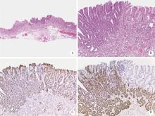
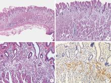
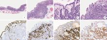
目的: 探讨幽门螺杆菌(Helicobacter pylori,Hp)阴性早期胃癌的临床病理特征。方法: 回顾性收集2009—2021年解放军联勤保障部队第九八九医院平顶山医疗区和首都医科大学附属北京朝阳医院共计30例Hp阴性早期胃癌的临床资料,观察其组织形态学特征和免疫表型,并结合文献进行探讨。结果: 30例患者的中位年龄58. 5岁(范围21~80岁),男性13例,女性17例;胃上部13例,胃中部9例,胃下部8例;肿瘤中位直径11 mm(范围1~30 mm);按巴黎分型0-Ⅱa型9例,0-Ⅱb型7例,0-Ⅱc型14例。内镜检查18例病变发红,12例病变呈退色调或发白,均可见微血管结构和微表面结构异常;所有病例胃体及胃角黏膜可见规律排列的集合小静脉。黏膜内高分化腺癌18例,肿瘤呈腺管样及乳头状结构,腺体密集,排列紊乱;细胞呈立方形或柱状,核染色质增多,核极性丢失,多表达胃型黏蛋白。印戒细胞癌7例,癌组织全部由印戒细胞构成,癌细胞主要分布在黏膜中表层内;胃泌酸腺瘤(局限于黏膜内的胃底腺型胃癌)2例、胃底腺型胃癌2例和胃底腺黏膜型胃癌1例。肿瘤组织由轻度大小不等的分枝管状腺构成,除1例黏膜表面上皮局部是肿瘤性外,其余4例黏膜表面上皮全为非肿瘤性;细胞单层排列,核靠近基底侧,细胞核仅有轻度的不典型性。5例胃底腺肿瘤免疫组织化学染色胃蛋白酶原Ⅰ和H+/K+ATPase阳性,其中1例黏膜表面和深处的小凹型肿瘤细胞MUC5AC阳性。所有的病例癌旁胃黏膜大致正常,无萎缩、肠上皮化生和Hp。结论: Hp阴性早期胃癌是一组异质性疾病群体,组织学类型多样,常见的有管状腺癌和印戒细胞癌。管状腺癌多发生于老年人和胃中上部,印戒细胞癌多发生于中青年人和胃下部,胃底腺型肿瘤相对罕见。
中图分类号:
- R735.2


| 1 |
Correa P , Piazuelo MB . The gastric precancerous cascade[J]. J Dig Dis, 2012, 13 (1): 2- 9.
doi: 10.1111/j.1751-2980.2011.00550.x |
| 2 |
Rugge M , Genta RM , Di Mario F , et al. Gastric cancer as preventable disease[J]. Clin Gastroenterol Hepatol, 2017, 15 (12): 1833- 1843.
doi: 10.1016/j.cgh.2017.05.023 |
| 3 |
Yamamoto Y , Fujisaki J , Omae M , et al. Helicobacter pylori-negative gastric cancer: Characteristics and endoscopic findings[J]. Dig Endosc, 2015, 27 (5): 551- 561.
doi: 10.1111/den.12471 |
| 4 | 侯卫华, 王新钊, 石中月, 等. 幽门螺杆菌根除后早期胃癌的临床病理特征分析[J]. 中华病理学杂志, 2022, 51 (8): 10- 16. |
| 5 |
Sato C , Hirasawa K , Tateishi Y , et al. Clinicopathological features of early gastric cancers arising in Helicobacter pylori uninfected patients[J]. World J Gastroenterol, 2020, 26 (20): 2618- 2631.
doi: 10.3748/wjg.v26.i20.2618 |
| 6 |
Japanese Gastric Cancer Association . Japanese classification of gastric carcinoma: 3rd English edition[J]. Gastric Cancer, 2011, 14 (2): 101- 112.
doi: 10.1007/s10120-011-0041-5 |
| 7 |
韩方海, 杨斌. 解读第15版日本胃癌处理规约[J]. 中华胃肠外科杂志, 2018, 21 (4): 409- 412.
doi: 10.3760/cma.j.issn.1671-0274.2018.04.010 |
| 8 | 九屿亮治. 胃癌病理分类: 日本国内实行的分类[M]//鹤田修. 胃与肠. 《胃与肠》翻译委员会, 译. 沈阳: 辽宁科学技术出版社, 2017: 15-26. |
| 9 | Yao T, Vieth M. Oxyntic gland adenoma[M]//WHO Classification of Tumours Editorial Board. WHO classification of tumours, digestive system tumours. 5th ed. Lyon: IARC Press, 2019: 83-84. |
| 10 |
Yemelyanova A , Vang R , Kshirsagar M , et al. Immunohistoche-mical staining patterns of p53 can serve as a surrogate marker for TP53 mutations in ovarian carcinoma: An immunohistochemical and nucleotide sequencing analysis[J]. Mod Pathol, 2011, 24 (9): 1248- 1253.
doi: 10.1038/modpathol.2011.85 |
| 11 |
Yamada A , Kaise M , Inoshita N , et al. Characterization of Helicobacter pylori-naive early gastric cancers[J]. Digestion, 2018, 98 (2): 127- 134.
doi: 10.1159/000487795 |
| 12 |
Kakinoki R , Kushima R , Matsubara A , et al. Re-evaluation of histogenesis of gastric carcinomas: A comparative histopathological study between Helicobacter pylori-negative and H. pylori-positive cases[J]. Dig Dis Sci, 2009, 54 (3): 614- 620.
doi: 10.1007/s10620-008-0389-5 |
| 13 |
Yoon H , Kim N , Lee HS , et al. Helicobacter pylori-negative gastric cancer in South Korea: Incidence and clinicopathologic cha-racteristics[J]. Helicobacter, 2011, 16 (5): 382- 388.
doi: 10.1111/j.1523-5378.2011.00859.x |
| 14 |
Kim HJ , Kim N , Yoon H , et al. Comparison between resectable Helicobacter pylori-negative and -positive gastric cancers[J]. Gut Liver, 2016, 10 (2): 212- 219.
doi: 10.5009/gnl14416 |
| 15 |
Mizutani T , Araki H , Saigo C , et al. Endoscopic and pathological characteristics of Helicobacter pylori infection-negative early gastric cancer[J]. Dig Dis, 2020, 38 (6): 474- 483.
doi: 10.1159/000506120 |
| 16 |
苏惠, 金鹏, 杨浪, 等. 幽门螺杆菌阴性早期胃癌的内镜及组织学特点分析[J]. 中华消化内镜杂志, 2021, 38 (7): 551- 555.
doi: 10.3760/cma.j.cn321463-20201031-00270 |
| 17 |
Takita M , Ohata K , Inamoto R , et al. Endoscopic and histological features of Helicobacter pylori-negative differentiated gastric adenocarcinoma arising in the antrum[J]. JGH Open, 2021, 5 (4): 470- 477.
doi: 10.1002/jgh3.12518 |
| 18 |
Nikaido M , Kakiuchi N , Miyamoto S , et al. Indolent feature of Helicobacter pylori-uninfected intramucosal signet ring cell carcinomas with CDH1 mutations[J]. Gastric Cancer, 2021, 24 (5): 1102- 1114.
doi: 10.1007/s10120-021-01191-8 |
| 19 |
Yorita N , Ito M , Boda T , et al. Potential of Helicobacter pylori-uninfected signet ring cell carcinoma to invade the submucosal layer[J]. J Gastroenterol Hepatol, 2019, 34 (11): 1955- 1962.
doi: 10.1111/jgh.14706 |
| 20 |
Tanaka M , Hoteya S , Kikuchi D , et al. Effect of Helicobacter pylori infection on malignancy of undifferentiated-type gastric cancer[J]. BMC Gastroenterol, 2022, 22 (1): 7.
doi: 10.1186/s12876-021-02034-7 |
| 21 |
Ushiku T , Kunita A , Kuroda R , et al. Oxyntic gland neoplasm of the stomach: Expanding the spectrum and proposal of terminology[J]. Mod Pathol, 2020, 33 (2): 206- 216.
doi: 10.1038/s41379-019-0338-1 |
| 22 |
Benedict MA , Lauwers GY , Jain D . Gastric adenocarcinoma of the fundic gland type: Update and literature review[J]. Am J Clin Pathol, 2018, 149 (6): 461- 473.
doi: 10.1093/ajcp/aqy019 |
| 23 |
Sato Y , Sato T , Matsushima J , et al. Histopathologic change of a case of gastric oxyntic neoplasm (gastric adenocarcinoma of fundic gland mucosa type) through 5 years with concurrent other oxyntic gland lesions[J]. Int J Surg Pathol, 2021, 29 (5): 557- 564.
doi: 10.1177/1066896920962574 |
| 24 |
Ueyama H , Yao T , Akazawa Y , et al. Gastric epithelial neoplasm of fundic-gland mucosa lineage: Proposal for a new classification in association with gastric adenocarcinoma of fundic-gland type[J]. J Gastroenterol, 2021, 56 (9): 814- 828.
doi: 10.1007/s00535-021-01813-z |
| 25 | Hou W , Li C , Shen M , et al. Endoscopic and clinicopathological features of gastric adenocarcinoma of fundic gland mucosa type: A case report and literature review[J]. Int J Clin Exp Med, 2019, 12 (12): 13993- 14000. |
| 26 |
Iwamuro M , Kusumoto C , Nakagawa M , et al. Endoscopic features of oxyntic gland adenoma and gastric adenocarcinoma of the fundic gland type differ between patients with and without Helicobacter pylori infection: A retrospective observational study[J]. BMC Gastroenterol, 2022, 22 (1): 294.
doi: 10.1186/s12876-022-02368-w |
| 27 |
Takatsuna M , Azumi R , Mizusawa T , et al. A case of Helicobacter pylori-negative early gastric adenocarcinoma with gastrointestinal phenotype[J]. Endosc Int Open, 2021, 9 (6): E863- E866.
doi: 10.1055/a-1396-3854 |
| 28 |
Sun QH , Zhang J , Shi YY , et al. Microbiome changes in the gastric mucosa and gastric juice in different histological stages of Helicobacter pylori-negative gastric cancers[J]. World J Gastroenterol, 2022, 28 (3): 365- 380.
doi: 10.3748/wjg.v28.i3.365 |
| [1] | 沈棋,刘亿骁,何群. 肾黏液样小管状和梭形细胞癌的临床病理特点及预后[J]. 北京大学学报(医学版), 2023, 55(2): 276-282. |
| [2] | 刘菊梅,梁丽,张继新,戎龙,张梓怡,吴悠,赵旭东,李挺. 411例早期胃癌及癌前病变内镜黏膜下剥离术标本的病理学评估[J]. 北京大学学报(医学版), 2023, 55(2): 299-307. |
| [3] | 农琳,王微,梁丽,李东,李鑫,李挺. 母细胞性浆样树突细胞肿瘤13例临床病理学特征[J]. 北京大学学报(医学版), 2023, 55(2): 308-314. |
| [4] | 哈雪梅,姚永正,孙莉华,辛春杨,熊焰. 实性肺胎盘样变形1例及文献复习[J]. 北京大学学报(医学版), 2023, 55(2): 357-361. |
| [5] | 宁博涵,张青霞,杨慧,董颖. 伴间质细胞增生、玻璃样变性及索状结构的子宫内膜样腺癌1例[J]. 北京大学学报(医学版), 2023, 55(2): 366-369. |
| [6] | 博尔术,洪鹏,张宇,邓绍晖,葛力源,陆敏,李楠,马潞林,张树栋. 乳头状肾细胞癌的临床病理特征和预后分析[J]. 北京大学学报(医学版), 2022, 54(4): 615-620. |
| [7] | 武颖超,蔡云龙,戎龙,张继新,刘金,汪欣. 早期胃癌淋巴结转移规律及内镜黏膜下剥离术治疗早期胃癌的疗效评价[J]. 北京大学学报(医学版), 2020, 52(6): 1093-1097. |
| [8] | 杨阳,刘毅强,王晓红,季科,李忠武,白健,杨爱蓉,胡颖,韩海勃,李子禹,步召德,吴晓江,张连海,季加孚. 单中心大样本Epstein-Barr病毒相关性胃癌亚型的临床病理及分子特征分析[J]. 北京大学学报(医学版), 2019, 51(3): 451-458. |
| [9] | 黄子雄,杜依青,张晓鹏,刘士军,徐涛. 肾细胞癌骨转移的临床与病理分析[J]. 北京大学学报(医学版), 2018, 50(5): 811-815. |
| [10] | 高翔,陈香梅,张婷,张静,陈茉,郭正阳,石岩岩,鲁凤民,丁士刚. 巨噬细胞加帽蛋白与胃癌细胞增殖及迁移能力的关系[J]. 北京大学学报(医学版), 2017, 49(3): 489-494. |
| [11] | 张贺军,刘琳娜,张超,石岩岩,丁士刚. 硫氧还蛋白-1高表达的幽门螺杆菌慢性感染蒙古沙土鼠模型的建立与评价[J]. 北京大学学报(医学版), 2016, 48(5): 766-770. |
| [12] | 李士杰,王警,李子禹,步召德,苏向前,李忠武,吴齐. 内镜黏膜下剥离术在早期胃癌治疗中的应用[J]. 北京大学学报(医学版), 2015, 47(6): 945-951. |
| [13] | 黄昊, 韩勇, 吴健, 田志华, 曲立科, 寿成超. 胃癌细胞MGC-803耐药细胞株的建立及分泌蛋白差异分析[J]. 北京大学学报(医学版), 2014, 46(2): 183-189. |
| [14] | 龚继芳,陆明,李洁,李燕,周军,鲁智豪,王晰程,李健,张小田,沈琳. 注射用紫杉醇(白蛋白结合型)治疗进展期胃癌[J]. 北京大学学报(医学版), 2014, 46(1): 144-148. |
| [15] | 刘琳娜, 张静, 丁士刚, 钟丽君, 李广川, 石岩岩, 王晔. 胃癌高、低发区胃癌与慢性胃炎患者幽门螺杆菌差异蛋白质的比较[J]. 北京大学学报(医学版), 2011, 43(6): 827-832. |
|
||
