北京大学学报(医学版) ›› 2025, Vol. 57 ›› Issue (5): 980-988. doi: 10.19723/j.issn.1671-167X.2025.05.025

• 论著 • 上一篇    下一篇

上颌骨前部和整体顺时针旋转改善骨性Ⅲ类牙颌面畸形患者鼻旁凹陷的对比

宋凤岐, 徐心雨, 刘筱菁, 李自力*()   

  1. 北京大学口腔医学院·口腔医院口腔颌面外科,国家口腔医学中心,国家口腔疾病临床医学研究中心,口腔生物材料和数字诊疗装备国家工程研究中心,北京 100081
  • 收稿日期:2022-10-09 出版日期:2025-10-18 发布日期:2024-04-03
  • 通讯作者: 李自力
  • 基金资助:
    国家自然科学基金(82171012); 首都卫生发展科研专项(CFH2022-2-4104)

Comparation of anterior maxilla and whole maxilla clockwise rotation to improve paranasal aesthetic defects of skeletal Class Ⅲ maxillofacial deformity

Fengqi SONG, Xinyu XU, Xiaojing LIU, Zili LI*()   

  1. Department of Oral and Maxillofacial Surgery, Peking University School and Hospital of Stomatology & National Center of Stomatology & National Clinical Research Center for Oral Diseases & National Engineering Research Center of Oral Biomaterials and Digital Medical Devices, Beijing 100081, China
  • Received:2022-10-09 Online:2025-10-18 Published:2024-04-03
  • Contact: Zili LI
  • Supported by:
    the National Natural Science Foundation of China(82171012); Capital' s Funds for Health Improvement and Research(CFH2022-2-4104)

RICH HTML


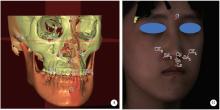
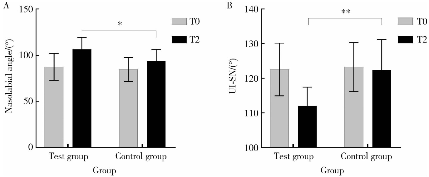
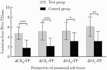
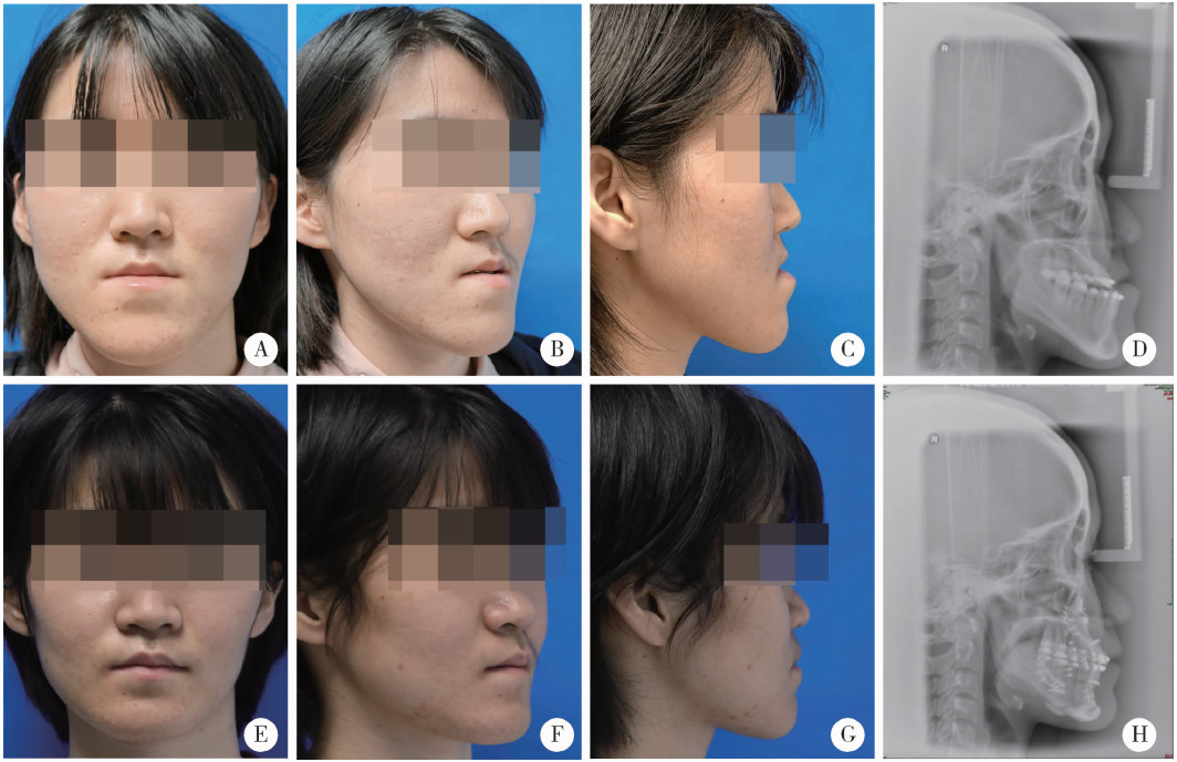
摘要: 目的: 比较上颌骨前部顺时针旋转与整体顺时针旋转两种手术方式改善骨性Ⅲ类牙颌面畸形患者鼻旁凹陷的美学效果,为临床手术方式的选择提供参考依据。方法: 设计非随机临床对照试验,纳入骨性Ⅲ类牙颌面畸形患者21例(试验组11例,对照组10例),试验组采用上颌Le Fort Ⅰ型分块截骨术配合上颌前部顺时针旋转,对照组采用上颌整体顺时针旋转。分别采集术前(T0期)、术后2周(T1期)、术后6个月(T2期)的大视野锥形束CT(cone beam computered tomography, CBCT)和三维面相,进行三维软硬组织测量,测量指标包括颊凸点(cheek mass, CK)和鼻翼基点(subalare, SA)的矢状向位移量、鼻唇角、咬合平面角、上前牙唇倾角等。采用独立样本t检验对比不同时间节点测量指标的组间差异。结果: T0期两组三维头影测量指标的差异无统计学意义,术中试验组上颌平均前徙距离为(-0.71±1.67) mm,明显小于对照组(2.26±1.68) mm,t=-4.052,P<0.05;试验组咬合平面顺时针旋转角度1.46°±2.38°,明显小于对照组4.31°±1.83°,t=-3.047,P<0.05。T0到T2期试验组CK点和SA点的矢状向前移距离分别为(4.96±1.18) mm和(5.19±1.17) mm,对照组分别为(2.01±1.50) mm和(2.69±1.45) mm,组间差异具有统计学意义(P<0.05)。T2期试验组的上前牙唇倾角度为112.15°±5.40°,显著小于对照组122.38°±8.83°(t=-3.237,P<0.05);鼻唇角为106.54°±12.82°,显著大于对照组93.90°±12.46°(t=2.288,P<0.05)。结论: 相较于上颌整体顺时针旋转,上颌Le Fort Ⅰ型分块截骨术后顺时针旋转上颌前部,可以在不改变上颌切牙矢状向位置和咬合平面角的前提下,增大鼻旁区软硬组织的前徙距离,纠正上前牙唇倾及鼻唇角过锐,更好地改善骨性Ⅲ类牙颌面畸形患者的鼻旁美学缺陷。

关键词: 骨性Ⅲ类牙颌面畸形, 鼻旁凹陷, 上颌Le Fort Ⅰ型分块截骨术, 顺时针旋转, 三维头影测量

Abstract: Objective: To compare the aesthetic effects of anterior maxilla clockwise rotation combined with segmental Le Fort Ⅰ osteotomy and whole maxilla clockwise rotation on improving paranasal concavity in patients with Class Ⅲ maxillofacial deformity. Methods: A non-randomized controlled trial was designed, and 21 patients diagnosed with skeletal Class Ⅲ maxillofacial deformity were included. In the study, 11 patients in the test group were treated by segmental Le Fort Ⅰ osteotomy combined with anterior maxilla clockwise rotation, and 10 patients in the control group were treated by whole maxilla clockwise rotation. The CBCT and 3D photography of preoperative (T0), 2 weeks postoperative (T1), and 6 months postoperative (T2) were collected respectively, and the three-dimensional cephalometry was carried out. The differences of specific parameters between the two groups were compared by independent sample t-test, including saggital displacement of the cheek mass point (CK) and subalare point (SA), nasolabial angle, occlusal plane angle and labial inclination angle of the upper incisor. Results: There were no significant differences of the parameters on T0 between the two groups. The average sagittal displacement of the upper incisors of the test group was (-0.71±1.67) mm and smaller than that of control group [(2.26±1.68) mm], t=-4.052, P < 0.05. The average angle of the occlusal plane clockwise rotation of the test group was 1.46°±2.38° and smaller than that of the control group (4.31°±1.83°), t=-3.047, P < 0.05. The angle of anterior maxilla clockwise rotation was 11.73°±2.81° during the surgery. The average saggital displacement of the paranasal soft tissue landmarks of the test group from T0 to T2 was larger than that of the control group [CK point, (4.96±1.18) mm vs. (2.01± 1.50) mm, P < 0.05;SA point, (5.19±1.17) mm vs. (2.69±1.45) mm, P < 0.05]. The labial inclination angle of the upper incisor of the test group was 112.15°±5.40° in T2 and significantly smaller than that of the control group (122.38°±8.83°), t=-3.237, P < 0.05. The nasolabial angle of the test group was 106.54°±12.82° in T2 and significantly larger than that of the control group (93.90°±12.46°), t=2.288, P < 0.05. Conclusion: Compared with whole maxilla clockwise rotation, anterior maxilla clockwise rotation combined with segmental Le Fort Ⅰ osteotomy can increase the saggital displacement of the paranasal soft tissue, correct labial inclination of the upper incisors and the acute naso-labial angle and better improve the paranasal aesthetic defects in patients with Class Ⅲ maxillofacial deformity with less changing on the saggital orientation of the upper incisors and the occlusal plane angle.

Key words: Skeletal Class Ⅲ maxillofacial deformity, Paranasal concavity, Segmental Le Fort Ⅰ osteo-tomy, Clockwise rotation, Three dimensional cephalometry

中图分类号: 

  • R782.1

图1

三维头影测量标志点示意图"

表1

三维头影测量中解剖标志点及参考平面的定义"

Parameters Abbreviation Definition
Bone and dental landmarks
    Sella S The center of the hypophyseal fossa
    Nasion N The midpoint of the frontonasal suture
    Orbitale OrL, OrR The most inferior point of each infraorbital rim
    Porion PoL, PoR The most superior point of each external acoustic meatus
    Subspinale A The point of maximum concavity in the midline of the dento-alveolar process of the maxilla
    Upper incisor UI The most mesial point of the tip of the crown of the right upper central incisor
    Upper incisor apex UIa The apex of the right upper central incisor
    Upper molar UML, UMR The mesial buccal cusp of the first upper molar
    Supramental B The point of maximum concavity in the midline of the dento-alveolar process of the mandible
    Point h hL, hR Point on the left/right maxilla directly below the soft tissue alar base on axial view
Soft tissue landmarks
    Glabella G The most anterior midpoint on the fronto-orbital soft tissue contour
    Subalare SAL, SAR Labial insertion of each alar base
    Cheek mass CKL, CKR The most anterior point on the mid-pupillary plane (MPP) under infraorbital area and ahead of cornea perpendicular plane (CPP)
    Subnasale Sn The midpoint of the angle at the columella base where the lower border of the nasal septum and the surface of the upper lip meet
    Labiale superius Ls Midpoint of the upper vermillion border
    Columella peak Cp Most superior point of the columella
    Cornea CL, CR The most anterior point of the cornea
Reference planes
    Frankfort horizontal plane FHP The plane passes bilateral Orbitale and the midpoint between bilateral porion
    Midsagittal plane MSP The plane perpendicular to the FHP plane and passing through sella and nasion
    Facial plane FP The plane perpendicular to the FHP and MSP planes and passing through glabella
    Occlusal plane OP The plane passes the midpoint between UI and the midpoint between UML and UMR
    Mid-pupillary plane MPP The plane perpendicular to the FHP and FP planes and passing through cornea
    Cornea perpendicular plane CPP The plane perpendicular to the FHP and MSP planes and passing through cornea

表2

试验组和对照组的基线特征和T0期三维头影测量值"

Parameters Test group(n=11) Control group(n=10) t P value
Female 9 (81.8) 8 (80.0) -0.101 0.921
Age/years 24.91±6.20 24.00±5.06 0.366 0.719
SNA/(°) 82.47±2.28 81.26±2.89 1.067 0.299
SNB/(°) 85.90±2.46 86.45±2.74 -0.489 0.631
OP-SN/(°) 8.68±5.17 8.33±3.19 0.182 0.857
UI-SN/(°) 122.59±7.56 123.32±7.11 0.229 0.821
SNhR/(°) 72.07±2.91 71.50±4.30 0.364 0.720
SNhL/(°) 71.66±2.68 70.92±4.41 0.468 0.645
CKR-FP/mm -2.22±2.68 -3.04±3.44 0.614 0.546
CKL-FP /mm -3.01±3.26 -3.79±2.93 0.580 0.569
SAR-FP /mm -3.23±3.15 -4.51±3.20 0.921 0.369
SAL-FP /mm -4.16±3.01 -4.87±3.38 0.512 0.615
NL angle/(°) 87.61±14.62 84.63±12.97 0.491 0.629
UI-FP/mm 0.37±4.06 -1.42±3.96 1.020 0.320

表3

T2期试验组和对照组的三维头影测量值对比"

Parameters Test group (n=11) Control group (n=10) t P value
SNA/(°) 84.29±2.66 84.02±2.78 0.225 0.824
SNB/(°) 82.69±1.85 82.84±2.73 -0.142 0.889
OP-SN/(°) 10.26±4.64 11.62±3.92 -0.723 0.479
UI-SN/(°) 112.15±5.40 122.38±8.83 -3.237 0.004**
SNhR/(°) 78.48±2.77 76.16±3.83 1.600 0.126
SNhL/(°) 78.44±2.55 75.97±4.18 1.657 0.114
CKR-FP/mm 2.78±2.92 -1.21±3.31 2.932 0.009**
CKL-FP /mm 2.33±3.20 -1.60±3.25 2.793 0.012*
SAR-FP/mm 2.03±3.17 -1.32±2.95 2.507 0.021
SAL-FP/mm 2.23±3.73 -1.71±2.91 2.682 0.015*
NL angle/(°) 106.54±12.82 93.90±12.46 2.288 0.034*
UI-FP/mm 0.84±4.87 1.79±3.89 -0.494 0.627

图2

T0期到T2期两组鼻唇角和上前牙唇倾角度的变化"

表4

T0期到T2期试验组和对照组的三维头影测量变化值对比"

Parameters Test group (n=11) Control group (n=10) t P value
SNA/(°) 1.82±1.59 2.76±1.87 -1.245 0.228
SNB/(°) -3.21±1.04 -3.62±2.44 0.495 0.629
OP-SN/(°) 1.58±2.91 3.29±1.81 -1.597 0.127
UI-SN/(°) -10.43±4.28 -0.94±4.89 -4.746 <0.001**
SNhR/(°) 6.4±1.88 4.67±1.91 2.098 0.049*
SNhL/(°) 6.79±1.48 5.05±2.23 2.122 0.047*
CKR-FP/mm 4.78±1.22 1.83±1.55 4.886 <0.001**
CKL-FP /mm 5.15±1.74 2.19±1.63 4.005 <0.001**
SAR-FP /mm 5.24±1.71 3.18±1.67 2.782 0.012*
SAL-FP /mm 6.1±1.94 3.17±1.99 3.421 0.003**
NL angle/(°) 12.01±8.17 9.27±13.28 0.576 0.571
UI-FP/mm -0.03±1.95 3.05±2.44 -3.210 0.005**

图3

T0期到T2期鼻旁区软组织标志点的矢状向位移"

图4

试验组患者术前(T0)和术后6个月(T2)对比"

图5

对照组患者术前(T0)和术后6个月(T2)对比"

1
Naini FB , Gill DS . Orthognathic surgery: Principles, planning and practice[M]. New York: John Wiley & Sons, 2017: 85- 86.
2
Catherine Z , Scolozzi P . Modified Le Fort Ⅰ step osteotomy for improvement of paranasal flatness in maxillary deficiency: Technical note and series of 24 cases[J]. J Stomatol Oral Maxillofac Surg, 2019, 120 (6): 559- 565.

doi: 10.1016/j.jormas.2019.07.003
3
Lee TY , Chung HY , Dhong ES , et al. Paranasal augmentation using multi-folded expanded polytetrafluorethylene (ePTFE) in the East Asian nose[J]. Aesthet Surg J, 2019, 39 (12): 1319- 1328.

doi: 10.1093/asj/sjz103
4
Park HM , Lee YK , Choi JY , et al. Maxillary incisor inclination of skeletal Class Ⅲ patients treated with extraction of the upper first premolars and two-jaw surgery: Conventional orthognathic surgery vs. surgery-first approach[J]. Angle Orthod, 2014, 84 (4): 720- 729.

doi: 10.2319/072113-529.1
5
Kim DK , Baek SH . Change in maxillary incisor inclination during surgical-orthodontic treatment of skeletal Class Ⅲ malocclusion: Comparison of extraction and nonextraction of the maxillary first premolars[J]. Am J Orthod Dentofacial Orthop, 2013, 143 (3): 324- 335.

doi: 10.1016/j.ajodo.2012.10.014
6
Choi JW , Lee JY , Oh TS , et al. Frontal soft tissue analysis using a 3 dimensional camera following two-jaw rotational orthognathic surgery in skeletal Class Ⅲ patients[J]. J Craniomaxillofac Surg, 2014, 42 (3): 220- 226.

doi: 10.1016/j.jcms.2013.05.004
7
Chemello PD , Wolford LM , Buschang PH . Occlusal plane alteration in orthognathic surgery. Part Ⅱ: Long-term stability of results[J]. Am J Orthod Dentofacial Orthop, 1994, 106 (4): 434- 440.

doi: 10.1016/S0889-5406(94)70066-4
8
Wolford LM , Chemello PD , Hilliard F . Occlusal plane alteration in orthognathic surgery. Part Ⅰ: Effects on function and esthetics[J]. Am J Orthod Dentofacial Orthop, 1994, 106 (3): 304- 316.

doi: 10.1016/S0889-5406(94)70051-6
9
林广贤, 宋震, 范飞. 鼻基底填充术矫正鼻翼基底凹陷的临床应用进展[J]. 中国美容整形外科杂志, 2022, 33 (5): 312- 314.
10
王兴. 正颌外科手术学[M]. 济南: 山东科学技术出版社, 1999: 199- 201.
11
Chen YR , Yeow VK . Multiple-segment osteotomy in maxillofacial surgery[J]. Plast Reconstr Surg, 1999, 104 (2): 381- 388.

doi: 10.1097/00006534-199908000-00009
12
Liu XJ , Li QQ , Pang YJ , et al. Modified method of recording and reproducing natural head position with a multicamera system and a laser level[J]. Am J Orthod Dentofacial Orthop, 2015, 147 (6): 781- 787.

doi: 10.1016/j.ajodo.2015.01.016
13
Wong R , Chau A , Hägg U . 3D CBCT McNamara' s cephalometric analysis in an adult southern Chinese population[J]. Int J Oral Maxillofac Surg, 2011, 40 (9): 920- 925.

doi: 10.1016/j.ijom.2011.03.011
14
Ho CT , Denadai R , Lai HC , et al. Computer-aided planning in orthognathic surgery: A comparative study with the establishment of burstone analysis-derived 3D norms[J]. J Clin Med, 2019, 8 (12): 2106.

doi: 10.3390/jcm8122106
15
Zhang L , Tang MY , Jin R , et al. Classification of nasolabial folds in Asians and the corresponding surgical approaches: By Shanghai 9th People' s Hospital[J]. J Plast Reconstr Aesthet Surg, 2015, 68 (7): 914- 919.

doi: 10.1016/j.bjps.2015.03.023
16
Lai HC , Denadai R , Ho CT , et al. Effect of Le Fort Ⅰ maxillary advancement and clockwise rotation on the anteromedial cheek soft tissue change in patients with skeletal Class Ⅲ pattern and midface deficiency: A 3D imaging-based prediction study[J]. J Clin Med, 2020, 9 (1): 262.

doi: 10.3390/jcm9010262
17
Han MD , Momin MR , Munaretto AM , et al. Three-dimensional cephalometric analysis of the maxilla: Analysis of new landmarks[J]. Am J Orthod Dentofacial Orthop, 2019, 156 (3): 337- 344.

doi: 10.1016/j.ajodo.2018.09.018
18
Lee TS , Park S . Clockwise rotation of the occlusal plane for aesthetic purposes by double jaw surgery without orthodontic treatment[J]. Plast Reconstr Surg, 2019, 144 (6): 1010e- 1013e.

doi: 10.1097/PRS.0000000000006248
19
Jeong S , Sung J , Kim S , et al. Upper airway morphologic changes after mandibular setback surgery in skeletal Class Ⅲ malocclusion patients measured using cone beam computed tomography super-imposition[J]. Int J Oral Maxillofac Surg, 2018, 47 (11): 1405- 1410.

doi: 10.1016/j.ijom.2018.05.002
20
Hernández-Alfaro F , Guijarro-Martínez R . On a definition of the appropriate timing for surgical intervention in orthognathic surgery[J]. Int J Oral Maxillofac Surg, 2014, 43 (7): 846- 855.

doi: 10.1016/j.ijom.2014.02.007
21
Ko EW , Hsu SS , Hsieh HY , et al. Comparison of progressive cephalometric changes and postsurgical stability of skeletal Class Ⅲ correction with and without presurgical orthodontic treatment[J]. J Oral Maxillofac Surg, 2011, 69 (5): 1469- 1477.

doi: 10.1016/j.joms.2010.07.022
22
Sawh-Martinez R , Lin AM , DeSesa CR , et al. Clockwise and counterclockwise Le Fort Ⅰ movements influence nasolabial mor-phology differently[J]. Plast Reconstr Surg, 2018, 142 (6): 1572- 1581.

doi: 10.1097/PRS.0000000000004988
[1] 金珉廷,刘怡. 三维颅面水平参考平面的确定方法[J]. 北京大学学报(医学版), 2019, 51(5): 937-943.
Viewed
Full text


Abstract

Cited

  Shared   
  Discussed   
No Suggested Reading articles found!