北京大学学报(医学版) ›› 2025, Vol. 57 ›› Issue (2): 360-368. doi: 10.19723/j.issn.1671-167X.2025.02.022

• 论著 • 上一篇    下一篇

基于下颌骨数据库和全连接神经网络的三维检索模型辅助下的下颌骨个性化重建

仇师禹1, 练洋2, 康一帆1, 张雷1, 蔡义望2, 单小峰1,*(), 蔡志刚1,*()   

  1. 1. 北京大学口腔医学院·口腔医院口腔颌面外科,国家口腔医学中心,国家口腔疾病临床医学研究中心,口腔生物材料和数字诊疗装备国家工程研究中心,口腔数字医学北京市重点实验室,北京 100081
    2. 北京百特康科技有限公司,北京 102629
  • 收稿日期:2021-10-20 出版日期:2025-04-18 发布日期:2025-04-12
  • 通讯作者: 单小峰,蔡志刚 E-mail:kqsxf@263.net;c2013xs@163.com
  • 基金资助:
    国家重点研发计划(2016YFC1102902);首都卫生发展科研专项(CFH2020-2-4102)

Personalized mandibular reconstruction assisted by three-dimensional retrieval model based on fully connected neural network and a database of mandibles

Shiyu QIU1, Yang LIAN2, Yifan KANG1, Lei ZHANG1, Yiwang CAI2, Xiaofeng SHAN1,*(), Zhigang CAI1,*()   

  1. 1. Department of Oral and Maxillofacial Surgery, Peking University School and Hospital of Stomatology & National Center for Stomatology & National Clinical Research Center for Oral Diseases & National Engineering Research Center of Oral Biomaterials and Digital Medical Devices & Beijing Key Laboratory of Digital Stomatology, Beijing 100081, China
    2. Byte-king Technology, Beijing 102629, China
  • Received:2021-10-20 Online:2025-04-18 Published:2025-04-12
  • Contact: Xiaofeng SHAN, Zhigang CAI E-mail:kqsxf@263.net;c2013xs@163.com
  • Supported by:
    the National Key Research and Development Program of China(2016YFC1102902);the Capital Health Development Research Project(CFH2020-2-4102)

RICH HTML


摘要:

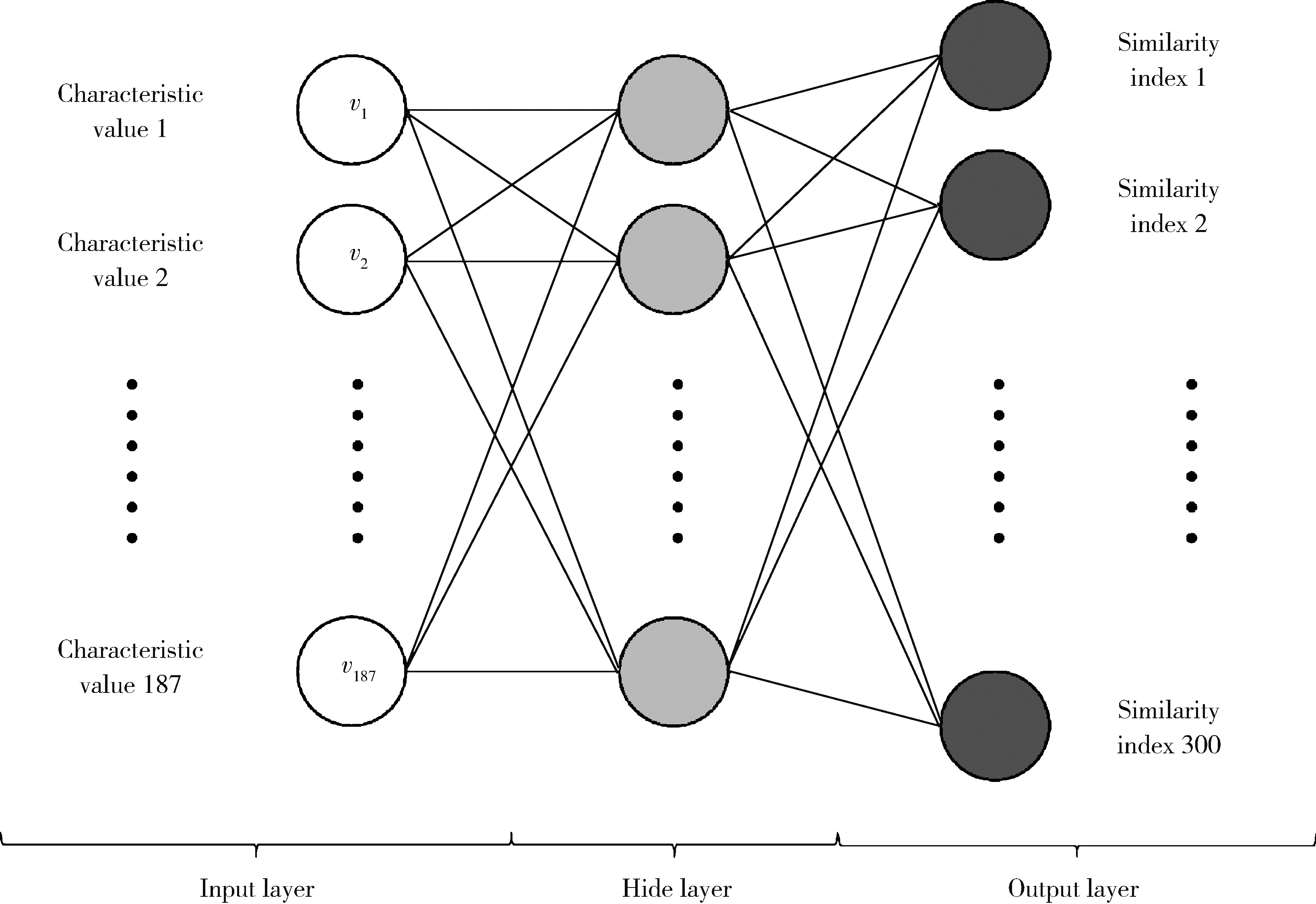
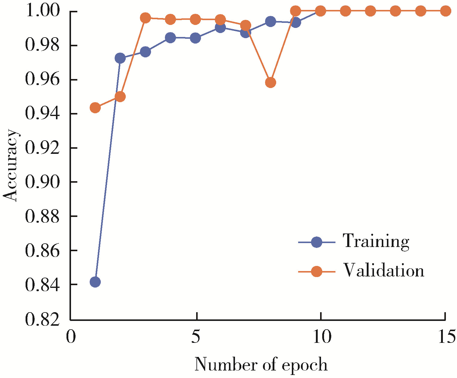
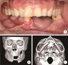
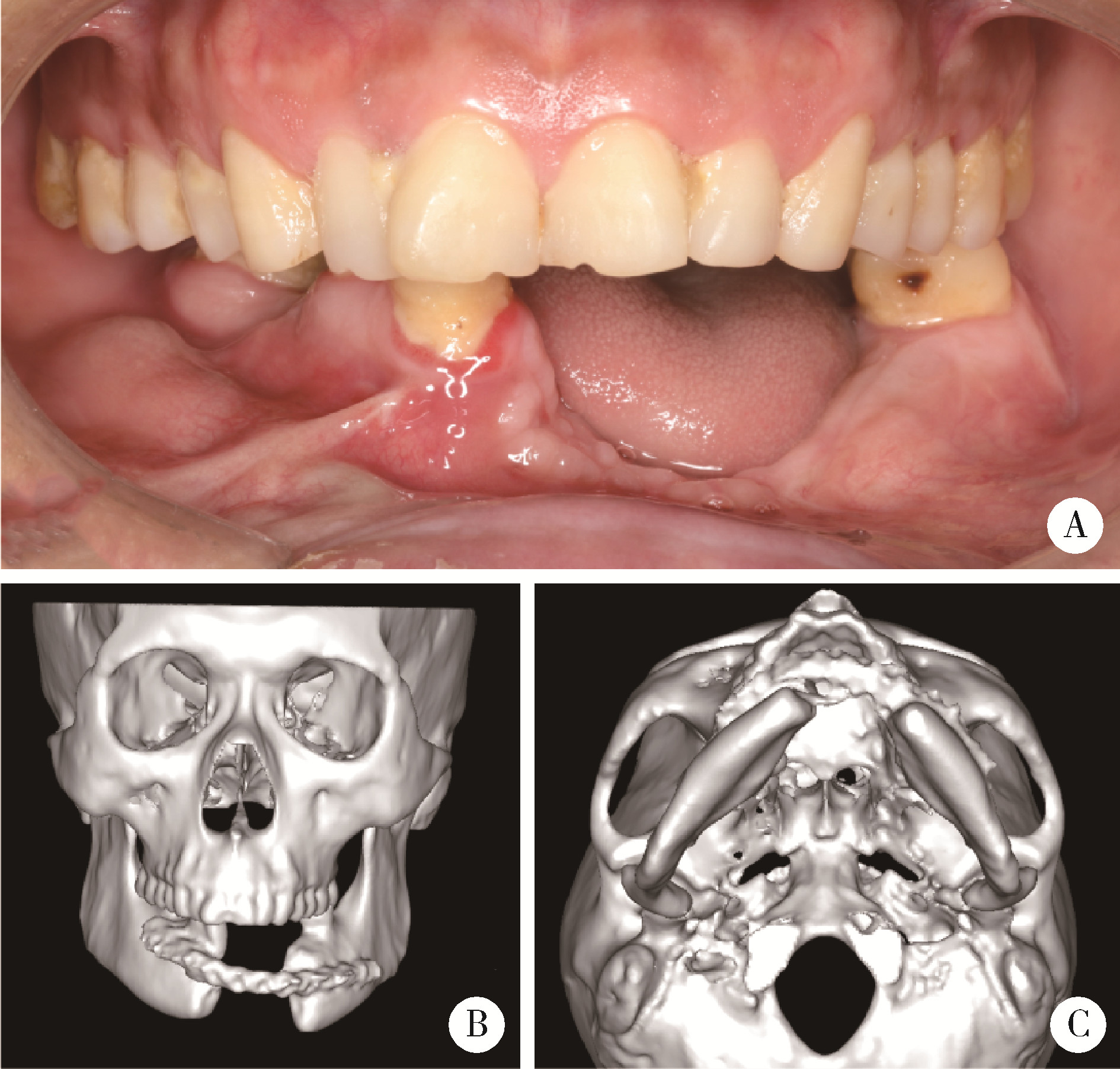
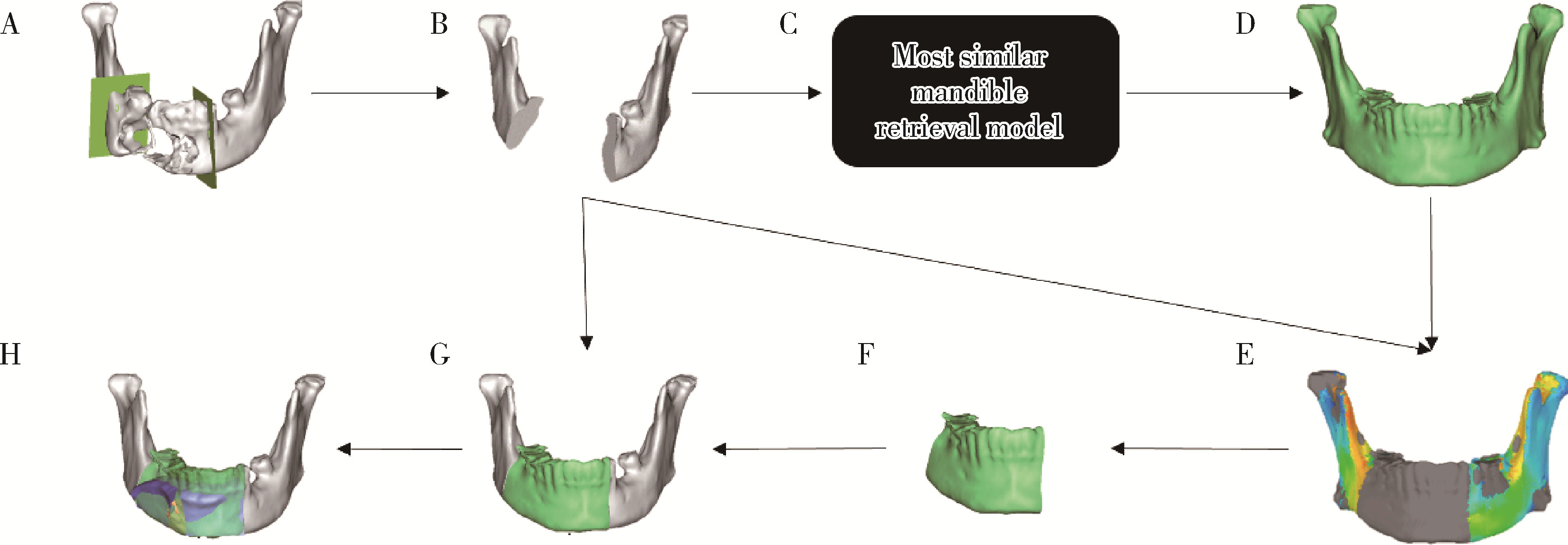
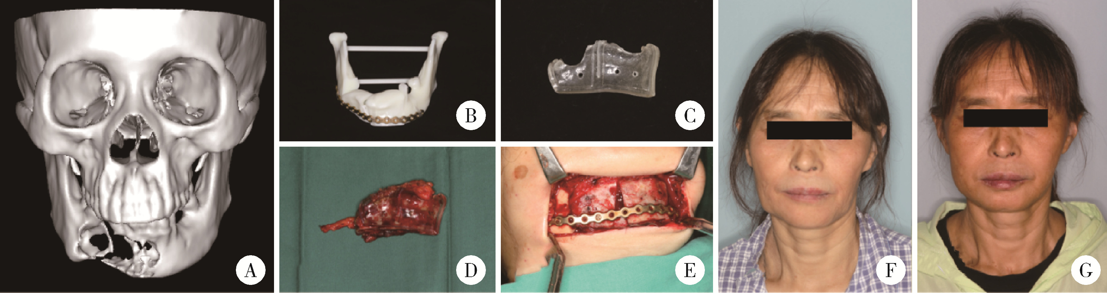
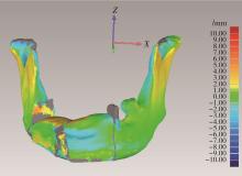
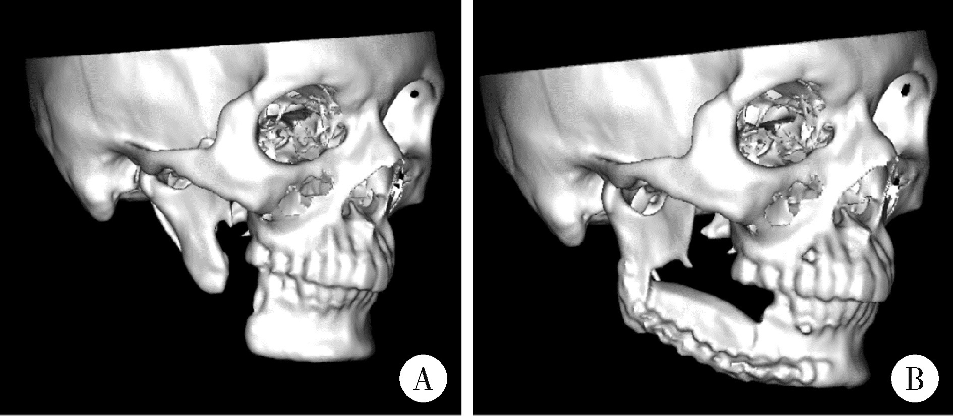
目的: 提出基于下颌骨数据库和全连接神经网络(fully connected neural network,FCNN) 的三维检索模型辅助下的下颌骨个性化重建方案,并验证该方案的临床可行性。方法: 建立一个300例正常中国北方汉族人下颌骨数据库,在头影测量的基础上,进一步筛选稳定性较好的下颌骨标志点,制定下颌骨标志点方案,提取下颌骨几何特征。开发三维检索算法,该算法能从上述数据库中检索出与待匹配下颌骨最相似的下颌骨。搭建FCNN训练上述算法以提高三维检索精度,使用Geomagic Control 2014软件评价基于上述下颌骨数据库和算法的三维检索模型匹配精度。从2019年12月到2021年3月,共有5例患者在北京大学口腔医院颌面外科接受了基于下颌骨数据库和FCNN的三维检索模型辅助下的下颌骨个性化重建手术。通过三维检索算法从上述下颌骨数据库中检索获得最相似下颌骨,使用最相似下颌骨恢复缺损区病变前形态和指导下颌骨重建。5例患者的下颌骨缺损均由髂骨瓣修复,使用个性化手术导板实现术前手术设计的转化和实施。结果: 通过筛选,可重复性和稳定性较高的下颌骨标志点被确定并组成下颌骨标志点方案。经过训练后,基于FCNN的三维检索模型在300例下颌骨数据库中检索获得的最相似下颌骨与待匹配下颌骨的平均偏差为(1.77±0.44) mm,均方根偏差为(2.58±0.86) mm。5例患者的下颌骨重建手术均成功,面部对称性和咬合功能得以恢复,所有患者都对术后外观恢复感到满意。三维比较显示,术后下颌骨与术前设计之间的平均偏差为(0.98±0.17) mm,偏差≤1 mm区域占比61.34%±14.13%,≤2 mm区域占比83.82%±7.35%,≤3 mm区域占比93.94%±2.87%。结论: 基于下颌骨数据库和FCNN的三维检索模型辅助下的下颌骨个性化重建具有临床可行性。

关键词: 下颌骨数据库, 全连接神经网络, 三维检索, 下颌骨重建

Abstract:

Objective: To propose a new protocol for personalized mandibular reconstruction assisted by three-dimensional (3D) retrieval model based on fully connected neural network (FCNN) and a database of mandibles, and to verify clinical feasibility of the protocol. Methods: A database of mandibles of 300 normal northern Chinese Han people was established. On the basis of cephalometry, the mandible landmarks with good stability were further screened. Mandibular landmarks were selected and geometric features of the mandible were extracted. A 3D retrieval algorithm was developed, which could retrieve the mandible most similar to a given mandible from the database. A FCNN was built to train the algorithm to improve accuracy of the 3D retrieval model. Using Geomagic Control 2014 software, matching accuracy of the 3D retrieval model was based on aforementioned mandible database and algorithm. From December 2019 to March 2021, a total of 5 patients underwent personalized mandibular reconstruction assisted by a 3D retrieval model based on mandible database and FCNN in the Department of Oral and Maxillofacial Surgery, Peking University School and Hospital of Stomatology. The most similar mandible was retrieved from mandible database through 3D retrieval algorithm. It was used to restore the premorbid morphology of defect area and guide mandibular reconstruction. For the 5 patients, mandible was reconstructed with iliac flap. Virtual surgical plan was transformed using individual surgical guides. Results: Through screening, mandibular landmarks with high reproducibility and stability were identified and composed of mandibular landmarker protocols. After training, the average deviation between most similar mandible retrieved from the 300-case mandible database through 3D retrieval model based on FCNN and given mandible was (1.77±0.44) mm. And the root-mean-square deviation between the most similar mandible retrieved from the database and given mandible was (2.58±0.86) mm. The mandibular reconstruction surgery was successful in all the 5 patients. Their facial symmetry and occlusion were restored. All the patients were satisfied with postoperative appearance. The mean deviation between postoperative mandible and preoperative design was (0.98±0.17) mm. The area with a deviation ≤1 mm accounted for 61.34%±14. 13%, ≤2 mm accounted for 83.82%±7.35%, and ≤3 mm accounted for 93.94%± 2.87%. Conclusion: The personalized mandibular reconstruction assisted by 3D retrieval model based on the 300-case mandible database and FCNN is feasible clinically.

Key words: Database of mandibles, Fully connected neural network, Three-dimensional retrieval, Mandibular reconstruction

中图分类号: 

  • R782.1

表1

下颌骨标志点"

Name Abbreviation Definition
Lower incisor point LIP Midpoint of mesio-incisal angle of bilateral lower first incisors
Lower first molar point LFP(L/R) Mesio-buccal cusp of lower first molar
Lower canine point LCP(L/R) Tip of lower canine
Infradentale In Most anterior point of inferior alveolar process on MSP
Supramental Sup Midpoint of In and Pogonion
Pogonion Po Most anterior point of mentum on MSP
Menton Me Lowest point of mentum on MSP
Gnathion Gn Midpoint of Po and Me
Post point of menton PPM Most posterior point of mentum on MSP
Condylar anterior point CAP(L/R) Most anterior point of condyle
Condylar posterior point CPP(L/R) Most posterior point of condyle
Condylar lateral point CLP(L/R) Most lateral point of condyle
Condylar medial point CMP(L/R) Most medial point of condyle
Condylar top point CTP(L/R) Top point of condyle
Coracoid process CP(L/R) Tip of coracoid process
Lowest point of sigmoid notch LPSN(L/R) Lowest point of sigmoid notch
Corresponding point of LPSN LPSNC(L/R) The same horizontal point of LPSN on the posterior edge of ramus
Gonion Go(L/R) Gonion point
Antegonial notch point AN Antegonial notch point
Lowest point of anterior edge of ramus LAR(L/R) Intersection of anterior edge of ramus and mandibular body
Corresponding point of LAR LARC(L/R) The same horizontal point of LAR on the posterior edge of ramus
Seven equal points of lower edge of mandible SLM(1-6) Seven equal points of mandibular lower edge
Superior prosthion SP Most anterior point of upper alveolar process on MSP
Subspinale Sub Midpoint of SP and anterior nasal spine
Upper incisor point UIP(L/R) Midpoint of mesio-incisal angle of bilateral upper first incisors
Upper canine point UCP(L/R) Tip of upper canine
Upper first molar point UFMP(L/R) Mesio-buccal cusp of upper first molar
TMJ eminence lowerest point ELP(L/R) Lowest point of TMJ eminence
TMJ fossa top point FTP(L/R) Top point of TMJ fossa

图1

全连接神经网络结构示意图"

图2

全连接神经网络训练和验证精度结果"

表2

5例患者的信息"

Case No. Gender Age Diagnosis Defect type (according to Jewer classification) Defect area (using FDI system for teeth)
1 Female 46 Fibromyxoma LC 37-41
2 Female 55 Ameloblastoma LCL 33-47
3 Male 25 Ossifying fibroma LC 31-right mandibular angle
4 Male 43 Mandibular defect secondary to resection for ameloblastoma 6 months ago LCL 35-43
5 Male 27 Mandibular defect secondary to resection for keratocyst 2 years ago L 47-right mandibular angle

图3

病例4影像资料"

图4

最相似下颌骨三维检索模型工作流程图"

图5

下颌骨重建手术流程图"

图6

术后三维色谱分析"

表3

新下颌骨标志点方案"

Name Abbreviation Definition
Lower incisor point LIP Midpoint of mesio-incisal angle of bilateral lower first incisors
Lower first molar point LFP(L/R) Mesio-buccal cusp of lower first molar
Lower canine point LCP(L/R) Tip of lower canine
Infradentale In Most anterior point of inferior alveolar process on MSP
Supramental Sup Midpoint of In and Pogonion
Pogonion Po Most anterior point of mentum on MSP
Menton Me Lowest point of mentum on MSP
Gnathion Gn Midpoint of Po and Me
Post point of menton PPM Most posterior point of mentum on MSP
Condylar anterior point CAP(L/R) Most anterior point of condyle
Condylar posterior point CPP(L/R) Most posterior point of condyle
Condylar lateral point CLP(L/R) Most lateral point of condyle
Condylar medial point CMP(L/R) Most medial point of condyle
Condylar top point CTP(L/R) Top point of condyle
Coracoid process CP(L/R) Tip of coracoid process
Lowest point of sigmoid notch LPSN(L/R) Lowest point of sigmoid notch
Gonion Go(L/R) Gonion point
Lowest point of anterior edge of ramus LAR(L/R) Intersection of anterior edge of ramus and mandibular body
Seven equal points of lower edge of mandible SLM(1-6) Seven equal points of mandibular lower edge
Superior prosthion SP Most anterior point of upper alveolar process on MSP
Subspinale Sub Midpoint of SP and anterior nasal spine
Upper incisor point UIP(L/R) Midpoint of mesio-incisal angle of bilateral upper first incisors
Upper canine point UCP(L/R) Tip of upper canine
Upper first molar point UFMP(L/R) Mesio-buccal cusp of upper first molar
TMJ eminence lowerest point ELP(L/R) Lowest point of TMJ eminence
TMJ fossa top point FTP(L/R) Top point of TMJ fossa

图7

病例5影像资料"

表4

术后3D色谱分析结果"

Case No. Avarege deviation /mm Devitation≤1 mm /% Devitation≤2 mm /% Devitation≤3 mm /%
1 0.72 75.44 90.85 97.31
2 1.19 42.33 71.52 89.36
3 1.02 62.48 85.73 94.33
4 0.95 73.97 87.21 94.09
5 1.01 52.50 83.79 94.59
1 Kakarala K , Shnayder Y , Tsue TT , et al. Mandibular reconstruction[J]. Oral Oncol, 2018, 77, 111- 117.
doi: 10.1016/j.oraloncology.2017.12.020
2 Hanasono MM , Skoracki RJ . Computer-assisted design and rapid prototype modeling in microvascular mandible reconstruction[J]. Laryngoscope, 2013, 123 (3): 597- 604.
doi: 10.1002/lary.23717
3 Zhang L , Liu Z , Li B , et al. Evaluation of computer-assisted mandibular reconstruction with vascularized fibular flap compared to conventional surgery[J]. Oral Surg Oral Med Oral Pathol Oral Radiol, 2016, 121 (2): 139- 148.
doi: 10.1016/j.oooo.2015.10.005
4 Ren W , Gao L , Li S , et al. Virtual planning and 3D printing modeling for mandibular reconstruction with fibula free flap[J]. Med Oral Patol Oral Cir Bucal, 2018, 98 (33): 2666- 2670.
5 Moiduddin K , Al-Ahmari A , Nasr ES , et al. A comparison study on the design of mirror and anatomy reconstruction technique in maxillofacial region[J]. Tech Health Care, 2016, 24 (3): 377- 389.
doi: 10.3233/THC-161136
6 陈全, 蔡志刚, 彭歆, 等. 下颌骨大范围缺损修复重建设计可变形模型的建立[J]. 中华口腔医学杂志, 2014, 49 (7): 414- 419.
doi: 10.3760/cma.j.issn.1002-0098.2014.07.008
7 Wang E , Tran KL , D'heygere E , et al. Predicting the premorbid shape of a diseased mandible[J]. Laryngoscope, 2021, 131 (3): E781- E786.
8 周子疌, 朱向阳, 韩婧, 等. 基于机器学习的颌骨特征点还原法辅助跨中线颌骨缺损重建[J]. 中国口腔颌面外科杂志, 2020, 18 (4): 323- 327.
9 Yao BC , He Y , Jie BM , et al. Reconstruction of bilateral post-traumatic midfacial defects assisted by three-dimensional craniomaxillofacial data in normal chinese people: A preliminary study[J]. J Oral Maxillofac Surg, 2019, 77 (11): 2302. e1- 2302. e13.
doi: 10.1016/j.joms.2019.04.030
10 傅民魁, 林久祥, 周彦恒, 等. 口腔正畸学[M]. 2版 北京: 北京大学医学出版社, 2014: 63- 78.
11 Shnayder Y , Lin D , Desai SC , et al. Reconstruction of the lateral mandibular defect: A review and treatment algorithm[J]. JAMA Facial Plast Surg, 2015, 17 (5): 367- 373.
doi: 10.1001/jamafacial.2015.0825
12 Cannon TY , Strub GM , Yawn RJ , et al. Oromandibular reconstruction[J]. Clin Anat, 2011, 25 (1): 108- 119.
13 Mazzola F , Smithers F , Cheng K , et al. Time and cost-analysis of virtual surgical planning for head and neck reconstruction: A matched pair analysis[J]. Oral Oncol, 2020, 100, 104491.
doi: 10.1016/j.oraloncology.2019.104491
14 Tarsitano A , Mazzoni S , Cipriani R , et al. The CAD/CAM technique for mandibular reconstruction: An 18 patients oncological case-series[J]. J Craniomaxillofac Surg, 2014, 42 (7): 1460- 1464.
doi: 10.1016/j.jcms.2014.04.011
15 Chang EI , Boukovalas S , Liu J , et al. Reconstruction of posterior mandibulectomy defects in the modern era of virtual planning and three-dimensional modeling[J]. Plast Reconstr Surg, 2019, 144 (3): 453- 462.
doi: 10.1097/PRS.0000000000005954
16 蔡志刚. 数字化外科技术在下颌骨缺损修复重建中的应用[J]. 中华口腔医学杂志, 2012, 47 (8): 474- 478.
17 Yu HB , Shen GF , Wang XD , et al. The indication and application of computer-assisted navigation in oral and maxillofacial surgery: Shanghai' s experience based on 104 cases[J]. J Craniomaxillofac Surg, 2013, 41 (8): 770- 774.
doi: 10.1016/j.jcms.2013.01.016
18 Davies JC , Chan HH , Jozaghi Y , et al. Analysis of simulated mandibular reconstruction using a segmental mirroring technique[J]. J Craniomaxillofac Surg, 2019, 47 (3): 468- 472.
doi: 10.1016/j.jcms.2018.12.016
[1] 康一帆, 葛严军, 吕晓鸣, 谢尚, 单小峰, 蔡志刚. 即刻种植体支持式义齿修复的血管化髂骨瓣重建下颌骨缺损[J]. 北京大学学报(医学版), 2025, 57(1): 78-84.
[2] 王聪伟,高敏,于尧,章文博,彭歆. 游离腓骨瓣修复下颌骨缺损术后义齿修复的临床分析[J]. 北京大学学报(医学版), 2024, 56(1): 66-73.
Viewed
Full text


Abstract

Cited

  Shared   
  Discussed   
No Suggested Reading articles found!