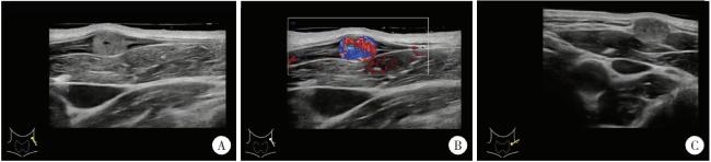
病例报告

左侧胸锁乳突肌间血管内乳头状内皮增生1例

  • 肖晓笛 1 ,
  • 夏有辰 1 ,
  • 柳剑英 2 ,
  • 付鹏 , 3, *
展开
  • 1. 北京大学第三医院成形外科, 北京 100191
  • 2. 北京大学第三医院病理科, 北京 100191
  • 3. 北京大学第三医院超声科, 北京 100191

收稿日期: 2022-12-09

  网络出版日期: 2025-09-05

版权

版权所有,未经授权,不得转载。

Left sided sternocleidomastoid interosseous intravascular papillary endothelial hyperplasia: A case report

  • Xiaodi XIAO 1 ,
  • Youchen XIA 1 ,
  • Jianying LIU 2 ,
  • Peng FU , 3, *
Expand
  • 1. Department of Plastic Surgery, Peking University Third Hospital, Beijing 100191, China
  • 2. Department of Pathology, Peking University Third Hospital, Beijing 100191, China
  • 3. Department of Ultrasound, Peking University Third Hospital, Beijing 100191, China
FU Peng, e-mail,

Received date: 2022-12-09

  Online published: 2025-09-05

Copyright

All rights reserved. Unauthorized reproduction is prohibited.

本文引用格式

肖晓笛 , 夏有辰 , 柳剑英 , 付鹏 . 左侧胸锁乳突肌间血管内乳头状内皮增生1例[J]. 北京大学学报(医学版), 2025 , 57(5) : 1002 -1004 . DOI: 10.19723/j.issn.1671-167X.2025.05.028

Abstract

Intravascular papillary endothelial hyperplasia (IPEH), also known as Masson tumor, is a rare vascular benign tumor of blood vessels. It may occur in any part of the body, especially the deep dermis and subcutaneous tissue of the head, neck, fingers and trunk. The imaging and histopathology of IPEH are similar to hemangiosarcoma, especially in the case of active vascular endothelial hyperplasia. IPEH is a reactive proliferative lesion of vascular intima. The etiology is still unclear. After some studies showed that IPEH was a benign lesion, few reports on the etiology of it were reported. IPEH is usually limited to the thrombotic vessels or lumens of vascular malformations, usually accompanied by a clear history of trauma. IPEH usually does not cause any symptoms. It looks like a slow-growing lump. Some cases have been reported with pain and swelling. Although IPEH is relatively rare, its accurate diagnosis is crucial because it may be similar to malignant angiogenic lesions in clinical practice. There were few reports of cases related to intravascular papillary endothelial hyperplasia located in the sternocleidomastoid muscle after reviewing the domestic and foreign literature in recent 10 years. This case reports that a young male, who was admitted to the hospital one month after finding a subcutaneous tumor in the left neck. After admittance, relevant preoperative examinations were completed. After multi-disciplinary discussion and elimination of surgical contraindications, a specific surgical plan was formulated. The tumor was removed under local anesthesia on the second day after admission. During the operation, it was found that the tumor was located between the sternocleidomastoid muscle bundles, and it was sent for pathologic examination. Paraffin section pathology was reported after operation. Histological examination showed that the morphology was consistent with vascular endothelial papillary hyperplasia. There were no related surgical complications and recurrence in the 3-month follow-up. The purpose of this paper is to provide clinicians with a certain understanding of this rare disease through the report of this case of IPEH, and to identify it in later clinical work, and at the same time, to avoid confusion with malignant diseases, such as hemangiosarcoma, leading to unnecessary treatment and increase the cost of treatment.

血管内乳头状内皮增生(intravascular papillary endothelial hyperplasia,IPEH)是一种少见的良性血管内病变[1],又被称为马松瘤(Masson tumor)、马松血管瘤、马松血管内血管内皮瘤和反应性乳头状内皮增生等。IPEH与血管肉瘤较为相似,临床上需要做出正确的鉴别诊断。本研究即对1例病变位于胸锁乳突肌内且病理提示非典型性改变的IPEH病例进行回顾和总结,希望引起临床医生的重视,以避免进行不必要的激进治疗。

1 病例资料

患者为男性,36岁,主因左颈部皮下肿物入院。患者1个月前无明显诱因发现左颈中部皮下一约黄豆大小肿物,于外院就诊时超声检查提示IPEH可能。随后患者于北京大学第三医院门诊就诊,以“左颈部IPEH待查”收入院。既往肾结石病史。查体:左颈部胸锁乳突肌中上段皮下扪及一约黄豆大小肿物,质韧,边界清,可活动,无红肿及压痛,表面无破溃,体位试验阴性,未闻及血管杂音。术前常规实验室检查均正常。彩色多普勒超声提示,左侧颈外触及肿物处局部浅静脉内可见一低回声实性结节,大小约0.8 cm×0.5 cm×1.0 cm,边界清,不可压缩,病灶内可见丰富血流信号,并可探及低频阻动脉频谱,峰值血流速度为38 cm/s,病变两端浅静脉管腔均可压,诊断为IPEH可能(图 1)。
图1 IPEH患者的超声影像

Figure 1 Ultrasound imaging of the patient with IPEH

A, a hypoechoic nodule with clear, incompressible borders was seen in a local superficial vein; B, color doppler imageing shows rich blood flow signal; C, adjacency of the nodule to the surrounding neck vessels.IPEH, intravascular papillary endothelial hyperplasia.
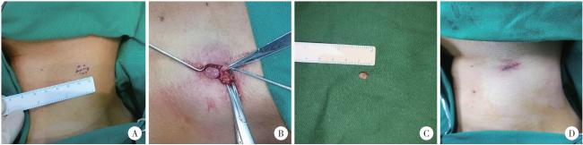
入院当日由血管瘤与脉管畸形多学科综合治疗协作组会诊,根据患者术前超声影像考虑为左颈部IPEH可能性大。入院后第2日在局部麻醉下行左颈外局部浅静脉肿物切除,依据术前切口设计线,逐层切开皮肤、皮下组织后,未见明显肿物,探查肿物位于胸锁乳突肌束间,顺肌束方向分离并离断少量肌束后,可见一边界光滑完整椭圆形肿物,大小约0.6 cm×0.4 cm,质韧,完整切除肿物后逐层减张缝合关闭伤口,术中出血量2 mL(图 2)。
图2 IPEH患者术中情况

Figure 2 Intraoperative situation of the patient with IPEH

A, tumor range and incision design line; B, herniation of a mass after blunt separation of the left sternocleidomastoid muscle; C, isolated mass specimen; D, wound condition after suture. IPEH, intravascular papillary endothelial hyperplasia.
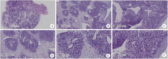
患者于术后第2日出院,出院时切口对合良好,未见明显红肿、渗出,颈部运动不受限。术后病理回报证实IPEH诊断:肉眼可见血管内灰白结节样物一枚,最大径0.6 cm;镜下可见血管内乳头状内皮增生,其内可见血管再通,血管壁纤维组织增生明显(图 3)。患者术后2周复诊可见一长度1.5 cm手术瘢痕,愈合情况尚可,稍红肿,无痛,未见渗出、渗血,颈部各方位运动自如。
图3 IPEH的病理特点

Figure 3 Pathological microscopic features of IPEH

A, full view of intravascular tumor (20×); B, vascular endothelial hyperplasia, fibrous tissue hyperplasia, fibrous interstitial hyperplasia and recanalized vessels (40×); C, papillary structure (200×); D, endothelial lining fissures and papillary hyperplasia (200×); E, no obvious polymorphism of endothelial cells (400×); F, papillary structure (400×).IPEH, intravascular papillary endothelial hyperplasia.

2 讨论

IPEH是一种良性血管内反应性增生性病变,其特征为血管内皮细胞在形成血栓时发生反应性增殖[2]。该病可发生在身体的任何部位,尤其是头部、颈部、手指和躯干的深层真皮和皮下组织[3]。其发病人群男女比例约为1 ∶ 1.3,可在9个月至80岁的任何年龄段发病[4]。根据组织学特征将IPEH分为原发型(56%)、继发型(40%)及血管外型(4%)三类,原发型为典型病变发生在扩张的静脉内,很少出现在动脉;继发型常存在于异常畸形血管内;血管外型最不常见,产生于血肿内[5-6]
自1923年Masson首次发现并报道这种血管内内皮细胞异常增生后,1976年Clearkin和Enzinger研究发现,显微镜下可见的内皮细胞簇状或乳头状增生常与血栓密切相关,由此认为IPEH是一种增生性病变,而非肿瘤性病变[7]。随后较少见有关IPEH病理发展过程的更新认知。1994年Levere等[8]通过综合DNA倍性分析、Northern杂交分析以及免疫印迹分析方法,提出IPEH最初可能是由轻微创伤或刺激使得募集到该区域的巨噬细胞释放内皮碱性成纤维细胞生长因子(basic fibroblast growth factors,bFGF)引起的。巨噬细胞释放bFGF可以刺激内皮细胞的增殖,而内皮细胞反过来会分泌更多的bFGF,从而建立一个正反馈回路,导致内皮细胞的级联增殖,但关于创伤与IPEH发生的关系,以及血栓是在内皮细胞增殖之前还是之后形成等问题,目前仍没有肯定的结论。
本病例未见明显血栓迹象,考虑可能处于后期血栓机化状态,或可能为血管壁内与血栓关系不大的IPEH。典型病例显微镜下可表现为以大量乳头内皮细胞为特征性结构的血管壁向内增生,伴有血栓或血栓碎片,无明显坏死及浸润性生长。应根据患者自然病史,与血管肉瘤进行鉴别,IPEH主要表现为血管腔内病变,而血管肉瘤则不然[9]。目前,IPEH首选的治疗方式仍为完整手术切除,切除后远期复发或恶变少有报道[10]
综上,本病例分析提示,IPEH存在不典型病理表现,诊疗过程中应多维度评估患者疾病类型,在避免延误治疗的同时也应避免过度医疗。

利益冲突  所有作者均声明不存在利益冲突。

作者贡献  肖晓笛:收集临床资料,完成文章撰写;夏有辰:文章审阅、加工、修改;柳剑英:提供病理图片和解读;付鹏:超声影像解读,总体把关;所有作者均对最终文稿进行审读并确认。

1
Han JW , Lee JH , Kim EK . Intravascular papillary endothelial hyperplasia (Masson' s hemangioma) of the face[J]. Arch Craniofac Surg, 2019, 20 (2): 109- 111.

DOI

2
Guledgud MV , Patil K , Saikrishna D , et al. Intravascular papillary endothelial hyperplasia: Diagnostic sequence and literature review of an orofacial lesion[J]. Case Rep Dent, 2014, 2014, 934593.

3
Yadav M , Popli K , Bisoi AK , et al. Masson ' s hemangioma mimicking as leaking aortic pseudoaneurysm: An extremely rare presentation[J]. Aorta (Stamford), 2019, 7 (2): 59- 62.

DOI

4
Mignogna C , Barca I , Di Vito A , et al. Extravascular type of intravascular papillary endothelial hyperplasia mimicking parotid gland neoplasia and the possible role of ferritin in the pathogenesis: A case report[J]. Mol Clin Oncol, 2017, 6 (2): 193- 196.

DOI

5
Soares AB , Altemani A , Furuse C , et al. Intravascular papillary endothelial hyperplasia: Report of 2 cases and immunohistochemical study[J]. Oral Surg Oral Med Oral Pathol Oral Radiol Endod, 2008, 106 (5): 708- 711.

DOI

6
Kim OH , Kim YM , Choo HJ , et al. Subcutaneous intravascular papillary endothelial hyperplasia: Ultrasound features and pathological correlation[J]. Skeletal Radiol, 2016, 45 (2): 227- 233.

DOI

7
Clearkin KP , Enzinger FM . Intravascular papillary endothelial hyperplasia[J]. Arch Pathol Lab Med, 1976, 100 (8): 441- 444.

8
Levere SM , Barsky SH , Meals RA . Intravascular papillary endothelial hyperplasia: A neoplastic "actor" representing an exag-gerated attempt at recanalization mediated by basic fibroblast growth factor[J]. J Hand Surg Am, 1994, 19 (4): 559- 564.

DOI

9
Hashimoto H , Daimaru Y , Enjoji M . Intravascular papillary endothelial hyperplasia: A clinicopathologic study of 91 cases[J]. Am J Dermatopathol, 1983, 5 (6): 539- 546.

DOI

10
Kim D , Israel H , Friedman M , et al. Intravascular papillary endothelial hyperplasia manifesting as a submandibular mass: An unusual presentation in an uncommon location[J]. J Oral Maxillofac Surg, 2007, 65 (4): 786- 790.

DOI

文章导航

/