
1 材料与方法
1.1 材料
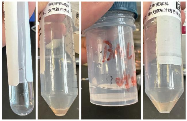
1.2 仪器和试剂
1.3 细胞转移技术操作流程
1.3.1 标记
1.3.2 剥离
1.3.3 转移
1.3.4 染色
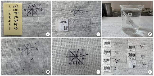
图2 细胞转移技术的操作流程Figure 2 The operation process of cell transfer technology A, divide the area and mark the directions; B, remove the cover glass and set it aside for later use, add Mount Quick gel; C, soaking in warm water; D, remove the gel film, place in the same direction as the cover glass; E, cut the gel film and make the marking copy; F, transfer to slides for different staining projects. |
1.3.5 质控
1.4 统计学分析
2 结果
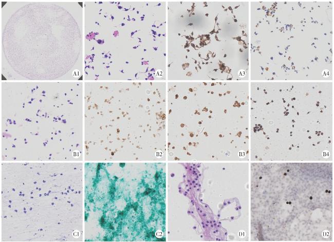
图3 细胞转移前后的染色图Figure 3 The staining images before and after cell transfer A, medullary thyroid carcinoma in fine needle aspiration fluid (A1, before, HE ×10; A2, after, HE ×200; A3, CgA+ + +, ICC ×200; A4, calcitonin+, ICC ×200); B, malignant melanoma in left inguinal lymph nodes fluid (B1, after, HE ×200; B2, HMB45+ +, ICC ×200; B3, malen-A+ +, ICC ×200; B4, S100+ +, ICC ×200); C, pulmonary infection in bronchoalveolar lavage fluid (C1, after, HE ×200; C2, GMS+, specific stain, ×200); D, brain metastasis of lung adenocarcinoma in cerebrospinal fluid (D1, after, HE ×200; D2, TTF-1+ + +, ICC ×200). HE, hematoxyllin-eosin; ICC, immunocytochemistry; CgA, chromogranin A; HMB45, human melanoma black 45; GMS, gomori methenamine silver; TTF-1, thyroid transcription factor-1. |
表1 细胞转移技术对诊断结果的提升情况Table 1 Enhancement of diagnostic results by cell transfer technology |
| Diagnostic results before cell transfer | Diagnostic results after cell transfer | Sample | n | Marker |
| Consider tumor cells; Individual epithelial cells with atypia | Adenocarcinoma of lung | CSF, BALF | 9 | CKPan (AE1/AE3), TTF-1, Pax-8, TFE3, p40 |
| A large number of cell nuclei with irregular shapes | T-cell lymphoblastic lymphoma | CSF | 2 | TdT, CD34, CD68, CKPan (AE1/AE3), CD3, CD20, Olig2, GFAP, Ki67 |
| Malignant cell | Malignant melanoma | Left inguinal lymph node FNA fluid | 1 | CKPan (AE1/AE3), S-100, LCA/CD45, vimentin, HMB45, melan-A |
| Scattered atypical cells are distributed, and the nucleoli can be seen | Cardia cancer | CSF | 1 | CEA, CKPan (AE1/AE3), LCA/CD45 |
| There are a large number of small lymphocytes and monocytes, as well as atypical cells with large nuclei and immature morphology | B lymphoblastic leukemia | CSF | 2 | TdT, Pax-5 |
| Clustered atypical glandular cells, with mucous present in the cytoplasm and prominent nucleoli | Endometrial cancer | Peritoneal wash | 1 | Pax-8 |
| A large number of mycelia were detected | Pneumocystis pneumonia; Pulmonary infection | Ebus needle aspiration fluid, BALF | 2 | GMS, PAS, AFB, WAFB |
| Malignant cell | Adenocarcinoma of stomach | Ascites (a little) | 1 | TTF-1, Pax-8, WT1, CK7, CK20, CDX-2 |
| Consider cells with unclear pathological significance that exhibit atypical changes (TBSRTC Ⅲ) | Medullary thyroid carcinoma | Thyroid FNA fluid | 1 | Congo red, calcitonin, Syn, CgA |
| Cells are scattered and distributed in clusters, with enlarged nuclei and an increased nuclear-cytoplasmic ratio; Some epithelial cells show atypia | Non-tumoral lesion | BALF, bronchial washing, CSF | 8 | GATA3, CEA, Ki67, TdT, CD34, CD20, CD117, MPO, CD68, TTF-1, SALL-4, Syn, CD1a, Olig2 |
TBSRTC, the Bethesda system for reporting thyroid cytopathology; CSF, cerebrospinal fluid; Ebus, endobronchial ultrasound; BALF, bronchoalveolar lavage fluid; FNA, fine-needle aspiration; CKPan, pan cytokeratin; AE, anion exchanger; TTF-1, thyriod transcription factor-1; Pax-8, paired box-8; TFE3, transcription factor enhancing 3; TdT, terminal deoxynucleotidyl transferase; CD, cluster of differentiation; Olig2, oligodendrocyte lineage gene 2; GFAP, glial fibrillary acidic protein; LCA, leukocyte common antigen; HMB45, human melanoma black 45; CEA, carcinoembryonic antigen; GMS, gomori methenamine silver; PAS, periodic acid-schiff; AFB, acid-fast bacillus; WAFB, wade-fite acid-fast bacillus; WT1, Wilms’ tumor 1; CK, cytokeratin; CDX2, caudal-type homeobox transcription factor 2; Syn, synapsin; CgA, chromogranin-A; GATA3, GATA-binding protein 3; MPO, myeloperoxidase; SALL-4, Sal-like protein-4. |



