病例报告

胸锁关节痛风1例

  • 甘地 1 ,
  • 付强 1 ,
  • 唐小惠 1 ,
  • 李楚炜 1 ,
  • 舒兆平 , 2, *
展开
  • 1. 南华大学附属第一医院风湿免疫科,湖南衡阳 421001
  • 2. 南华大学附属第一医院医院感染管理科,湖南衡阳 421001

收稿日期: 2024-07-31

  网络出版日期: 2026-01-07

基金资助

湖南省教育厅科学研究项目(22C0214)

湖南省自然科学基金项目(2023JJ40582)

版权

版权所有,未经授权,不得转载。

Gout of the manubriosternal joints: A case report

  • Di GAN 1 ,
  • Qiang FU 1 ,
  • Xiaohui TANG 1 ,
  • Chuwei LI 1 ,
  • Zhaoping SHU , 2, *
Expand
  • 1. Department of Rheumatology and Immunology, The First Affiliated Hospital, Hengyang Medical School, University of South China, Hengyang 421001, Hunan, China
  • 2. Department of Healthcare-associated Infection Management, The First Affiliated Hospital, Hengyang Medical School, University of South China, Hengyang 421001, Hunan, China
SHU Zhaoping, e-mail,

Received date: 2024-07-31

  Online published: 2026-01-07

Supported by

the Scientific Research Fund of Hunan Provincial Education Department(22C0214)

the Hunan Provincial Natural Science Foundation(2023JJ40582)

Copyright

All rights reserved. Unauthorized reproduction is prohibited.

摘要

痛风是嘌呤代谢异常和/或尿酸排泄障碍所致的代谢性疾病,以单钠尿酸盐结晶沉积于关节或软组织引发的复发性急性关节炎为特征,根据病因可分为原发性和继发性。多数病例与尿酸排泄障碍相关,其自然病程包括无症状期、急性关节炎期与间歇期、痛风石及慢性关节炎期。急性痛风性关节炎常表现为突发性关节剧痛,伴受累关节及周围软组织红肿、发热、压痛,大关节受累时可出现关节积液,发作常于夜间或清晨,具有自限性,多在两周内自行缓解。急性痛风发作最常见的部位是第一跖趾关节,其次为中足、踝、膝、腕、指和肘关节,极少累及中轴关节。疼痛常伴有活动受限,严重影响患者的日常生活与生活质量。若累及罕见部位或血尿酸正常时易致误诊,因此需结合病史、查体、实验室及影像学检查综合判断。本例为19岁男性,因反复前胸痛入院,外院曾疑诊强直性脊柱炎。查体可见双侧胸锁关节压痛、红肿,皮温增高伴活动受限。骶髂关节MRI无异常,血清尿酸正常。胸部双能量CT显示胸骨、双侧锁骨、肩胛骨、多根肋骨及胸骨边缘多处痛风石。依据患者病史、体征及影像学检查确诊为痛风,给予降尿酸、镇痛及饮食调整治疗。随访两年,患者胸痛未再发作,多次血尿酸检查均低于300 μmol/L,复查双能量CT显示胸锁关节等处的痛风石明显减少。

本文引用格式

甘地 , 付强 , 唐小惠 , 李楚炜 , 舒兆平 . 胸锁关节痛风1例[J]. 北京大学学报(医学版), 2026 , 58(3) : 670 -673 . DOI: 10.19723/j.issn.1671-167X.2026.03.030

Abstract

Gout is a metabolic disorder caused by abnormalities in purine metabolism and/or impaired uric acid excretion. It is characterized by the deposition of monosodium urate crystals in joints or connective tissues, leading to recurrent episodes of acute arthritis. Gout can be categorized into two major types based on its etiology: primary and secondary. The majority of gout cases are associated with impaired uric acid excretion. The natural course of gout can be divided into three stages: (1) asymptomatic phase; (2) acute arthritis phase and intercritical phase; (3) chronic gouty arthritis and tophaceous phase. Acute gouty arthritis typically presents with sudden, severe joint pain, accompanied by redness, swelling, warmth, and tenderness of the affected joint and surrounding soft tissues. When large joints are involved, joint effusion may occur. Acute gouty arthritis often develops at night or in the early morning, is self-limiting, and usually resolves within two weeks. The most common site of acute gout flare-ups is the first metatarsophalangeal joint, followed by the midfoot, ankle, knee, wrist, fingers, and elbow joints. Acute gout attacks rarely affect the axial joints. Pain in these affected areas is often accompanied by limited function, significantly impacting a patient' s daily activities and quality of life. Uncommon sites of gout involvement, as well as gout with normal serum uric acid levels, can easily lead to misdiagnosis. Therefore, a comprehensive diagnosis requires a detailed medical history, physical examination, laboratory tests, and imaging studies. This case report describes a 19-year-old male who was admitted with recurrent anterior chest pain. The diagnosis at another hospital suggested ankylosing spondylitis. Physical examination revealed significant tenderness, redness, and swelling of the bilateral sternoclavicular joints, with local warmth and mildly restricted movement. MRI of the sacroiliac joints showed no significant abnormalities, and serum uric acid levels were normal. Chest dual-energy CT scans revealed multiple tophi in the sternum, bilateral clavicles, scapulae, multiple ribs, and along the edges of the sternum. Based on the patient' s medical history, physical findings, and imaging studies, a diagnosis of gout was confirmed. Treatment included urate-lowering therapy, pain management, and dietary adjustments. During a two-year follow-up period, the patient did not experience recurrent anterior chest pain, and multiple serum uric acid tests consistently showed levels below 300 μmol/L. Repeat chest dual-energy CT scans indicated significant reduction of tophi in the sternoclavicular joints and other affected areas.

痛风是嘌呤代谢异常和/或尿酸排泄障碍所致的代谢性疾病,以单钠尿酸盐结晶沉积于关节或软组织引发的复发性急性关节炎为特征,根据病因可分为原发性和继发性。多数病例与尿酸排泄障碍相关,其自然病程包括无症状期、急性关节炎期与间歇期、痛风石及慢性关节炎期。急性痛风性关节炎常表现为突发性关节剧痛,伴受累关节及周围软组织红肿、发热、压痛,大关节受累时可出现关节积液,发作常于夜间或清晨,具有自限性,多在两周内自行缓解。急性痛风发作最常见的部位是第一跖趾关节,其次为中足、踝、膝、腕、指和肘关节,极少累及中轴关节[1]。疼痛常伴有活动受限,严重影响患者的日常生活与生活质量。若累及罕见部位或血尿酸正常时易致误诊,因此需结合病史、查体、实验室及影像学检查综合判断。
本文报道1例反复前胸痛的19岁男性,血清尿酸水平正常,行胸部双能量CT检查显示胸骨、双侧锁骨、肩胛骨、多根肋骨及胸骨边缘多处痛风石,而得以确诊为痛风的患者,现将诊疗过程报道如下。

1 临床资料

患者,男性,19岁,主因“反复胸前区疼痛2月”就诊于南华大学附属第一医院风湿免疫科,患者于2个月前无明显诱因出现胸前区疼痛,伴有疼痛部位肿胀,无发热、畏寒,无第一跖趾关节等关节肿痛,无对称性多关节肿痛,无口干、眼干,无腰痛,无反复口腔溃疡,无光过敏,无面部红斑,无雷诺现象(Raynaud phenomenon),无皮肤黏膜出血点。患者否认既往高尿酸血症、冠心病、高血压、糖尿病、慢性肾病、乙型肝炎、结核等病史,否认既往长期使用药物史。患者曾于外院检查人类白细胞抗原B27(human leukocyte antigen B27, HLA-B27)阳性,诊断考虑强直性脊柱炎,给予止痛药治疗后,疼痛症状较前好转,但病情仍有反复。患者为求进一步诊治,就诊于我院风湿免疫科门诊。
体格检查:双侧胸锁关节明显压痛、红肿,局部皮温升高,活动轻度受限,脊柱无畸形,活动可,骶髂关节无压痛,“4字征”阴性。
实验室辅助检查:红细胞沉降率34 mm/h,C反应蛋白5.73 mg/L,血尿酸282.6 μmol/L,血常规、肝肾功能、抗核抗体谱、免疫球蛋白、补体、抗链球菌溶血素O等未见异常。
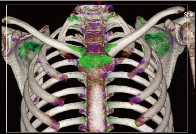
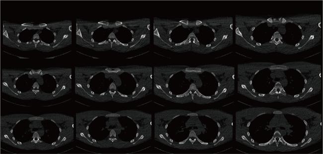
其他检查:胸部双能量CT检查提示,胸骨、双侧锁骨、肩胛骨、双侧多根肋骨、胸骨边缘多发痛风石,胸骨柄骨质密度减低(图 1)。常规CT图像见图 2。心电图、心脏彩超、骶髂关节MRI未见明显异常。
图1 双能量CT显示胸骨、双侧锁骨、肩胛骨、双侧多根肋骨、胸骨边缘多处有尿酸盐结晶沉积(绿色显示)

Figure 1 Dual-energy CT demonstrated the urate crystal deposition (shown in green) in the sternum, bilateral clavicles, scapulae, multiple ribs on both sides, and along the edges of the sternum

图2 患者胸部CT

Figure 2 Chest CT of the patient

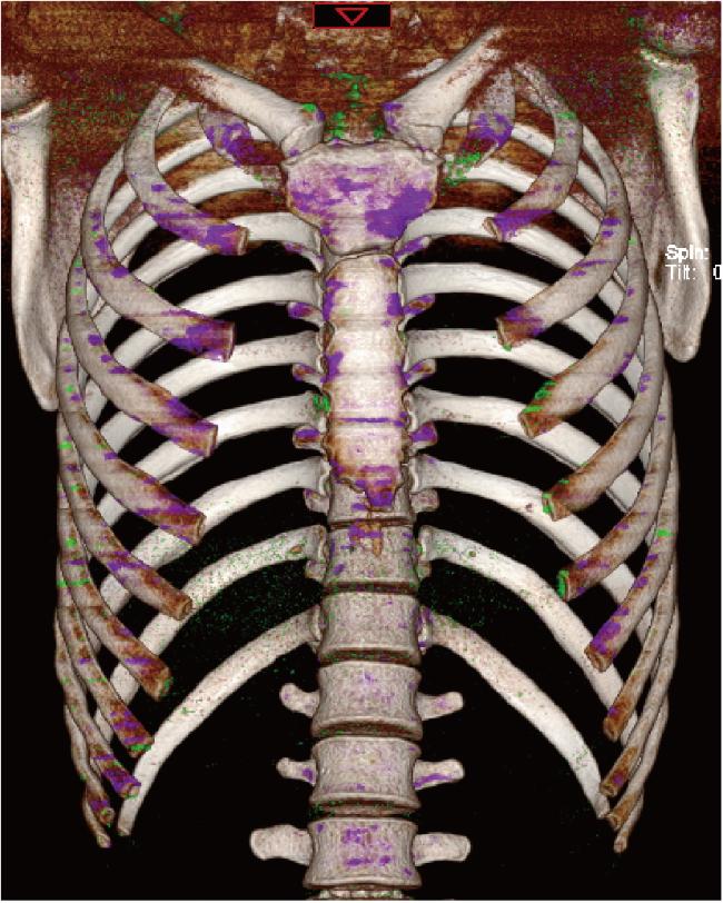
结合患者病史、临床表现及双能量CT检查结果,根据2015年美国风湿病学会/欧洲抗风湿病联盟发布的痛风分类标准[2],本例患者的痛风诊断明确。治疗上给予非布司他40 mg、每日一次,洛索洛芬钠60 mg、每日三次。患者出院后2年内多次随访,血尿酸水平分别为270.0、271.1、226.0、265.7 μmol/L,未再发胸锁关节疼痛,双能量CT检查提示胸锁关节及其他部位的痛风石较前明显减少(图 3)。
图3 患者2年后随访复查双能量CT见尿酸盐结晶沉积较前明显较少

Figure 3 Follow-up after two years with dual-energy CT showed significantly fewer urate crystal deposits compared to before

2 分析与讨论

本例患者的血尿酸水平并不高,由于患者受累关节的特殊性,无法行关节腔穿刺抽取关节液,排除其他相关疾病后,根据2015年美国风湿病学会/欧洲抗风湿病联盟发布的痛风分类标准:(1)患者受累关节不能忍受触摸、按压(2分);(2)反复典型症状发作,且发作间期完全缓解(2分);(3)双能量CT证实尿酸盐沉积(4分);患者总得分为8分,符合痛风分类诊断标准,故该患者诊断为痛风。
2018年欧洲抗风湿病联盟更新的痛风循证建议中指出,不应仅根据高尿酸血症来诊断痛风[3]。高尿酸血症应被视为痛风的一个主要危险因素,而不是作为其诊断的主要指标,没有高尿酸血症并不能完全排除痛风的诊断。一项研究发现,有14%的痛风患者在痛风急性发作期间血尿酸水平低于6 mg/dL(360 μmol/L)[4]。在痛风急性发作期,血尿酸水平的诊断价值相对有限。因此,对于血尿酸水平正常,但有反复关节疼痛的患者,临床医生也需考虑痛风的诊断。
目前,关节液或痛风石在偏振光显微镜下呈针状和负性双折光是诊断痛风的金标准,但由于该检查的侵入性,在临床上很少进行。近些年,双能量CT在临床上的使用越来越普遍,其原理为使用不同能量谱获得的衰减测量值,并利用两种谱之间已知的衰减变化来区分和量化材料,然后通过彩色编码技术,使痛风石呈现出来[5]。双能量CT诊断痛风的敏感度为90%~100%,特异度为83%~89%[6],但由于其潜在的辐射危险和价格偏高,使其在临床上并未成为检查首选。
本例青年男性患者因反复胸前区疼痛2个月就诊,血尿酸检查正常,HLA-B27阳性,外院曾误诊为强直性脊柱炎,且并没有完善骶髂关节CT或MRI进一步明确诊断,仅给予止痛等对症治疗,虽然症状较前有所缓解,但病情仍有反复,患者无典型的第一跖趾关节红肿热痛,也是导致患者误诊的原因之一。虽然这些不典型部位的痛风比较少见,但临床医生仍不能掉以轻心。关于不典型部位痛风的既往文献报道发现,不仅在胸锁关节,脊柱、颈髓、支气管、阴囊、心脏二尖瓣、肝脏、中耳和乳房等部位均有报道[7-14]
目前,难治性痛风越来越受到临床医生的重视,根据2019年中华医学会内分泌学会发布的相关指南,难治性痛风定义为:(1)单用或联用常规降尿酸药物足量、足疗程,但血尿酸仍≥360 μmol/L;(2)接受规范化治疗,痛风仍发作≥2次/年;(3)存在多发性和/或进展性痛风石[15]。长期的高尿酸血症如果不能得到控制,最终会导致关节骨质破坏、痛风性肾病甚至是急性肾衰竭,目前该指南建议使用普瑞凯希来控制难治性痛风。普瑞凯希是一种尿酸氧化酶,能催化尿酸盐氧化为尿囊素。一项重复、随机、双盲、安慰剂对照试验提示,在患有慢性痛风、血清尿酸水平升高和别嘌呤醇不耐受或难治性的患者中,普瑞凯希能降低尿酸水平[16]。近些年,白细胞介素1(interleukin 1, IL-1)拮抗剂(如阿纳白滞素、卡那单抗和利纳西普)也被批准用于痛风的治疗与预防。
综上所述,随着目前诊断与治疗手段的增多,对于临床医生而言,需要精准地做出诊断,避免误诊和漏诊一些不常见部位的痛风以及血尿酸水平正常的痛风。

利益冲突   所有作者均声明不存在利益冲突。

作者贡献声明  甘地:总结病例资料,撰写论文;付强:提出研究思路和诊断思路,指导论文撰写;唐小惠、李楚炜:查阅、整理文献;舒兆平:总体把关和审定论文。所有作者均参与论文修改,并对最终文稿进行审读和确认

1
Dalbeth N , Gosling AL , Gaffo A , et al. Gout[J]. Lancet, 2021, 397 (10287): 1843- 1855.

DOI

2
Neogi T , Jansen TLTA , Dalbeth N , et al. 2015 Gout classification criteria: An American College of Rheumatology/European League Against Rheumatism collaborative initiative[J]. Ann Rheum Dis, 2015, 74 (10): 1789- 1798.

DOI

3
Richette P , Doherty M , Pascual E , et al. 2018 updated European League Against Rheumatism evidence-based recommendations for the diagnosis of gout[J]. Ann Rheum Dis, 2020, 79 (1): 31- 38.

DOI

4
Schlesinger N , Norquist JM , Watson DJ . Serum urate during acute gout[J]. J Rheumatol, 2009, 36 (6): 1287- 1289.

DOI

5
Singh JA , Budzik JF , Becce F , et al. Dual-energy computed tomography vs ultrasound, alone or combined, for the diagnosis of gout: A prospective study of accuracy[J]. Rheumatology, 2021, 60 (10): 4861- 4867.

DOI

6
McCollough CH , Leng S , Yu L , et al. Dual- and multi-energy CT: Principles, technical approaches, and clinical applications[J]. Radiology, 2015, 276 (3): 637- 653.

DOI

7
Ng W , Sin CH , Wong CH , et al. Unusual presentation of spinal gout: 2 cases report and literature review[J]. J Orthop Case Rep, 2017, 7 (6): 50- 54.

8
Park EH , Yoo WH , Choi Y . Tophaceous gout: An unusual cause of cervical cord compression[J]. Rheumatology (Oxford), 2021, 61 (1): 467.

DOI

9
Adamson R , Lacy JM , Cheng AM , et al. Tophus causing bronchial obstruction[J]. Am J Respir Crit Care Med, 2013, 188 (12): e72- e73.

DOI

10
Montelongo-Rodríguez FA , Madero-Morales PA , Martínez-Fernández AM , et al. Gouty tophus in the scrotum: An unusual extra-articular manifestation of gout[J]. Ochsner J, 2021, 21 (4): 413- 415.

DOI

11
Rohani A , Chamanian S , Hosseinzade P , et al. A case of mitral valve tophus in a patient with severe gout tophaceous arthritis[J]. J Clin Imaging Sci, 2012, 2, 68.

DOI

12
Varinot J , Cazejust J , Wendum D . A gouty tophus appearing as an atypical liver nodule in a cirrhotic patient[J]. Clin Res Hepatol Gastroenterol, 2011, 35 (12): 855- 856.

DOI

13
Mutlu A , Dündar E , İşeri M , et al. An unusual presentation of gout: Tophi in the middle ear[J]. J Int Adv Otol, 2016, 12 (2): 216- 218.

14
Green R , Sensarma K , Jain M , et al. An unusual breast lump presenting as a malignancy found to be a mammary gouty tophus[J]. Breast J, 2011, 17 (5): 528- 529.

DOI

15
中华医学会内分泌学分会. 中国高尿酸血症与痛风诊疗指南(2019)[J]. 中华内分泌代谢杂志, 2020, 36 (1): 1- 13.

16
Sundy JS , Baraf HSB , Yood RA , et al. Efficacy and tolerability of pegloticase for the treatment of chronic gout in patients refractory to conventional treatment: Two randomized controlled trials[J]. JAMA, 2011, 306 (7): 711- 720.

DOI

文章导航

/