
1 资料与方法
1.1 病例纳入与排除标准
1.2 一般资料
1.3 手术方法及术后处理
1.4 随访及疗效评定
1.5 统计学分析
2 结果
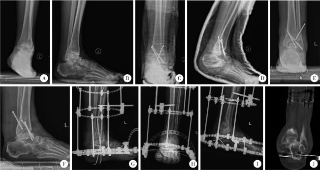
图1 踝关节融合术后感染行一期清创、胫距跟融合、Ilizarov外固定架固定病例Figure 1 A case of ankle arthrodesis infection treated with primary debridement, tibiotalocalcaneal arthrodesis, and Ilizarov external fixation Patient, male, 71 years old, with postoperative infection following left ankle arthrodesis. A and B, weight-bearing anteroposterior and lateral radiographs of the left ankle before the initial ankle arthrodesis demonstrated ankle osteoarthritis, ankle varus, and anterior dislocation of the talus relative to the tibia; C and D, immediate postoperative radiographs after the initial ankle arthrodesis showed fixation of the ankle with three screws; E and F, six months after the initial ankle arthrodesis, weight-bearing anteroposterior and lateral radiographs of the left ankle revealed nonunion, loosening and backing out of the screws, and erosion and destruction of the periarticular bone; G, following removal of the three screws and thorough debridement, one-stage tibiotalocalcaneal arthrodesis with Ilizarov external fixation was performed; H to J, six months postoperatively, radiographs and CT scan indicated bony union. |
表1 患者术前及末次随访时AOFAS踝-后足评分、VAS评分的比较Table 1 Comparison of AOFAS ankle-hindfoot scores and VAS scores preoperatively and at the final follow-up |
| Parameters | Preoperative | Final follow-up | Statistic | P value |
| AOFAS ankle-hindfoot score, | 42.8±10.4 | 77.6±10.2 | t=-6.952 | <0.001 |
| VAS score,M(P25, P75) | 7.0 (5.3, 7.8) | 1.0 (0, 2.0) | Z=-3.074 | 0.002 |
AOFAS, American Orthopaedic Foot and Ankle Society; VAS, visual analogue score. |
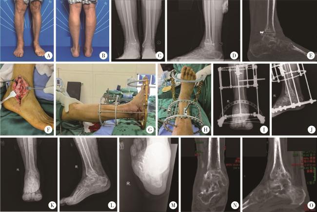
图2 踝关节骨折术后感染行内固定取出、一期清创、踝关节融合、Ilizarov外固定架固定的典型病例Figure 2 Typical case: Postoperative infection of an ankle fracture treated with removal of internal fixation, one-stage debridement, ankle arthrodesis, and Ilizarov external fixation Patient, male, 77 years old, presented with pain and swelling in the right ankle for over 1 year following an ankle fracture surgery. A and B, preoperative weight-bearing clinical photographs of both ankles showing swelling in the right ankle, with satisfactory hindfoot alignment; C to E, preoperative weight-bearing anteroposterior and lateral radiographs and weight-bearing sagittal CT scan of the right ankle revealed postoperative changes from the internal fixation, demonstrating a moth-eaten appearance of bone destruction in the ankle joint; F, intraoperative view via a lateral approach to the ankle joint, showing accumulation of purulent fluid within the right ankle joint; G and H, the ankle joint internal fixation was removed, and after thorough debridement, primary ankle arthrodesis with Ilizarov external fixation was performed; I and J, immediate postoperative anteroposterior and lateral radiographs of the right ankle; K to O, at the final follow-up, radiographs and CT scans confirmed bony union with satisfactory hindfoot alignment. |



