北京大学学报(医学版) ›› 2026, Vol. 58 ›› Issue (4): 842-847. doi: 10.19723/j.issn.1671-167X.2026.04.022

• 论著 • 上一篇    下一篇

一期融合治疗踝及后足感染性关节炎

王执宇1, 李文菁1,2, 张保宙1, 李莹1,*(), 杜辉1, 王岩1, 武勇1   

  1. 1. 首都医科大学附属北京积水潭医院足踝外科,北京 102208
    2. 北京市创伤骨科研究所,北京 100035
  • 收稿日期:2025-10-27 出版日期:2026-08-18 发布日期:2026-05-22
  • 通讯作者: 李莹
  • 基金资助:
    北京市卫生健康科技成果和适宜技术推广项目(BHTPP2024038)

Single-stage arthrodesis for septic arthritis of the ankle and hindfoot

Zhiyu WANG1, Wenjing LI1,2, Baozhou ZHANG1, Ying LI1,*(), Hui DU1, Yan WANG1, Yong WU1   

  1. 1. Department of Foot and Ankle Surgery, Beijing Jishuitan Hospital, Capital Medical University, Beijing 102208, China
    2. Beijing Institute of Trauma Orthopedics, Beijing 100035, China
  • Received:2025-10-27 Online:2026-08-18 Published:2026-05-22
  • Contact: Ying LI
  • Supported by:
    the Beijing Health Scientific and Technological Achievements and Appropriate Technologies Promotion Project(BHTPP2024038)

RICH HTML


摘要:

目的: 评估一期清创、关节融合、Ilizarov外固定架固定治疗踝及后足感染性关节炎的临床效果。方法: 选择2015年4月至2023年6月北京积水潭医院足踝外科由同一术者行一期清创、关节融合、Ilizarov外固定架固定手术治疗的12例踝关节及后足感染性关节炎患者的临床资料进行回顾性分析,其中男8例,女4例;手术时平均年龄为(53.6±16.2)岁,均为单侧手术。记录患者一般临床特征、细菌培养结果和拆除外固定架时间,记录并比较患者术前及末次随访时美国足踝外科协会(American Orthopaedic Foot and Ankle Society, AOFAS)踝-后足评分和疼痛视觉模拟评分(visual analogue scale,VAS),记录患者对手术的满意度以及并发症发生情况。结果: 所有患者术后平均随访(51.3±34.0)个月,其中细菌培养阳性7例。所有患者感染均得到控制,骨愈合率100%,中位外固定架拆除时间16(12,24)周。术前,中位VAS评分7(5.3,7.8)分,平均AOFAS踝-后足评分(42.8±10.4)分;末次随访时,中位VAS评分1(0,2)分,平均AOFAS踝-后足评分(77.6±10.2)分,均较术前显著改善(P<0.01)。并发症方面,针道感染1例,肢体短缩3例,肢体麻木2例。12例患者9例对手术非常满意,3例对手术满意。结论: 一期清创、关节融合、Ilizarov外固定架固定是治疗踝及后足感染性关节炎的可靠方法,能有效控制感染,缩短治疗时间,且患者术后功能恢复及影像学结果良好。

关键词: 感染性关节炎, 踝关节, 后足, 一期手术, 融合

Abstract:

Objective: To evaluate the clinical outcomes of one-stage debridement, arthrodesis, and Ilizarov external fixation for the treatment of septic arthritis of the ankle and hindfoot. Methods: A retrospective analysis was conducted on 12 patients with septic arthritis of the ankle or hindfoot who underwent one-stage debridement, arthrodesis, and Ilizarov external fixation performed by the same surgeon between April 2015 and June 2023. The study included 8 males and 4 females, with a mean age of (53.6±16.2) years at surgery; all procedures were unilateral. General clinical characteristics, bacterial culture results, and the time to external fixator removal were recorded. The American Orthopaedic Foot and Ankle Society (AOFAS) ankle-hindfoot score and the visual analogue scale (VAS) for pain were recorded and compared between the preoperative period and the final follow-up. Patient satisfaction with the surgery and the occurrence of complications were also documented. Results: All patients were followed up for a mean of (51.3±34.0) months postoperatively. Positive bacterial cultures were found in 7 cases. Infection control was achieved in all patients, with a 100% bone fusion rate confirmed by radiographs or CT. The median time to external fixator removal was 16 (12, 24) weeks. Preoperatively, the median VAS score was 7 (5.3, 7.8) points, and the mean AOFAS ankle-hindfoot score was (42.8±10.4) points. At the final follow-up, the median VAS score was 1 (0, 2) point, and the mean AOFAS score was (77.6±10.2) points, both showing significant improvement compared with preoperative values (P < 0.01). Regarding complications, there was 1 case of pin tract infection, 3 cases of limb shortening, and 2 cases of skin numbness. Nine patients were very satisfied with the surgery, and 3 were satisfied. Conclusion: One-stage debridement, arthrodesis, and Ilizarov external fixation are a reliable method for treating septic arthritis of the ankle and hindfoot. It effectively controls infection, shortens treatment duration, and yields favorable functional and imaging outcomes, with high patient satisfaction and acceptable complication rates.

Key words: Septic arthritis, Ankle, Hindfoot, Single-stage procedure, Arthrodesis

中图分类号: 

  • R684.3

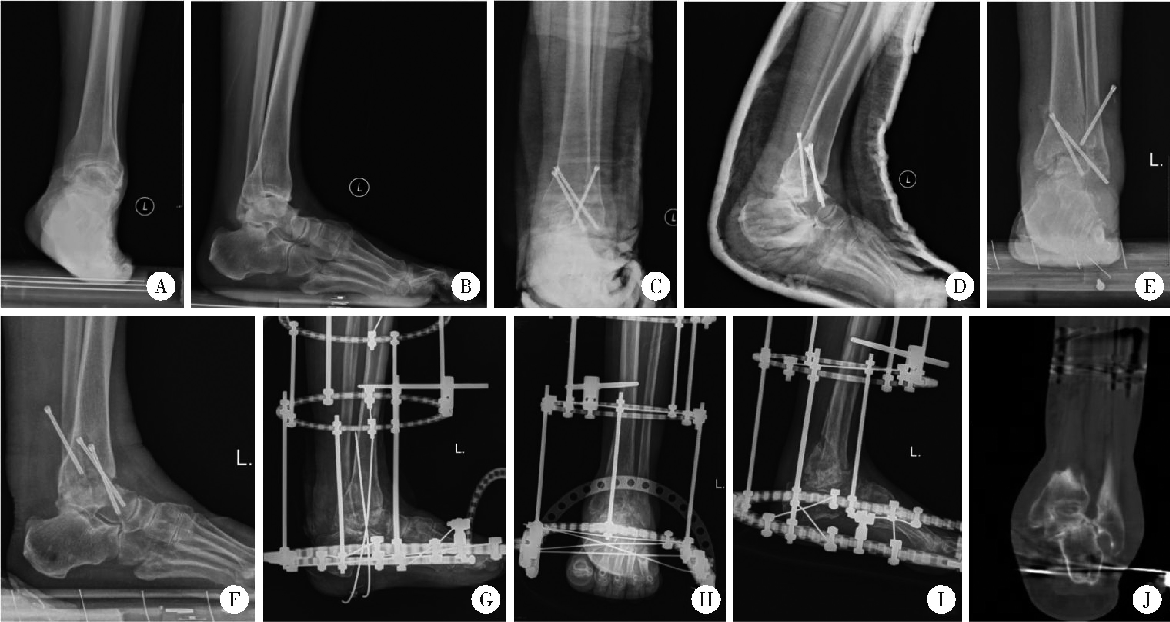
图1

踝关节融合术后感染行一期清创、胫距跟融合、Ilizarov外固定架固定病例"

表1

患者术前及末次随访时AOFAS踝-后足评分、VAS评分的比较"

Parameters Preoperative Final follow-up Statistic P value
AOFAS ankle-hindfoot score,$\bar{x} \pm s$ 42.8±10.4 77.6±10.2 t=-6.952 <0.001
VAS score,M(P25, P75) 7.0 (5.3, 7.8) 1.0 (0, 2.0) Z=-3.074 0.002

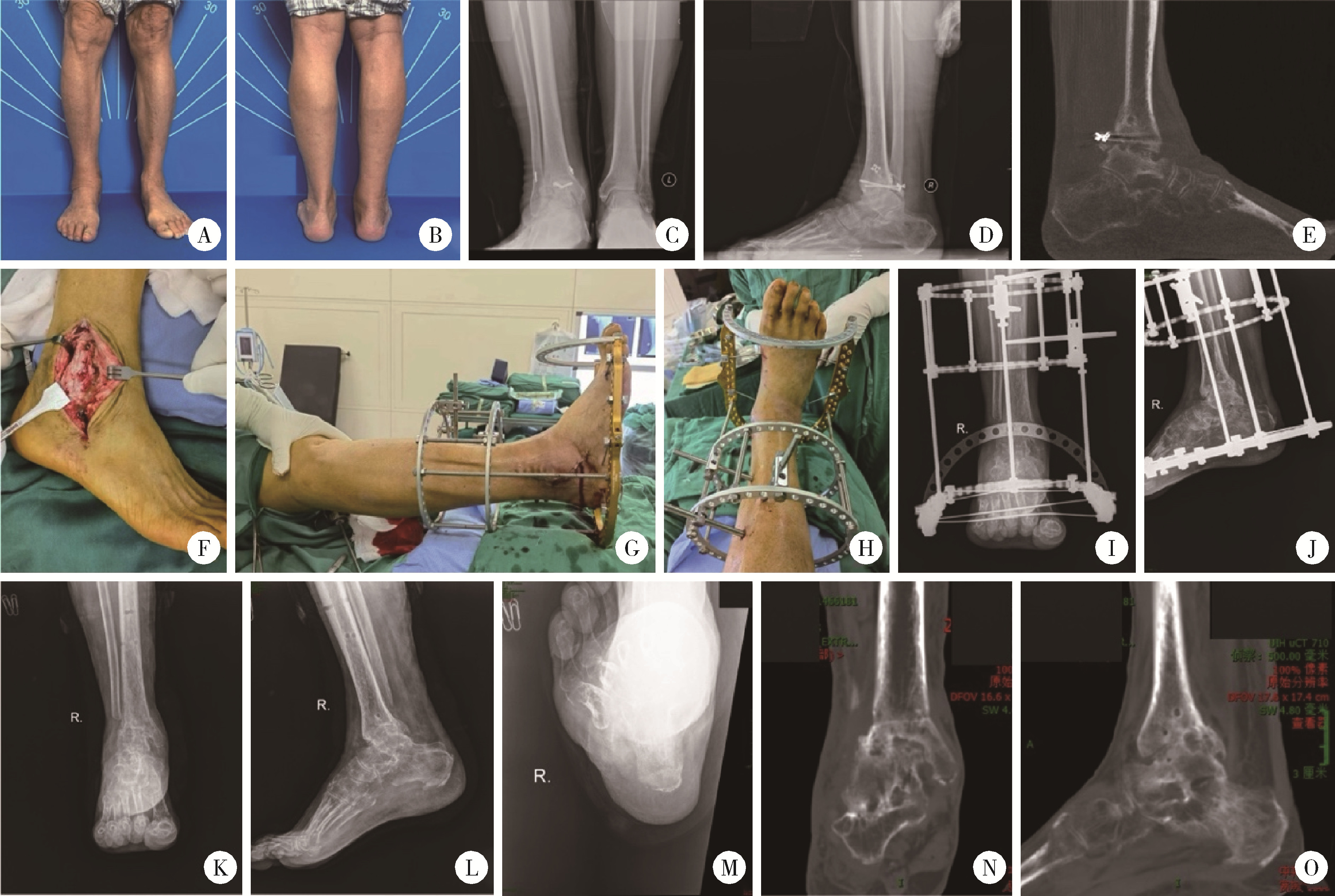
图2

踝关节骨折术后感染行内固定取出、一期清创、踝关节融合、Ilizarov外固定架固定的典型病例"

1
Simoni G , Maccauro G , Fenga D , et al. Arthrodesis of the ankle joint in septic osteoarthritis: Six years long term outcomes in authors' personal experience[J]. Eur Rev Med Pharmacol Sci, 2019, 23 (Suppl 2): 139- 144.
2
Hong YC , Jung KJ , Chang HJ , et al. Staged joint arthrodesis in the treatment of severe septic ankle arthritis sequelae: A case report[J]. Int J Environ Res Public Health, 2021, 18 (23): 12473.

doi: 10.3390/ijerph182312473
3
Kienast B , Kiene J , Gille J , et al. Posttraumatic severe infection of the ankle joint-long term results of the treatment with resection arthrodesis in 133 cases[J]. Eur J Med Res, 2010, 15 (2): 54.

doi: 10.1186/2047-783X-15-2-54
4
Mankovecky MR , Roukis TS . Arthroscopic synovectomy, irrigation, and debridement for treatment of septic ankle arthrosis: A systematic review and case series[J]. J Foot Ankle Surg, 2014, 53 (5): 615- 619.

doi: 10.1053/j.jfas.2013.10.012
5
Hulscher JBF , te Velde EA , Schuurman AH , et al. Arthrodesis after osteosynthesis and infection of the ankle joint[J]. Injury, 2001, 32 (2): 145- 152.

doi: 10.1016/S0020-1383(00)00156-X
6
Suda AJ , Richter A , Abou-Nouar G , et al. Arthrodesis for septic arthritis of the ankle: Risk factors and complications[J]. Arch Orthop Trauma Surg, 2016, 136 (10): 1343- 1348.

doi: 10.1007/s00402-016-2520-y
7
Pérez-Prieto D , Portillo ME , González-Lucena G , et al. Foot and ankle infections: Debridement, early fixation and rifampicin provide earlier recovery of function and quality of life[J]. Foot Ankle Surg, 2019, 25 (1): 13- 18.

doi: 10.1016/j.fas.2017.07.644
8
Kollig E , Esenwein SA , Muhr G , et al. Fusion of the septic ankle: Experience with 15 cases using hybrid external fixation[J]. J Trauma Inj Infect Crit Care, 2003, 55 (4): 685- 691.

doi: 10.1097/01.TA.0000051933.83342.E4
9
Klouche S , El-Masri F , Graff W , et al. Arthrodesis with internal fixation of the infected ankle[J]. J Foot Ankle Surg, 2011, 50 (1): 25- 30.

doi: 10.1053/j.jfas.2010.10.011
10
Hartmann R , Grubhofer F , Waibel FWA , et al. Treatment of hindfoot and ankle infections with Ilizarov external fixator or spacer, followed by secondary arthrodesis[J]. J Orthop Res, 2021, 39 (10): 2151- 2158.

doi: 10.1002/jor.24938
11
Richter I , Krähenbühl N , Ruiz R , et al. Mid- to long-term outcome in patients treated with a mini-open sinus-tarsi approach for calcaneal fractures[J]. Arch Orthop Trauma Surg, 2021, 141 (4): 611- 617.

doi: 10.1007/s00402-020-03530-3
12
Movassaghi K , Wakefield C , Bohl DD , et al. Septic arthritis of the native ankle[J]. JBJS Rev, 2019, 7 (3): e6.

doi: 10.2106/JBJS.RVW.18.00080
13
Mercurio M , Castioni D , Porco E , et al. Periprosthetic ankle infection: Eradication rate, complications, and limb salvage. A systematic review[J]. Foot Ankle Surg, 2022, 28 (5): 550- 556.

doi: 10.1016/j.fas.2021.07.009
14
Solow M , Sarraj M , Johal H , et al. A case report of pneumococcal septic arthritis following a respiratory and gastrointestinal prodrome with accompanying literature review[J]. J Foot Ankle Surg, 2019, 58 (6): 1293- 1297.

doi: 10.1053/j.jfas.2018.12.043
15
Gee C , Tandon T , Avasthi A , et al. Primary meningococcal septic arthritis of the ankle joint: A case report[J]. J Foot Ankle Surg, 2014, 53 (2): 216- 218.

doi: 10.1053/j.jfas.2013.12.019
16
Rabinovich RV . Complex ankle arthrodesis: Review of the literature[J]. World J Orthop, 2015, 6 (8): 602.

doi: 10.5312/wjo.v6.i8.602
17
Ellington K , Hirose CB , Bemenderfer TB . What is the treatment "algorithm" for infection after ankle or hindfoot arthrodesis[J]. Foot Ankle Int, 2019, 40 (Suppl 1): 64S- 70S.
18
Perlman MH , Thordarson DB . Ankle fusion in a high risk population: An assessment of nonunion risk factors[J]. Foot Ankle Int, 1999, 20 (8): 491- 496.

doi: 10.1177/107110079902000805
19
Cierny G , Cook WG , Mader JT . Ankle arthrodesis in the presence of continuous sepsis[J]. Clin Podiatric Med Surg, 1990, 7 (3): 545- 563.

doi: 10.1016/S0891-8422(23)00387-7
20
Meyer C , Badillo K , Burns P . Safety and complication rate of ring external fixation in the foot and ankle[J]. J Foot Ankle Surg, 2023, 62 (6): 991- 995.

doi: 10.1053/j.jfas.2023.08.002
[1] 陈炼炼, 蔡张滢, 叶琳娜, 李洁. 腰椎间盘突出患者腰椎融合术后恐动症现状及其影响因素[J]. 北京大学学报(医学版), 2025, 57(2): 317-322.
[2] 李文菁,张保宙,李恒,赖良鹏,杜辉,孙宁,龚晓峰,李莹,王岩,武勇. 胫距跟融合治疗终末期踝和后足病变的中短期临床结果[J]. 北京大学学报(医学版), 2024, 56(2): 299-306.
[3] 汪大伟,王华东,李利,尹欣,黄伟,郭继东,杨亚锋,刘义灏,郑扬. 自体下关节突骨块应用于骨质疏松患者腰椎椎间融合术的疗效分析[J]. 北京大学学报(医学版), 2023, 55(5): 899-909.
[4] 熊士凯,史尉利,王安鸿,谢兴,郭秦炜. 腓骨远端撕脱骨折的影像学诊断:踝关节X线与CT三维重建的比较[J]. 北京大学学报(医学版), 2023, 55(1): 156-159.
[5] 张梅香,史文芝,刘建新,王春键,李燕,王蔚,江滨. MLL-AF6融合基因阳性急性髓系白血病的临床特征及预后[J]. 北京大学学报(医学版), 2021, 53(5): 915-920.
[6] 侯宗辰,敖英芳,胡跃林,焦晨,郭秦炜,黄红拾,任爽,张思,谢兴,陈临新,赵峰,皮彦斌,李楠,江东. 慢性踝关节不稳患者足底压力特征及相关因素分析[J]. 北京大学学报(医学版), 2021, 53(2): 279-285.
[7] 江东,胡跃林,焦晨,郭秦炜,谢兴,陈临新,赵峰,皮彦斌. 慢性踝关节不稳合并后踝撞击同期手术中长期疗效及影响因素分析[J]. 北京大学学报(医学版), 2019, 51(3): 505-509.
[8] 杨榕,李庆祥,毛驰,彭歆,王洋,郭玉兴,郭传瑸. 多模态影像融合技术与颅底-颞下区肿瘤的诊断和治疗[J]. 北京大学学报(医学版), 2019, 51(1): 53-58.
[9] 彭俐,王祖华,孙玉春,渠薇,韩扬,梁宇红. 根尖切除手术导板的计算机辅助设计及三维打印[J]. 北京大学学报(医学版), 2018, 50(5): 905-910.
[10] 王荣丽,周志浩,席宇诚,王启宁,王宁华,黄真. 机器人辅助脑瘫儿童踝关节康复临床初步研究[J]. 北京大学学报(医学版), 2018, 50(2): 207-212.
[11] 刘洪,越雷,陈顺伦,胡博,李淳德△,邑晓东,李宏,卢海霖,王宇,于峥嵘,孙浩林,. 前路椎间盘切除椎间融合术治疗伴有交感神经症状的颈椎病[J]. 北京大学学报(医学版), 2018, 50(2): 347-351.
[12] 孙宇,刘鑫,樊东升,傅渝,潘胜发,张凤山,张立,王少波,刁垠泽,陈欣,周非非,赵衍斌. 平山病外科治疗的中期临床效果及影像学结果[J]. 北京大学学报(医学版), 2017, 49(6): 1019-1026.
[13] 赵一姣,刘怡,孙玉春,王勇. 一种基于曲率连续算法的冠、根三维数据融合方法[J]. 北京大学学报(医学版), 2017, 49(4): 719-723.
[14] 龚晓峰,吕艳伟,王金辉,王岩,武勇,王满宜. 踝关节CT与踝关节骨折分型的相关性研究[J]. 北京大学学报(医学版), 2017, 49(2): 281-285.
[15] 姬洪全,周方,田耘,张志山,郭琰,吕扬,杨钟玮,侯国进. 手术治疗Maisonneuve骨折失误1例报告[J]. 北京大学学报(医学版), 2017, 49(2): 354-356.
Viewed
Full text


Abstract

Cited

  Shared   
  Discussed   
No Suggested Reading articles found!