Journal of Peking University(Health Sciences) ›› 2019, Vol. 51 ›› Issue (1): 86-92. doi: 10.19723/j.issn.1671-167X.2019.01.016

Previous Articles     Next Articles

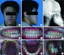
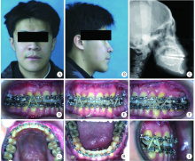
Orthodontic-orthognathic treatment stability in skeletal class Ⅲ malocclusion patients

Xiu-jing WANG1,Yi-mei ZHANG1,Yan-heng ZHOU2,()   

  1. 1. First Clinical Division, Peking University School and Hospital of Stomatology & National Clinical Research Center for Oral Diseases & National Engineering Laboratory for Digital and Material Technology of Stomatology & Beijing Key Laboratory of Digital Stomatology, Beijing 100081, China
    2. Department of Orthodontics, Peking University School and Hospital of Stomatology, Beijing 100081, China
  • Received:2018-10-07 Online:2019-02-18 Published:2019-02-26
  • Contact: Yan-heng ZHOU E-mail:yanhengzhou@vip.163.com
  • Supported by:
    Supported by the National Natural Science Foundation of China(81801014)

Abstract:

Objective:To investigate stability of skeletal hard tissues, dental hard tissues and soft tissues after orthodontic-orthognathic treatment in a long term. This study reviewed longitudinal changes in orthodontic-orthognathic patients of skeletal class Ⅲ malocculsion, using lateral cephalometric radiographs in 3-12 years after treatment in comparison to treatment finishing. Methods: Twenty-two patients with skeletal Class Ⅲ malocclusion following orthodontic-orthognathic surgery in Peking University School and Hospital of Stomatology from January 1, 2000 to January 1, 2009 were observed. The lateral cephalometric radiographs of the following stages were collected: treatment finishing (T1), 3 to 12 years after treatment (T2). Statistical analyses of cephalometrics were evaluated. Paired student t test was performed by SPSS 17.0. Results: Data of all the 22 patients were studied in longitudinal timeline after treatment and 3-12 years after treatment. From T1 to T2, we evaluated 11-SN (angle between the upper incisors axis and SN plane), 11-NA angle (angle between the upper incisors axis and NA plane), 11-NA mm (perpendicular distance from upper incisors to NA plane), 11-41(angle between the upper incisors axis and lower incisors axis), 41-NB angle (angle between lower incisors and NB plane), 41-NB(perpendicular distance from lower incisors to NB plane), 41-MP angle (angle between lower incisors and GoGn plane), and IMPA [angle between lower incisor and mandibular plane (tangent line to submandibular border)]. Most hard tissues of the teeth remained stable but upper anterior teeth angulations decreased, indicating by significantly reducing 11-SN (T1:110.98°±6.77°; T2: 109.21°±5.80°; P=0.005); reducing 11-NA(T1: 28.31°±6.80°;T2: 26.49°±6.18°; P=0.002); increasing 11-41 (T1:123.51°±8.14°;T2:125.7°±10.01°;P=0.035). From T1 to T2, we also evaluated SNA (angle of sella-nasion-A-point ), SNB (angle of sella-nasion-B-point), ANB (angle of A-point-nasion-B-point), GoGn-SN(angle between GoGn and SN plane), GoGn-FH (angle between GoGn and Frankfort plane), Y axis(angel between Sella-Gn and Frankfort plane), N-ANS (distance from nasion point to ANS point), ANS-Me (distance from ANS point to Menton point), N-Me (distance from nasion point to Menton point), ANS-Me/N-Me% (proportion of ANS-Me to N-Me), and FMA (angle between Frankfort and mandibular plane), Wits appraisal (horizontal distance between points A and B on functional occlusal plane). Skeletal hard tissues also remained relatively stable, only N-Me value changed significantly with a decreasing facial height (T1:124.98°±11.98°; T2:122.4°±11.05°; P=0.024). From T1 to T2, we finally evaluated FH-NsPg angle (angle between NsPg and Frankfort plane), H angle (angel between H line and NB), FH-A’UL angle (angle between A’UL and Frankfort plane), FH-B’LL angle (angle between B’LL and Frankfort plane), UL-LL (angle between UL and LL), UL-EP (distance between UL and E line), LL-EP (distance between LL and E line), Sn-H(perpendicular distance between Sn point and H line), Nls-H (distance of nose-lip-sulcus to H line), Li-H (lower lip to H line), Si-H (lower lip sulcus to H line), and NLA (nasolabial angle, angle of Cm-Sn-UL-point).Soft tissues changes were observed in decreasing UL-EP [T1: (-2.78±2.20) mm; (-3.29±2.44) mm; P=0.02] and H angle(T1: 8.27°±3.71°; 7.32°±3.83°; P=0.006).Other soft tissues remained relatively stable by retruding upper lip position and chin changes with no statistical significance. Conclusion: Orthodontic-orthognathic treatment can improve esthetics and occlusal function in patients of skeletal class Ⅲ malocclusion with a stable long-term outcome.

Key words: Malocclusion, Maxillary retrognathism, Mandibular prognathism, Orthognathic surgery

CLC Number: 

  • R783.5

Table 1

Comparison of skeletal measurements after treatment (Tfinished) and 3-12 years after treatment (Tfinished 3-12) in skeletal class Ⅲ patients"

Variables Tfinished Tfinished 3-12 t P
SNA/(°) 82.87±4.62 82.87±4.54 -0.008 0.990
SNB/(°) 81.76±4.80 81.91±4.33 -0.320 0.752
ANB/(°) 1.10±2.26 0.97±2.35 0.657 0.518
GoGn-SN/(°) 37.17±7.77 36.52±7.44 1.124 0.274
GoGn-FH/(°) 33.61±8.00 33.70±6.36 -0.096 0.925
Y axis/(°) 70.08±4.81 69.83±4.34 0.660 0.517
N-ANS/mm 55.88±5.22 54.84±5.49 2.003 0.058
ANS-Me/mm 69.34±7.91 67.76±6.93 1.981 0.061
N-Me/mm 124.98±11.98 122.40±11.05 2.437 0.024*
ANS-Me/N-Me/% 55.60±2.48 55.50±2.42 0.260 0.797
FMA/(°) 34.72±9.51 35.16±7.93 -0.432 0.671
Wits value/mm -4.71±2.48 -5.11±2.74 1.455 0.160

Table 2

Comparison of dentition measurements after treatment (Tfinished) and 3-12 years after treatment (Tfinished 3-12) in skeletal class Ⅲ patients"

Variables Tfinished Tfinished 3-12 t P
11-SN/(°) 110.98±6.77 109.21±5.80 3.085 0.005#
11-NA angle/(°) 28.31±6.80 26.49±6.18 3.471 0.002#
11-NA/mm 10.04±8.63 8.87±6.07 0.870 0.394
11-41/(°) 123.51±8.14 125.70±10.01 -2.251 0.035*
41-NB angle/(°) 27.13±6.95 26.80±8.15 0.425 0.675
41-NB/(mm) 7.56±5.74 6.94±4.59 0.782 0.442
41-MP/(°) 88.24±9.04 88.33±10.18 -0.097 0.923
IMPA/(°) 87.38±8.90 87.45±9.92 -0.078 0.928

Table 3

Comparison of soft tissue measurements after treatment (Tfinished) and 3 to 12 years after treatment (Tfinished 3-12) in skeletal class Ⅲ patients"

Variables Tfinished Tfinished 3-12 t P
FH-NsPg/(°) 89.22±8.63 88.64±7.69 0.723 0.478
H angle/(°) 8.27±3.71 7.32±3.83 3.031 0.006#
FH-A’UL/(°) 70.62±8.89 72.24±8.67 -1.279 0.215
FH-B’LL/(°) 47.61±19.11 46.26±17.88 1.422 0.170
UL-LL/(°) 110.64±25.60 115.30±12.98 -0.960 0.348
UL-EP/mm -2.78±2.20 -3.29±2.44 2.511 0.020*
LL-EP/mm 0.03±2.17 -0.26±2.44 1.113 0.278
Sn-H/mm 4.22±3.52 4.99±3.73 -2.051 0.053
Nls-H/mm 7.77±2.33 10.09±14.49 -0.757 0.457
Li-H/mm 1.49±1.42 1.35±1.79 0.601 0.555
Si-H/mm 5.51±1.51 5.47±1.33 0.199 0.845
NLA/(°) 90.66±12.55 92.66±10.01 -1.387 0.180

Figure 1

Intraoral photographs and radiographs before treatment A,frontal view of face;B,lateral profile of face;C,cephalometric radiograph;D,occlusion of right;E, occlusion of frontal view;F,occlusion of left;G,occlusion of upper arch;H,occlusion of lower arch;I,overjet."

Figure 2

Intraoral photographs and radiographs during treatment A,frontal view of face;B,lateral profile of face;C,cephalometric radiograph;D,occlusion of right;E,occlusion of frontal view;F,occlusion of left;G,occlusion of upper arch;H,occlusion of lower arch;I,overjet."

Figure 3

Intraoral photographs and radiographs finishing treatment A,frontal view of face;B,lateral profile of face;C,cephalometric radiograph;D,occlusion of right;E,occlusion of frontal view;F,occlusion of left;G,occlusion of upper arch;H,occlusion of lower arch;I,overjet."

Figure 4

Intraoral photographs and radiographs of 9.4 years after treatment A,frontal view of face;B,lateral profile of face;C,cephalometric radiograph;D,occlusion of right;E,occlusion of frontal view;F,occlusion of left;G,occlusion of upper arch;H,occlusion of lower arch;I,overjet."

[1] Bell WH, Jacobs JD, Quejada JG . Simultaneous repositioning of the maxilla, mandible, and chin. Treatment planning and analysis of soft tissues[J]. Am J Orthod, 1986,89(1):28-50.
doi: 10.1016/0002-9416(86)90110-7
[2] Hack GA, de Mol van Otterloo JJ, Nanda R . Long-term stability and prediction of soft tissue changes after Lefort Ⅰ surgery[J]. Am J Orthod Dentofacial Orthop, 1993,104(6):544-555.
doi: 10.1016/S0889-5406(05)80438-X pmid: 8249930
[3] 王友山, 杨学文, 东耀峻 . 正颌外科术后畸形复发的生物学因素及其防治[J]. 中华口腔医学杂志, 1996,31(3):188-190.
doi: 10.1007/BF02951625
[4] Bailey LJ, Dover AJ, Proffit WR . Long-term soft tissue changes after orthodontic and surgical corrections of skeletal class Ⅲ malocclusions[J]. Angle Orthod, 2007,77(3):389-396.
doi: 10.2319/0003-3219(2007)077[0389:LSTCAO]2.0.CO;2 pmid: 3740712
[5] 林久祥 . 现代口腔正畸学[M]. 北京: 北京大学医学出版社, 2011: 196-220.
[6] 琚泽程, 徐宝华 . 外科-正畸联合矫治骨性下颌前突[J]. 中华口腔医学杂志, 1996,31(3):176-178.
[7] Joss CU, Thüer UW . Stability of the hard and soft tissue profile after mandibular advancement in sagittal split osteotomies: a longitudinal and long-term follow-up study[J]. Eur J Orthod, 2008,30(1):16-23.
doi: 10.1093/ejo/cjm080 pmid: 17962316
[8] den Besten CA, Mensink G, van Merkesteyn JP . Skeletal stability after mandibular advancement in bilateral sagittal split osteotomies during adolescence[J]. J Craniomaxillofac Surg, 2013,41(5):e78-e82.
doi: 10.1016/j.jcms.2012.11.012 pmid: 23253633
[9] Costa F, Robiony M, Zorzan E , et al. Stability of skeletal class Ⅲ malocclusion after combined maxillary and mandibular procedures[J]. J Oral Maxillofac Surg, 2006,64(4):642-651.
doi: 10.1016/j.joms.2005.11.043 pmid: 16546644
[10] Proffit WR, Phillips C, Turvey TA . Long-term stability of adole-scent versus adult surgery for treatment of mandibular deficiency[J]. Int J Oral Maxillofac Surg, 2010,39(4):327-332.
doi: 10.1016/j.ijom.2010.01.012 pmid: 20181460
[11] Joss CU, Vassalli IM . Stability after bilateral sagittal split osteotomy setback surgery with rigid internal fixation: a systematic review[J]. J Oral Maxillofac Surg, 2009,67(2):301-313.
doi: 10.1016/j.joms.2008.01.046 pmid: 19138603
[12] Mansour S, Burstone C, Legan H . An evaluation of soft-tissue changes resulting from Lefort Ⅰ maxillary surgery[J]. Am J Or-thod, 1983,84(1):37-47.
doi: 10.1016/0002-9416(83)90146-X pmid: 6575616
[13] Ayoub AA, Khambay AB, Mcdonald JX , et al. State of the art analysis of soft tissue changes in response to Lefort Ⅰ maxillary advancement[J]. Brit J Oral Maxillofac Surg, 2016,54(7):812-817.
doi: 10.1016/j.bjoms.2016.05.023 pmid: 27325452
[14] Proffit WR, Phillips C, Prewitt JW , et al. Stability after surgical-orthodontic correction of skeletal class iii malocclusion. 2. maxillary advancement[J]. Int J Adult Orthodon Orthognath Surg, 1991,6(2):71-80.
pmid: 1811032
[1] Lei HOU,Guo-hua YE,Xiao-jing LIU,Zi-li LI. Evaluation of mandibular stability and condylar volume after orthognathic surgery in patients with severe temporomandibular joint osteoarthrosis [J]. Journal of Peking University(Health Sciences), 2020, 52(1): 113-118.
[2] Ming-xin MAO,Li XU,Wu-di JING,Xiao XU,Jian-xia HOU,Xiao-tong LI,Xiao-xia WANG. Alveolar crest and relevant analysis of labial side of anterior teeth on skeletal Angle class Ⅲ patients [J]. Journal of Peking University(Health Sciences), 2020, 52(1): 77-82.
[3] Xian-tao SUN,Wei HE,Xiao-jing LIU,Zi-li LI,Xing WANG. Feasibility of Delaire cephalometric analysis to predict the ideal sagittal position of the maxilla and chin for surgery-first patients in orthognathic surgery [J]. Journal of Peking University(Health Sciences), 2020, 52(1): 90-96.
[4] Ren-jie DU,Jian JIAO,Yan-heng ZHOU,Jie SHI. Occlusal changes before and after orthodontic treatment in patients with aggressive periodontitis [J]. Journal of Peking University(Health Sciences), 2019, 51(5): 919-924.
[5] WANG Yi-ran, ZHOU Yan-heng, WANG Xue-dong, WEI Song, LIU Wei-tao. Evaluation of maxillary three-dimensional changes in maxillary protraction with alternating rapid palatal expansion and constriction based on the cone-beam computed tomography [J]. Journal of Peking University(Health Sciences), 2018, 50(4): 685-693.
[6] WU Ling, LIU Xiao-jing, LI Zi-li, WANG Xing. Evaluation of accuracy of virtual occlusal definition in Angle class Ⅰ molar relationship [J]. Journal of Peking University(Health Sciences), 2018, 50(1): 154-159.
[7] XU Xiao, XU Li, JIANG Jiu-hui, WU Jia-qi, LI Xiao-tong, JING Wu-di. Accuracy analysis of alveolar dehiscence and fenestration of maxillary anterior teeth of Angle class Ⅲ by cone-beam CT [J]. Journal of Peking University(Health Sciences), 2018, 50(1): 104-109.
[8] MA Jing, JIANG Jiu-hui. Morphological analysis of alveolar bone of anterior mandible in high-angle skeletal class Ⅱ and class Ⅲ malocclusions assessed with cone-beam computed tomography [J]. Journal of Peking University(Health Sciences), 2018, 50(1): 98-103.
[9] FAN Shao-qing, ZHOU Yan-heng. Evaluation of the correction of the skeletal class Ⅲ malocclusion by distalization of the whole mandible dentition with micro-implant anchorage [J]. Journal of Peking University(Health Sciences), 2017, 49(3): 531-539.
[10] ZHENG Xu, HU Xing-Xue, MA Ning, CHEN Xiao-Hong. A new method to orthodontically correct dental occlusal plane canting: waveshaped arch [J]. Journal of Peking University(Health Sciences), 2017, 49(1): 176-180.
[11] WANG Zhe, ZHU Liu-ning, ZHOU Lin, YI Biao. Feasibility of integrating 3D photos and cone-beam computed tomography images used to evaluate changes of soft and hard tissue after orthognathic surgery [J]. Journal of Peking University(Health Sciences), 2016, 48(3): 544-549.
[12] ZHANG Jie, LI Xiao-tong. Study of anterior alveolar bone thickness in skeletal class Ⅲ malocclusion patients with orthognathic surgery [J]. Journal of Peking University(Health Sciences), 2016, 48(1): 111-115.
[13] LI Min, WANG Si-wei, ZHAO Yi-jiao, LIU Yi. A cone-beam computed tomography study on crown-root morphology of maxillary anterior teeth in Class Ⅱ, division 2 malocclusion [J]. Journal of Peking University(Health Sciences), 2016, 48(1): 105-110.
[14] HE Wei, XIE Xiao-yan, WANG Xing, WANG Xiao-xia1, FU Kai-yuan2, LI Zi-li. Effect of segmental Le FortⅠosteotomy and bilateral sagittal split ramus osteotomy on the condyle position in skeletal class Ⅲ malocclusion patients [J]. Journal of Peking University(Health Sciences), 2015, 47(5): 829-833.
[15] HE Ying, GUO Chuan-Bin, DENG Xu-Liang, WANG Xing, WANG Xiao-Xia. Three-dimensional measurement and facial symmetry analysis for the craniofacial structure of people in North China with normal occlusion [J]. Journal of Peking University(Health Sciences), 2015, 47(4): 708-713.
Viewed
Full text


Abstract

Cited

  Shared   
  Discussed   
[1] . [J]. Journal of Peking University(Health Sciences), 2009, 41(4): 456 -458 .
[2] . [J]. Journal of Peking University(Health Sciences), 2009, 41(2): 125 -128 .
[3] . [J]. Journal of Peking University(Health Sciences), 2009, 41(2): 135 -140 .
[4] . [J]. Journal of Peking University(Health Sciences), 2009, 41(2): 158 -161 .
[5] . [J]. Journal of Peking University(Health Sciences), 2009, 41(2): 217 -220 .
[6] . [J]. Journal of Peking University(Health Sciences), 2009, 41(1): 52 -55 .
[7] . [J]. Journal of Peking University(Health Sciences), 2009, 41(3): 297 -301 .
[8] . [J]. Journal of Peking University(Health Sciences), 2009, 41(5): 599 -601 .
[9] . [J]. Journal of Peking University(Health Sciences), 2009, 41(5): 516 -520 .
[10] . [J]. Journal of Peking University(Health Sciences), 2007, 39(3): 304 -309 .