Journal of Peking University (Health Sciences) ›› 2026, Vol. 58 ›› Issue (4): 754-761. doi: 10.19723/j.issn.1671-167X.2026.04.011

Previous Articles     Next Articles

Analysis of endoscopic surgical strategies for duplex kidney and ureteral malformation complicated with upper urinary tract calculi

Xinji YANG1, Xiushi LIN1, Zhifei XIE2, Yuqing LIU1, Chunlei XIAO1, Shudong ZHANG1, Xiaofei HOU1,*(), Min QIU1,*()   

  1. 1. Department of Urology, Peking University Third Hospital, Beijing 100191, China
    2. Department of Urology, Tianjin Wuqing District Second People' s Hospital, Tianjin 301700, China
  • Received:2026-02-22 Online:2026-08-18 Published:2026-05-19
  • Contact: Xiaofei HOU, Min QIU
  • Supported by:
    the Wu Jieping Medical Foundation Special Fund for Clinical Research(320.6750.2022-03-45)

RICH HTML


Abstract:

Objective: To investigate the endoscopic surgical management strategies for duplex kidney and ureteral duplication anomalies complicated by upper urinary tract calculi, to summarize individualized diagnostic and therapeutic experiences for different anomaly subtypes, and to provide a reference for standardized clinical management of this condition. Methods: The clinical data of patients with duplex kidney and ureteral duplication anomalies and upper urinary tract calculi who underwent endoscopic surgery at the Department of Urology, Peking University Third Hospital from November 2012 to December 2025 were retrospectively analyzed. Baseline characteristics, imaging findings, perioperative parameters, complications, and changes in renal function were collected. Endoscopic surgical techniques and key diagnostic and therapeutic points for different anomaly subtypes were summarized. Statistical analysis was performed using SPSS 22.0 and R 3.6.1 software. Results: A total of 38 patients were enrolled, including 16 males and 22 females, with a mean age of (49.92±14.27) years. The median follow-up duration was 17.80 months (1.60-41.90). The predominant clinical symptom was flank pain (32 cases, 84.21%). The most common anomaly subtype was unilateral incomplete duplex kidney and ureteral duplication (24 cases, 63.16%). Based on the anomaly subtype and stone characteristics, all the patients underwent precise preoperative assessment using CT urography and other imaging modalities, and indivi-dualized surgical approaches were selected: In the study, 15 patients underwent ureteroscopic lithotripsy (URL), 15 underwent flexible ureteroscopic lithotripsy (FURL), 7 underwent percutaneous nephrolithotomy (PCNL), and 1 patient received PCNL combined with FURL. The median operative time was 74.00 (64.75-92.50) min, the median intraoperative blood loss was 2.00 (1.00-4.25) mL, and the median postoperative hospital stay was 3.00 (2.00-5.75) d. The overall postoperative stone-free rate was 94.74%. No severe perioperative complications occurred. Postoperative fever occurred in 14 patients (36.84%), all of whom recovered after symptomatic treatment. Postoperative serum creatinine levels were significantly lower than preoperative levels (P < 0.05), indicating significant improvement in renal function. Conclusion: Endoscopic surgery is safe and effective for the treatment of duplex kidney and ureteral duplication anomalies complicated by upper urinary tract calculi, offering advantages of minimal trauma, rapid recovery, and a high stone-free rate. The strategy of "precise diagnosis-individualized surgical approach selection" allows the formulation of tailored treatment plans for different anomaly subtypes and is worthy of clinical promotion and application.

Key words: Duplex kidney and ureteral duplication, Upper urinary tract calculi, Ureteroscopy, Percutaneous nephrolithotomy, Endoscopic surgery

CLC Number: 

  • R693.1

Table 1

Demographic and baseline clinical characteristics of patients with duplex kidney and ureteral malformation complicated with upper urinary tract calculi"

Variables Patients (n=38)
Gender, n (%)
  Male 16 (42.11)
  Female 22 (57.89)
Age/years, $\bar x \pm s$ 49.92±14.27
BMI/(kg/m2), M (P25P75) 25.01 (22.63, 26.47)
Symptoms, n (%)
  Low back pain 32 (84.21)
  Lower urinary tract irritation symptoms 10 (26.32)
  Lower urinary tract obstruction symptoms 3 (7.89)
  Hematuria 12 (31.58)
  Fever 6 (15.79)
  Nausea and vomiting 7 (18.42)
  Detected during physical examination 3 (7.89)
Duration of symptoms/d, M (P25P75) 30.00 (21.50, 165.00)
Basic medical history, n (%)
  Hypertension 13 (34.21)
  Diabetes mellitus 6 (15.79)
  Coronary heart disease 4 (10.53)

Table 2

Preoperative imaging evaluation of duplex kidney and ureteral malformation types, stone characteristics and hydronephrosis"

Variables Patients (n=38)
Side of Ureteral duplication, n (%)
  Left 12 (31.58)
  Right 17 (44.74)
  Bilateral 9 (23.68)
Type of Ureteral duplication, n (%)
  Unilateral incompletely 24 (63.16)
  Unilateral completely 4 (10.53)
  Bilateral incompletely 1 (2.63)
  Bilateral completely 3 (7.89)
  Bilateral completely and contralateral incompletely 5 (13.16)
  Unilateral inverted Y 1 (2.63)
Stone location, n (%)
  Calices renales of the upper kidney 10 (26.32)
  Calices renales of the lower kidney 8 (21.05)
  Ureteropelvic junction 6 (15.79)
  Ureter 11 (28.95)
  Multiple locations 3 (7.89)
Number of stones, n (%)
  1 21 (55.26)
  2 2 (5.26)
  Multiple 15 (39.48)
Maximum stone diameter/cm, M (P25P75) 1.06 (0.84, 1.48)
Stone CT value/HU, $\bar x \pm s$ 897.82±348.62
Pelvic hydronephrosis, n (%) 23 (60.53)
Hydronephrosis size/cm, M (P25P75) 3.43 (2.96, 3.79)

Figure 1

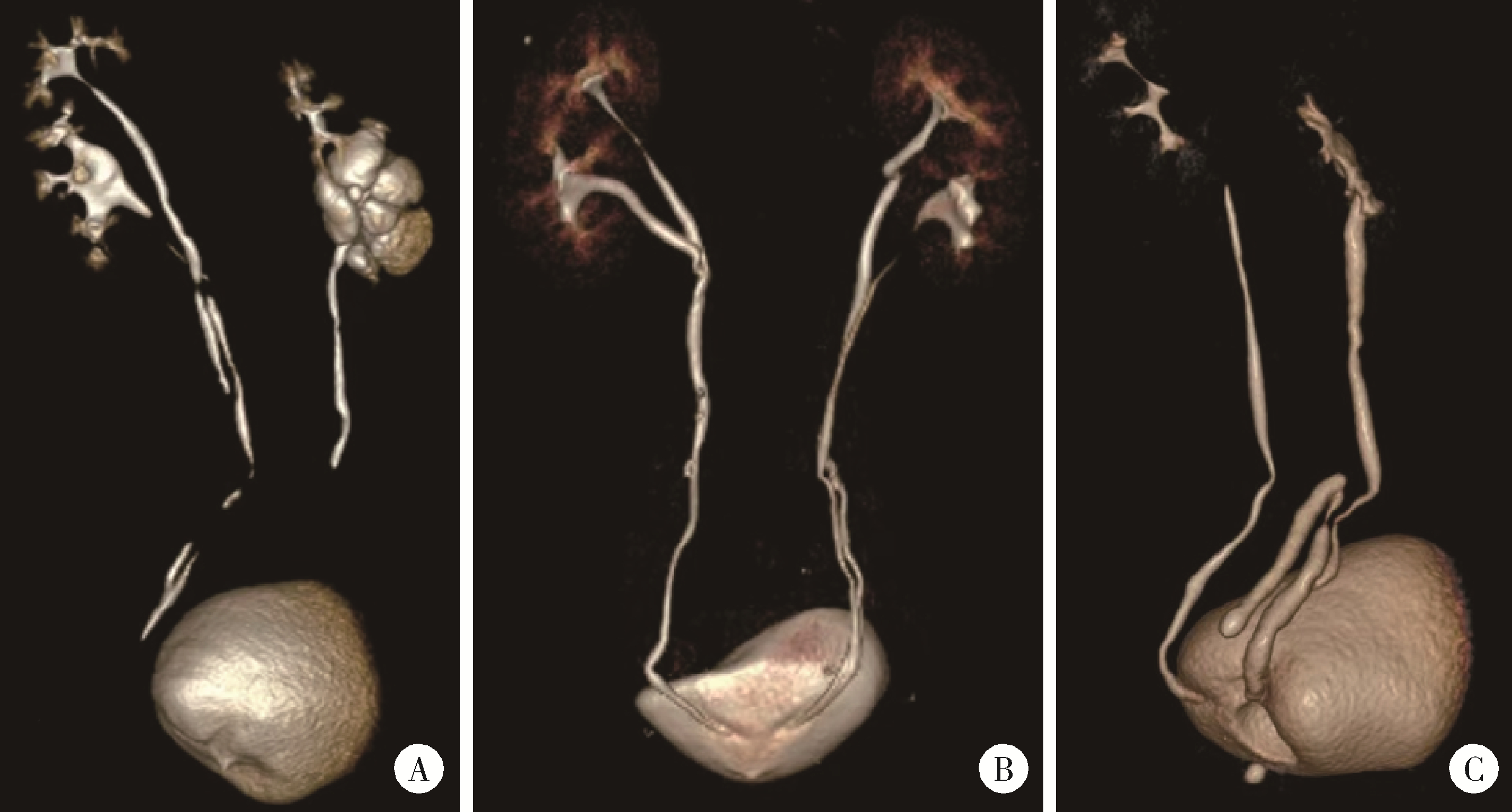
CT three-dimensional reconstruction showing different types of renal pelvis and ureter duplication malformations A, incomplete duplication of the left renal pelvis and ureter; B, incomplete duplication of the left renal pelvis and ureter, with complete duplication of the right renal pelvis and ureter; C, inverted Y-shaped duplicated ureter on the right side."

Table 3

Summary of endoscopic surgical approaches, intraoperative management and special situations"

Variables Patients (n=38)
ASA grade, n (%)
  Ⅰ 21 (55.26)
  Ⅱ 17 (44.74)
Anesthesia method, n (%)
  General anesthesia 25 (65.79)
  Spinal anesthesia 13 (34.21)
Surgical method, n (%)
  URL 15 (39.47)
  FURL 15 (39.47)
  PCNL 7 (18.43)
  PCNL+FURL 1 (2.63)
Intraoperative special conditions, n (%)
  Ureteral stricture 19 (50.00)
  Ureteral orifice cyst 3 (7.89)
  Complete impaction 2 (5.30)
  Stricture incision 3 (7.90)
  Cyst incision 1 (2.63)
  Balloon dilation 2 (5.26)
Surgical time/min, M (P25P75) 74.00 (64.75, 92.50)
Estimated blood loss/mL, M (P25P75) 2.0 (1.00, 4.25)

Figure 2

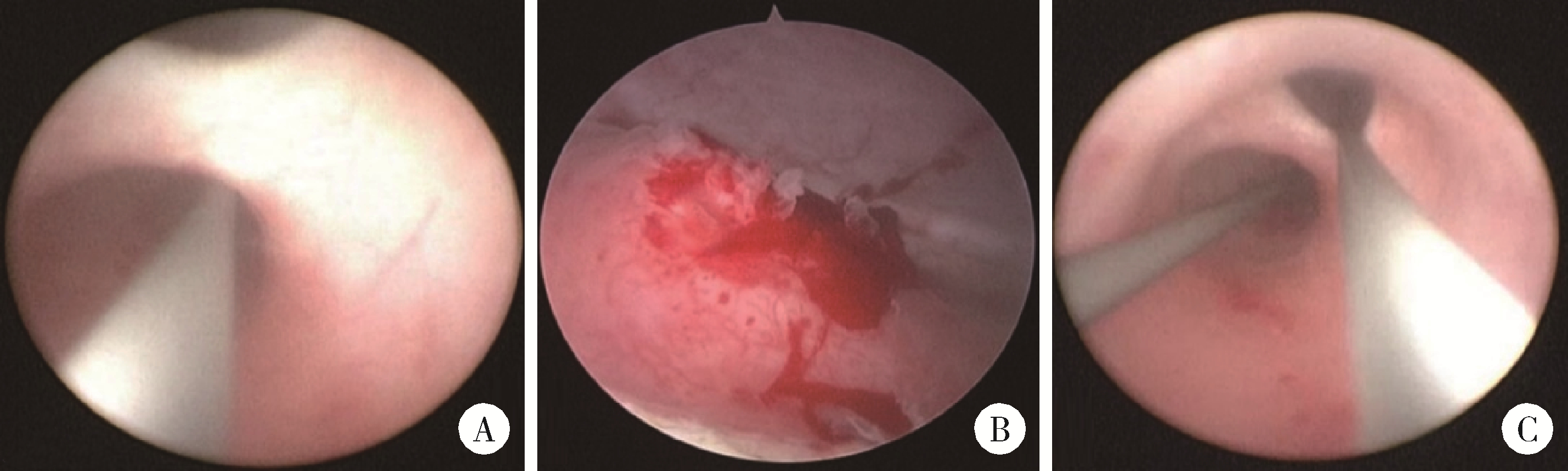
Intraoperative ureteroscopic views of duplex kidney and ureter malformation A, ureteroscopically, two ureteral tracts can be visualized; B, a cyst is visible at the orifice of the duplicated ureter; C, inverted Y-shaped duplicated ureter."

Table 4

Postoperative stone clearance, complications and renal function improvement"

Variables Patients (n=38)
Postoperative fever, n (%) 14 (36.84)
Complete stone clearance, n (%) 36 (94.74)
Staged Surgery, n (%) 1 (2.63)
Follow-up time/months, M (P25P75) 17.80 (1.60, 41.90)
Postoperative extubation time/d, M (P25P75) 37.00 (31.25, 42.25)
Postoperative hospital stay/d, M (P25P75) 3.00 (2.00, 5.75)
Postoperative creatinine decline, n (%) 32 (84.21)

Table 5

Comparison of renal function related laboratory parameters before and after surgery"

Variable Preoperative Postoperative V P value
Creatinine/(μmol/L), M (P25P75) 75.50 (65.50, 89.70) 70.00 (57.00, 80.00) 43 0.007
Glomerular filtration rate/[mL/(min·1.73 m2)], M (P25P75) 92.77 (82.79, 102.75) 100.00 (93.00, 113.00) 135 0.033
1
Horst M , Smith GHH . Pelvi-ureteric junction obstruction in duplex kidneys[J]. BJU Int, 2008, 101(12): 1580- 1584.

doi: 10.1111/j.1464-410X.2007.07386.x
2
Pioli Lamêgo de Faria C , Gomes Botelho L , Santos Vidal I , et al. Urinary incontinence due to complete ureteral duplication with an ectopic vulvar ureteral orifice: A case report[J]. Cureus, 2023, 15(7): e42114.
3
Lakshman G , Krishnamoorthy S , Kumaresan N . Duplex kidney with calculus obstruction of lower moiety lessons learnt[J]. J Med Pharm Allied Sci, 2022, 11(2): 4718- 4720.

doi: 10.55522/jmpas.V11I2.2335
4
Katims A , Rosen DC , Chandhoke R , et al. Extravesical ectopic ureteral calculus obstruction in a completely duplicated collecting system[J]. J Endourol Case Rep, 2020, 6(1): 42- 44.

doi: 10.1089/cren.2019.0108
5
Zheng Z , Hu W , Ji C , et al. A study of the difference in biochemical metabolism between patients with unilateral and bilateral upper urinary tract stones[J]. Sci Rep, 2024, 14(1): 30154.

doi: 10.1038/s41598-024-81454-3
6
梁月有, 郑克立, 戴宇平, 等. 先天性重复肾盂、输尿管畸形合并结石的诊断与治疗: 附32例报告[J]. 新医学, 2002, 33(12): 725- 726.
7
唐翠松, 汤光宇, 李伟, 等. 64排容积CT尿路成像在肾盂输尿管重复畸形诊断中的应用价值及优势[J]. 临床放射学杂志, 2010, 29(11): 1504- 1507.
8
冀竞辉, 林秀石, 潘大猛, 等. 输尿管硬镜联合勺状阻石篮激光碎石术治疗输尿管1.5 cm及以上结石的效果[J]. 北京大学学报(医学版), 2025, 57(4): 676- 683.

doi: 10.19723/j.issn.1671-167X.2025.04.008
9
余霄腾, 黄奕瑄, 李新飞, 等. 结石相关输尿管狭窄的上尿路修复手术技术与临床结局[J]. 北京大学学报(医学版), 2025, 57(4): 670- 675.

doi: 10.19723/j.issn.1671-167X.2025.04.007
10
李志军, 李勇, 钱坤, 等. 重复肾输尿管畸形合并上尿路结石的腔内外科治疗体会[J]. 中国内镜杂志, 2018, 24(5): 90- 94.
11
Pan Y , Chen G , Chen H , et al. The left ureterocele and stone of calyceal diverticulum in the patient with bilateral incomplete duplex kidneys managed by flexible ureteroscopy: A case report and literature review[J]. BMC Urol, 2020, 20(1): 35.

doi: 10.1186/s12894-020-00604-7
12
花梅免, 杨庆, 张振声, 等. 经尿道输尿管口囊肿电切术在重复肾输尿管畸形合并异位输尿管口囊肿患者中的应用(附9例报告)[J]. 海军军医大学学报, 2022, 43(12): 1481- 1484.
13
张韬, 赵贵成, 沈军, 等. 重复肾输尿管畸形12例诊治体会[J]. 实用医学杂志, 2015, 31(19): 3228- 3231.
14
Ye WX , Ren LG , Chen L . Inverted Y ureteral duplication with an ectopic ureter and multiple urinary calculi: A case report[J]. World J Clin Cases, 2022, 10(4): 1326- 1332.

doi: 10.12998/wjcc.v10.i4.1326
15
Falahatkar R , Falahatkar S , Khajavi Gaskarei MA , et al. The global, prevalence, and risk factors of postoperative fever after percutaneous nephrolithotomy: A systematic review and meta-analysis[J]. Asian J Urol, 2024, 11(2): 253- 260.

doi: 10.1016/j.ajur.2022.04.008
16
Bohare U , Gupta HL , Gupta M , et al. Evaluation of risk factors for fever and sepsis after ureteroscopic lithotripsy: A prospective observational study[J]. Int J Anat Radiol Surg, 2023, 12(6): 13- 16.
[1] Xiaoteng YU, Yixuan HUANG, Xinfei LI, Changfu CHEN, Fangzhou ZHAO, Honggang YING, Zihao TAO, Yiming ZHANG, Liqing XU, Zhihua LI, Kunlin YANG, Liqun ZHOU, Xuesong LI, Zheng ZHAO. Surgical techniques and clinical outcomes of upper urinary tract reconstruction for stone-related ureteral strictures [J]. Journal of Peking University (Health Sciences), 2025, 57(4): 670-675.
[2] Lei WANG,Tian-dong HAN,Wei-xing JIANG,Jun LI,Dao-xin ZHANG,Ye TIAN. Comparison of safety and effectiveness of active migration technique and in situ lithotripsy technique in the treatment of 1-2 cm upper ureteral calculi by flexible ure-teroscopy [J]. Journal of Peking University (Health Sciences), 2023, 55(3): 553-557.
[3] DAI Xiang,ZUO Mei-ni,ZHANG Xiao-peng,HU Hao,XU Tao. Comparison of long-term outcomes in different managements of diverticular neck in percutaneous nephrolithotomy for diverticular calculi [J]. Journal of Peking University (Health Sciences), 2021, 53(4): 704-709.
[4] Jun-hui ZHANG,Yi-hang JIANG,Yu-guang JIANG,Ji-qing ZHANG,Ning KANG. Clinical outcomes of simultaneous bilateral endoscopic surgery for bilateral upper urinary tract calculi [J]. Journal of Peking University (Health Sciences), 2020, 52(4): 672-677.
[5] Ming-rui WANG,Qi WANG,Hao HU,Jin-hui LAI,Yong-xin HE,Jie XIONG,Xian-hui LIU,Shi-jun LIU,Ke-xin XU,Tao XU. Long-term analysis of safety and efficacy of standard percutaneous nephrolithotomy in patients with solitary kidneys [J]. Journal of Peking University (Health Sciences), 2020, 52(4): 663-666.
[6] Run-zhuo MA,Hai-zhui XIA,Min LU,Zhi-ying ZHANG,Qi-ming ZHANG,Jian LU,Guo-liang WANG,Lu-lin MA. Impact of diagnostic ureteroscopy and biopsy on radical nephroureterectomy of upper tract urothelial carcinoma [J]. Journal of Peking University(Health Sciences), 2019, 51(4): 665-672.
[7] HAO Yi-chang, CHEN Kun, LIU Yu-qing, LU Jian, XIAO Chun-lei, MA Lu-lin. Transurethral flexible ureteroscopic holmium laser resection for tumors of renal pelvis: 6 cases report and literature review [J]. Journal of Peking University(Health Sciences), 2018, 50(5): 816-821.
[8] SHEN Cheng, ZHANG Bo, HAN Wen-ke, LIN Jian, WANG Gang, ZHANG Xiao-chun, SONG Yi, ZHAO Zheng, ZHANG Zhong-yuan, JIN Jie, YU Wei. Percutaneous renal access for percutaneous nephrolithotomy guided by contrast enhanced ultrasound: a single-center preliminary experience in China [J]. Journal of Peking University(Health Sciences), 2017, 49(6): 1071-1075.
[9] MA Run-zhuo, QIU Min, HE Wei, YANG Bin, XIA Hai-zhui, ZOU Da, LU Min, MA Lu-lin1, LU Jian. Ureteroscope can assist risk stratification in upper tract urothelial carcinoma [J]. Journal of Peking University(Health Sciences), 2017, 49(4): 632-637.
[10] YU Lu-ping, XU Tao. Analysis of development, safety and efficacy of percutaneous nephrolithotomy for management of upper urinary tract calculi in pediatric patients [J]. Journal of Peking University(Health Sciences), 2017, 49(4): 626-631.
[11] MA Kai, HUANG Xiao-Bo, XIONG Liu-Lin, XU Qing-Quan, XU Tao, YE Hai-Yun, YU Lu-Ping, WANG Xiao-Feng. Treatment of upper urinary calculi through novel modular flexible ureteroscope: a report of 36 cases [J]. Journal of Peking University(Health Sciences), 2015, 47(4): 615-617.
[12] ZHANG Li-Jie, YE Xiong-Jun, HUANG Xiao-Bo, XIONG Liu-Lin, MA Kai, LI Jian-Xing, WANG Xiao-Feng. Comparison of tubeless-percutaneous nephrolithotomy and ureteroscopic lithotripsy in treatment of upper-ureteral calculi sized ≥1.5 cm [J]. Journal of Peking University(Health Sciences), 2015, 47(1): 170-174.
[13] YANG Bo, HU Wei-Guo, HU Hao, CHEN Liang, LI Jian-Xing, WANG Xiao-Feng. Causes and remediations of retrograde intrarenal surgery failure in renal calculi treatment [J]. Journal of Peking University(Health Sciences), 2014, 46(5): 794-797.
Viewed
Full text


Abstract

Cited

  Shared   
  Discussed   
No Suggested Reading articles found!