Journal of Peking University (Health Sciences) ›› 2026, Vol. 58 ›› Issue (2): 290-300. doi: 10.19723/j.issn.1671-167X.2026.02.011

Previous Articles     Next Articles

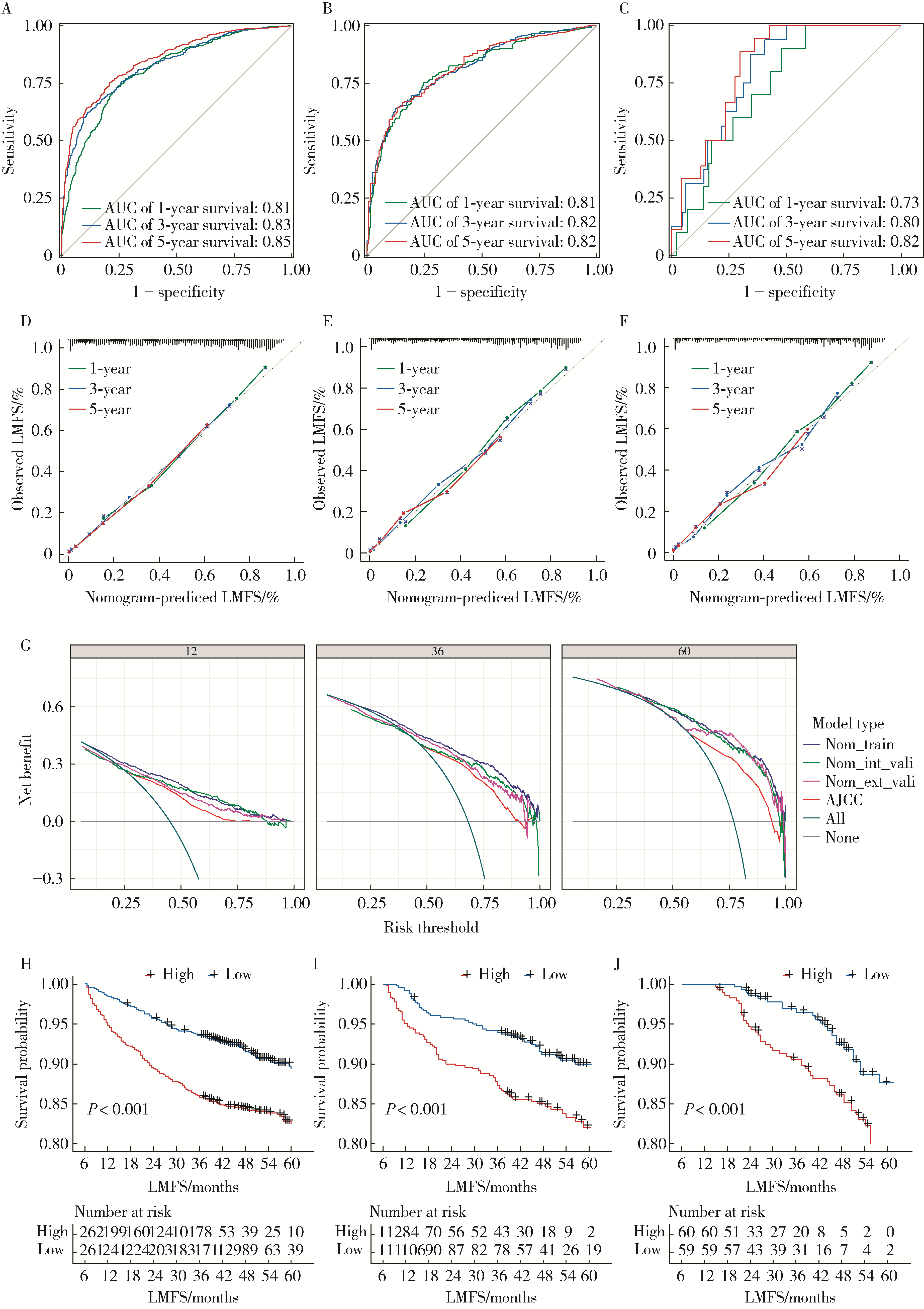
Development and validation of a multicenter nomogram predicting the risk of liver metastasis after curative resection of colorectal cancer

Nannan WANG1,2, Dajin YUAN1, Yubing ZHU1, Lei DING1,*()   

  1. 1. Department of Gastrointestinal Surgery, Beijing Shijitan Hospital, Capital Medical University, Beijing 100038, China
    2. Department of Colorectal Surgery, The Affiliated Cancer Hospital of Nanjing Medical University & Jiangsu Cancer Hospital & Jiangsu Institute of Cancer Research, Nanjing 210009, China
  • Received:2025-11-25 Online:2026-04-18 Published:2026-02-25
  • Contact: Lei DING
  • Supported by:
    the Youth Research Fund of Beijing Shijitan Hospital(2023-q19)

RICH HTML


Abstract:

Objective: To identify independent clinicopathological and molecular risk factors for metachronous liver metastasis and to construct a novel multicenter nomogram for predicting 1-, 3-, and 5-year liver metastasis-free survival (LMFS). Methods: In this multicenter retrospective cohort study, we analyzed clinical data from 865 patients with stages Ⅰ-Ⅲ CRC who underwent curative resection between January 2020 and December 2024. The population was derived from two institutions: Beijing Shijitan Hospital (n=746) and Jiangsu Cancer Hospital (n=119). Patients from the primary center were randomly assigned to a training cohort (n=523) and an internal validation cohort (n=223) at a 7 ∶3 ratio, while patients from the second center served as an independent external validation cohort (n=119). Candidate variables included demographics, tumor markers, pathological features, and molecular biomarkers [KRAS/BRAF mutation and microsatellite instability (MSI)]. Multivariable Cox proportional hazards regression analyses were utilized to identify independent predictors. Model performance was evaluated using the concordance index (C-index), time-dependent area under the receiver operating characteristic curve (AUC), calibration curves, and decision curve analysis (DCA). Results: Baseline characteristics were balanced across cohorts (P>0.05). Multivariable analysis identified nine independent prognostic factors: age, differentiation, T stage, N stage, vascular invasion, perineural invasion, and molecular markers. Notably, KRAS mutation (HR=1.42, 95%CI: 1.27-1.63) and BRAF mutation (HR=1.53, 95%CI: 1.29-1.84) were associated with significantly increased risk, whereas micro-satellite instability-high (MSI-H) status (HR=0.71, 95%CI: 0.54-0.92) served as a protective factor. The nomogram demonstrated robust discrimination with C-indices of 0.85 (95%CI: 0.82-0.89) in the training cohort, 0.81 (95%CI: 0.77-0.83) in the internal validation cohort, and 0.75 (95%CI: 0.71-0.79) in the external validation cohort. In the training set, AUCs for predicting 1-, 3-, and 5-year LMFS were 0.81 (95%CI: 0.77-0.86), 0.83 (95%CI: 0.80-0.89), and 0.85 (95%CI: 0.78-0.92), respectively. Calibration curves showed excellent agreement, and DCA indicated higher net clinical benefit than the American Joint Committee on Cancer (AJCC) tumor node metastasis (TNM) staging system. Conclusion: We established and externally validated a nomogram integrating clinicopathological features with KRAS, BRAF, and MSI status. This model exhibited enhanced predictive accuracy and generalizability compared with conventional staging systems. It serves as a valuable tool for identifying high-risk patients and guiding individualized postoperative surveillance strategies to improve long-term survival outcomes.

Key words: Colorectal cancer, Curative resection, Metachronous liver metastasis, Liver metastasis-free survival, Predictive model, Nomogram

CLC Number: 

  • R735.34

Figure 1

CONSORT flow diagram of patient selection and study design"

Table 1

Comparison of baseline clinicopathologic characteristics of CRC patients across multiple centers"

Variables SJTH cohort JSCH cohort P
Training set (n=523) Internal validation set (n=223) External validation set (n=119)
Age/years, n (%) 0.246
   < 65 298 (57.0) 138 (61.9) 77 (64.7)
  ≥65 225 (43.0) 85 (38.1) 42 (35.3)
Gender, n (%) 0.867
  Male 325 (62.1) 136 (61.0) 71 (59.7)
  Female 198 (37.9) 87 (39.0) 48 (40.3)
BMI/(kg/m2), n (%) 0.115
   < 18.5 43 (8.2) 17 (7.6) 15 (12.6)
  18.5 - < 24 251 (48.0) 109 (48.9) 55 (46.2)
  24 - < 28 206 (39.4) 87 (39.0) 44 (37.0)
  ≥28 23 (4.4) 10 (4.5) 5 (4.2)
CEA/(μg/L), n (%) 0.372
   < 5.0 240 (45.9) 95 (42.6) 53 (44.5)
  ≥5.0 283 (54.1) 128 (57.4) 66 (55.5)
CA199/(U/mL), n (%) 0.391
   < 37 245 (46.8) 97 (43.5) 47 (39.5)
  ≥37 278 (53.2) 126 (56.5) 72 (60.5)
Tumor site, n (%) 0.092
  Ascending colon 103 (19.7) 38 (17.0) 31 (26.1)
  Transverse colon 13 (2.5) 13 (5.8) 4 (3.4)
  Descending colon 29 (5.5) 14 (6.3) 12 (10.1)
  Sigmoid colon 155 (29.6) 61 (27.4) 28 (23.5)
  Rectum 223 (42.6) 97 (43.5) 44 (37.0)
Differentiation grade, n (%) 0.238
  High 78 (14.9) 26 (11.7) 21 (17.6)
  Moderate 333 (63.7) 151 (67.7) 66 (55.5)
  Low 112 (21.4) 46 (20.6) 32 (26.9)
Tumor size/cm, n (%) 0.155
   < 5 352 (67.3) 148 (66.3) 82 (68.9)
  ≥5 171 (32.7) 75 (33.6) 37 (31.1)
T stage, n (%) 0.054
  T1 47 (9.0) 18 (8.1) 6 (5.0)
  T2 93 (17.8) 31 (13.9) 15 (12.6)
  T3 348 (66.5) 166 (74.4) 85 (71.4)
  T4 35 (6.7) 8 (3.6) 13 (10.9)
N stage, n (%) 0.875
  N0 321 (61.4) 133 (59.6) 74 (62.2)
  N1 128 (24.5) 58 (26.0) 32 (26.9)
  N2 74 (14.1) 32 (14.3) 13 (10.9)
KRAS, n (%) 0.779
  Mutant type 256 (48.9) 112 (50.2) 55 (46.2)
  Wild type 267 (51.1) 111 (49.8) 64 (53.8)
NRAS, n (%) 0.425
  Mutant type 85 (16.3) 45 (20.2) 20 (16.8)
  Wild type 438 (83.7) 178 (79.8) 99 (83.2)
BRAF, n (%) 0.309
  Mutant type 79 (15.1) 40 (17.9) 14 (11.8)
  Wild type 444 (84.9) 183 (82.1) 105 (88.2)
MSI status, n (%) 0.287
  MSI-H 110 (21.0) 49 (22.0) 18 (15.1)
  MSS 413 (79.0) 174 (78.0) 101 (84.9)
Vascular invasion, n (%) 0.296
  Yes 170 (32.5) 71 (31.8) 30 (25.2)
  No 353 (67.5) 152 (68.2) 89 (74.8)
Nerve invasion, n (%) 0.313
  Yes 174 (33.3) 71 (31.8) 31 (26.1)
  No 349 (66.7) 152 (68.2) 88 (73.9)
Chemotherapy, n (%) 0.197
  Yes 258 (49.3) 120 (53.8) 52 (43.7)
  No 265 (50.7) 103 (46.2) 67 (56.3)
Targeted therapy, n (%) 0.112
  Yes 46 (8.8) 21 (9.4) 4 (3.4)
  No 477 (91.2) 202 (90.6) 115 (96.6)

Table 2

Cox regression for liver metastasis-free survival in patients with CRC"

Variables Univariate analysis Multivariate analysis
P HR (95%CI) P HR (95%CI)
Age/years
   < 65 Reference Reference
  ≥65 0.039 1.55 (1.12-1.95) 0.041 1.52 (1.22-1.74)
Gender
  Female Reference
  Male 0.610 1.12 (0.72-1.73)
BMI/(kg/m2)
   < 18.5 Reference
  18.5 - < 24 0.571 1.00 (0.94-1.05)
  24 - < 28 0.173 1.23 (0.94-1.45)
  ≥28 0.119 1.35 (0.95-1.59)
CEA/(μg/L)
   < 5.0 Reference
  ≥5.0 0.422 1.19 (0.78-1.82)
CA199/(U/mL)
   < 37 Reference
  ≥37 0.237 1.35 (0.82-2.27)
Tumor site
  Ascending colon Reference
  Transverse colon 0.352 0.38 (0.05-2.90)
  Descending colon 0.751 0.84 (0.27-2.54)
  Sigmoid colon 0.420 1.30 (0.69-2.47)
  Rectum 0.652 1.15 (0.63-2.11)
Differentiation grade
  High Reference Reference
  Moderate 0.019 1.85 (1.68-2.11) 0.005 1.19 (1.04-1.28)
  Low 0.001 2.41 (2.24-2.70) 0.005 2.54 (2.30-2.97)
Tumor size/cm
   < 5 Reference
  ≥5 0.119 1.26 (0.95-1.54)
T stage
  T1 Reference Reference
  T2 0.005 2.89 (1.97-3.42) 0.038 1.59 (1.03-2.38)
  T3 0.001 4.75 (3.55-5.97) 0.021 3.55(2.20-4.68)
  T4 < 0.001 8.22 (7.41-10.24) < 0.001 5.21(3.58-6.04)
N stage
  N0 Reference Reference
  N1 0.001 2.17 (1.69-2.8) 0.003 1.62 (1.18-2.02)
  N2 < 0.001 3.96 (2.55-4.80) < 0.001 2.88 (1.96-3.14)
KRAS
  Wild type Reference Reference
  Mutant type 0.012 1.56 (1.35-1.88) 0.005 1.42 (1.27-1.63)
NRAS
  Wild type Reference Reference
  Mutant type 0.105 1.24 (0.89-1.45) 0.275 1.27 (0.93- 1.43)
BRAF
  Wild type Reference Reference
  Mutant type 0.031 1.43 (1.20-1.93) 0.029 1.53 (1.29-1.84)
MSI status
  MSS Reference Reference
  MSI-H 0.024 0.60 (0.47-0.81) 0.035 0.71 (0.54-0.92)
Vascular invasion
  No Reference Reference
  Yes < 0.001 2.78 (1.82-4.25) 0.017 1.90 (1.67-2.28)
Nerve invasion
  No Reference Reference
  Yes < 0.001 2.80 (1.83-4.28) 0.046 1.12 (1.08-1.38)
Chemotherapy
  No Reference Reference
  Yes < 0.001 4.29 (2.55-7.21) < 0.001 2.65 (1.49-4.72)
Targeted therapy
  No Reference Reference
  Yes < 0.001 9.26 (5.16-15.43) < 0.001 8.35 (4.91-14.20)

Figure 2

Nomogram predicting 1-, 3- and 5-year probabilities for LMFS in patients with CRC"

Figure 3

Comprehensive evaluation of the nomogram for predicting liver metastasis-free survival (LMFS) in patients with colorectal cancer (CRC)"

Table 3

Comparison of representative prediction models for colorectal cancer liver metastasis published from 2015 to 2025"

Study Data source Sample size Prediction endpoint Methodology Variables Model performance Validation strategy
Yan, et al. (2019)[10] SEER database 32 819 LM & LNM Logistic regression Age, CEA, tumor size, grade, N stage AUC: 0.74-0.81 Internal random split
Li, et al. (2020)[12] SEER database 9 958 OS & CSS Cox regression Age, marital status, race, tumor location, pathological grade, histological type, T stage, N stage, colectomy, hepatic surgery, CEA C-index: 0.749 Internal validation
Hao, et al. (2022)[14] Single left 623 Metachronous LM Lasso regression Age, CEA, vascular invasion, T stage, N stage, family history of cancer, KRAS mutation C-index: 0.787 Internal validation
Xiao, et al. (2022)[15] Single left 611 Metachronous LM Deep learning (ResNet-50)+Cox HE image risk score, VELIPI, pT, pN C-index: 0.807 Internal validation split (7 ∶3)
Shao, et al. (2024)[16] SEER database 4 981 OS & CSS Cox & competing risk models Age, race, grade, T stage, N stage, surgery, chemotherapy, CEA, tumor deposits, lung metastasis, tumor size C-index: 0.74- 0.79 Internal random split (7 ∶3)
Jing, et al. (2025)[17] Single left 212 Postoperative liver metastasis Random forest (fusion model) CT radiomics features + Clinical data AUC: 0.751 Internal random split
1
Siegel RL , Miller KD , Wagle NS , et al. Cancer statistics, 2023[J]. CA Cancer J Clin, 2023, 73 (1): 17- 48.
2
Zheng R , Zhang S , Zeng H , et al. Cancer incidence and mortality in China, 2016[J]. J Natl Cancer Cent, 2022, 2 (1): 1- 9.
3
Morris VK , Kennedy EB , Baxter NN , et al. Treatment of metastatic colorectal cancer: ASCO guideline[J]. J Clin Oncol, 2023, 41 (3): 678- 700.

doi: 10.1200/JCO.22.01690
4
Reboux N , Jooste V , Goungounga J , et al. Incidence and survival in synchronous and metachronous liver metastases from colorectal cancer[J]. JAMA Netw Open, 2022, 5 (10): e2236666.

doi: 10.1001/jamanetworkopen.2022.36666
5
Carconi C , Cerreti M , Roberto M , et al. The management of oligometastatic disease in colorectal cancer: Present strategies and future perspectives[J]. Crit Rev Oncol, 2023, 186, 103990.

doi: 10.1016/j.critrevonc.2023.103990
6
Benson AB , Venook AP , Al-Hawary MM , et al. Colon cancer, version 2. 2021, NCCN clinical practice guidelines in oncology[J]. J Natl Compr Cancer Netw, 2021, 19 (3): 329- 359.

doi: 10.6004/jnccn.2021.0012
7
Tan EK , Ooi LL . Colorectal cancer liver metastases-understanding the differences in the management of synchronous and metachronous disease[J]. Ann Acad Med Singap, 2010, 39 (9): 715- 719.
8
Horie T , Kanemitsu Y , Takamizawa Y , et al. Prognostic differences between oligometastatic and polymetastatic disease after resection in patients with colorectal cancer and hepatic or lung metastases: Retrospective analysis of a large cohort at a single institution[J]. Surgery, 2023, 173 (2): 328- 334.

doi: 10.1016/j.surg.2022.10.014
9
Balachandran VP , Gonen M , Smith JJ , et al. Nomograms in oncology: More than meets the eye[J]. Lancet Oncol, 2015, 16 (4): e173- e180.

doi: 10.1016/S1470-2045(14)71116-7
10
Yan Y , Liu H , Mao K , et al. Novel nomograms to predict lymph node metastasis and liver metastasis in patients with early colon carcinoma[J]. J Transl Med, 2019, 17 (1): 193.

doi: 10.1186/s12967-019-1940-1
11
Li G , Li S , Cao L , et al. Nomogram development and validation for predicting minimally invasive step-up approach failure in infected necrotizing pancreatitis patients: A retrospective cohort study[J]. Int J Surg, 2023, 109 (6): 1677- 1687.

doi: 10.1097/JS9.0000000000000415
12
Li Y , Liu W , Zhao L , et al. Nomograms predicting overall survival and cancer-specific survival for synchronous colorectal liver-limited metastasis[J]. J Cancer, 2020, 11 (21): 6213.

doi: 10.7150/jca.46155
13
Collins GS , Reitsma JB , Altman DG , et al. Transparent reporting of a multivariable prediction model for individual prognosis or diagnosis (TRIPOD): The TRIPOD statement[J]. BMJ, 2014, 350, g7594.
14
Hao M , Li H , Wang K , et al. Predicting metachronous liver metastasis in patients with colorectal cancer: Development and assessment of a new nomogram[J]. World J Surg Oncol, 2022, 20 (1): 80.

doi: 10.1186/s12957-022-02558-6
15
Xiao C , Zhou M , Yang X , et al. Accurate prediction of metachronous liver metastasis in stage Ⅰ-Ⅲ colorectal cancer patients using deep learning with digital pathological images[J]. Front Oncol, 2022, 12, 844067.

doi: 10.3389/fonc.2022.844067
16
Shao S, Tian D, Li M, et al. Survival prediction in sigmoid-colon-cancer patients with liver metastasis: A prospective cohort study[J/OL]. JNCI Cancer Spectr, 2024, 8(5): pkae080[2025-11-01]. https://doi.org/10.1093/jncics/pkae080.
17
Jing HH , Hao D , Liu XJ , et al. Development and validation of a radiopathomics model for predicting liver metastases of colorectal cancer[J]. Eur Radiol, 2025, 35 (6): 3409- 3417.
18
Zhang Q , Zhou X , Cheng S , et al. Prognostic nomogram for colorectal cancer liver metastasis treated with tumor resection and chemotherapy based on SEER database[J]. J Gastrointest Oncol, 2025, 16 (5): 2067- 2083.

doi: 10.21037/jgo-2025-415
19
Pitroda SP , Khodarev NN , Huang L , et al. Integrated molecular subtyping defines a curable oligometastatic state in colorectal liver metastasis[J]. Nat Commun, 2018, 9, 1793.

doi: 10.1038/s41467-018-04278-6
20
Bi F , Dong J , Jin C , et al. Iparomlimab (QL1604) in patients with microsatellite instability-high (MSI-H) or mismatch repair-deficient (dMMR) unresectable or metastatic solid tumors: A pivotal, single-arm, multicenter, phase Ⅱ trial[J]. J Hematol Oncol, 2024, 17 (1): 109.

doi: 10.1186/s13045-024-01627-5
21
Margonis GA , Buettner S , Andreatos N , et al. Association of BRAF mutations with survival and recurrence in surgically treated patients with metastatic colorectal liver cancer[J]. JAMA Surg, 2018, 153 (7): e180996.

doi: 10.1001/jamasurg.2018.0996
22
Li Y , Solis-Ruiz J , Yang F , et al. NGS-defined measurable residual disease (MRD) after initial chemotherapy as a prognostic biomarker for acute myeloid leukemia[J]. Blood Cancer J, 2023, 13, 59.

doi: 10.1038/s41408-023-00833-7
23
Hamfjord J , Guren TK , Dajani O , et al. Total circulating cell-free DNA as a prognostic biomarker in metastatic colorectal cancer before first-line oxaliplatin-based chemotherapy[J]. Ann Oncol, 2019, 30 (7): 1088- 1095.

doi: 10.1093/annonc/mdz139
24
Luo X , Deng H , Xie F , et al. Prognostication of colorectal cancer liver metastasis by CE-based radiomics and machine learning[J]. Transl Oncol, 2024, 47, 101997.

doi: 10.1016/j.tranon.2024.101997
25
Lee HS , Kwon HW , Lim SB , et al. FDG metabolic parameter-based models for predicting recurrence after upfront surgery in synchronous colorectal cancer liver metastasis[J]. Eur Radiol, 2023, 33 (3): 1746- 1756.
26
Okimoto S , Kobayashi T , Tashiro H , et al. Significance of the Glasgow Prognostic Score for patients with colorectal liver metastasis[J]. Int J Surg, 2017, 42, 209- 214.

doi: 10.1016/j.ijsu.2017.04.068
[1] Bowen LI, Qiang ZHANG, Yixin SUN. Establishment and validation of a risk prediction model for scoliosis after Nuss procedure in children and young adults with pectus excavatum [J]. Journal of Peking University (Health Sciences), 2025, 57(5): 941-946.
[2] Zhicun LI, Tianyu WU, Lei LIANG, Yu FAN, Yisen MENG, Qian ZHANG. Risk factors analysis and nomogram model construction of postoperative pathological upgrade of prostate cancer patients with single core positive biopsy [J]. Journal of Peking University (Health Sciences), 2024, 56(5): 896-901.
[3] Yuanmei LIU, Yicheng FU, Jingxin HAO, Fuchun ZHANG, Huilin LIU. Construction and validation of a nomogram for predicting in-hospital postoperative heart failure in elderly patients with hip fracture [J]. Journal of Peking University (Health Sciences), 2024, 56(5): 874-883.
[4] Zezhen ZHOU,Shaohui DENG,Ye YAN,Fan ZHANG,Yichang HAO,Liyuan GE,Hongxian ZHANG,Guoliang WANG,Shudong ZHANG. Predicting the 3-year tumor-specific survival in patients with T3a non-metastatic renal cell carcinoma [J]. Journal of Peking University (Health Sciences), 2024, 56(4): 673-679.
[5] Hailong HE,Qing LI,Tao XU,Xiaowei ZHANG. Construction of a predictive model for postoperative pain relief after microscopic spermatic cord surgery for spermatic cord pain [J]. Journal of Peking University (Health Sciences), 2024, 56(4): 646-655.
[6] Junqi SU,Xiaoying WANG,Zhiqiang SUN. Establishment and verification of a prognostic nomogram for survival of tongue squamous cell carcinoma patients who underwent cervical dissection [J]. Journal of Peking University (Health Sciences), 2024, 56(1): 120-130.
[7] Hai MAO,Fan ZHANG,Zhan-yi ZHANG,Ye YAN,Yi-chang HAO,Yi HUANG,Lu-lin MA,Hong-ling CHU,Shu-dong ZHANG. Predictive model of early urinary continence recovery based on prostate gland MRI parameters after laparoscopic radical prostatectomy [J]. Journal of Peking University (Health Sciences), 2023, 55(5): 818-824.
[8] DING Ting-ting,ZENG Chu-xiong,HU Li-na,YU Ming-hua. Establishment of a prediction model for colorectal cancer immune cell infiltration based on the cancer genome atlas (TCGA) database [J]. Journal of Peking University (Health Sciences), 2022, 54(2): 203-208.
[9] MU Dong-liang,XUE Cheng,AN Bin,WANG Dong-xin. Epidural block associated with improved long-term survival after surgery for colorectal cancer: A retrospective cohort study with propensity score matching [J]. Journal of Peking University (Health Sciences), 2021, 53(6): 1152-1158.
[10] Wen-peng WANG,Jie-fu WANG,Jun HU,Jun-feng WANG,Jia LIU,Da-lu KONG,Jian LI. Clinicopathological features and prognosis of colorectal stromal tumor [J]. Journal of Peking University (Health Sciences), 2020, 52(2): 353-361.
[11] Xue-hua ZHU,Ming-yu YANG,Hai-zhui XIA,Wei HE,Zhi-ying ZHANG,Yu-qing LIU,Chun-lei XIAO,Lu-lin MA,Jian LU. Application of machine learning models in predicting early stone-free rate after flexible ureteroscopic lithotripsy for renal stones [J]. Journal of Peking University(Health Sciences), 2019, 51(4): 653-659.
[12] Le GAO,Shu-qing YU,Ji-chun YANG,Jun-ling MA,Si-yan ZHAN,Feng SUN. Quality assessment of global guidelines on colorectal cancer screening [J]. Journal of Peking University(Health Sciences), 2019, 51(3): 548-555.
[13] LIU Yu-qing, LU Jian, HAO Yi-chang, XIAO Chun-lei, MA Lu-lin. Predicting model based on risk factors for urosepsis after percutaneous nephrolithotomy [J]. Journal of Peking University(Health Sciences), 2018, 50(3): 507-513.
Viewed
Full text


Abstract

Cited

  Shared   
  Discussed   
No Suggested Reading articles found!