Journal of Peking University (Health Sciences) ›› 2021, Vol. 53 ›› Issue (1): 34-39. doi: 10.19723/j.issn.1671-167X.2021.01.006
Previous Articles Next Articles
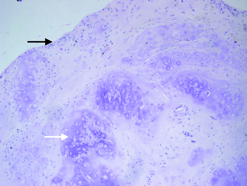
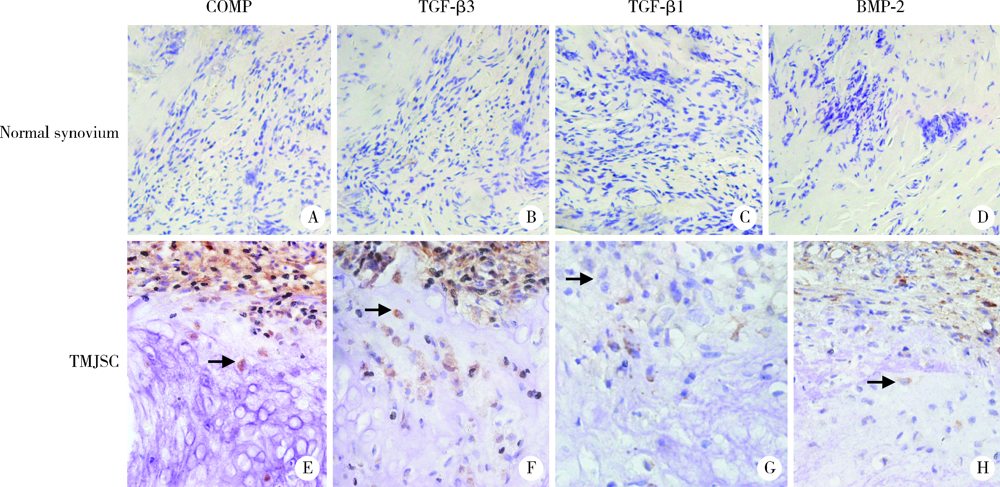
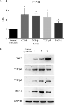
Expression of cartilage oligomeric matrix protein in the synovial chondromatosis of the temporomandibular joint
HAN Wei-hua1,LUO Hai-yan2,GUO Chuan-bin1,NING Qi1,MENG Juan-hong1,Δ( )
)
- 1. Department of Oral and Maxillofacial Surgery, Peking University School and Hospital of Stomatology & National Clinical Research Center for Oral Diseases & National Engineering Laboratory for Digital and Material Technology of Stomatology & Beijing Key Laboratory of Digital Stomatology, Beijing 100081, China
2. Department of Oral Pathology, Peking University School and Hospital of Stomatology & National Clinical Research Center for Oral Diseases & National Engineering Laboratory for Digital and Material Technology of Stomatology & Beijing Key Laboratory of Digital Stomatology, Beijing 100081, China
CLC Number:
- R782.6


| [1] |
Meng JH, Guo CB, Yi B, et al. Clinical and radiologic findings of synovial chondromatosis affecting the temporomandibular joint[J]. Oral Surg Oral Med Oral Pathol Oral Radiol Endod, 2010,109(3):441-448.
doi: 10.1016/j.tripleo.2009.09.036 pmid: 20097104 |
| [2] | Coles MJ, Tara HH. Synovial chondromatosis: a case study and brief review[J]. Am J Orthop (Belle Mead NJ), 1997,26(1):37-40. |
| [3] | 韩方凯, 马绪臣. 颞下颌关节滑膜软骨瘤病[J]. 现代口腔医学杂志, 2006,20(4):425-428. |
| [4] |
Helmy ES, Bays RA, Sharawy MM. Synovial chondromatosis associated with experimental osteoarthritis in adult monkeys[J]. J Oral Maxillofac Surg, 1989,47(8):823-827.
doi: 10.1016/s0278-2391(89)80041-2 pmid: 2746392 |
| [5] |
Li YJ, Cai HX, Fang W, et al. Fibroblast growth factor 2 involved in the pathogenesis of synovial chondromatosis of temporomandibular joint[J]. J Oral Pathol Med, 2014,43(5):388-394.
doi: 10.1111/jop.12146 |
| [6] |
Li Y, El Mozen LA, Cai H, et al. Transforming growth factor beta 3 involved in the pathogenesis of synovial chondromatosis of temporomandibular joint[J]. Sci Rep, 2015,5:8843.
doi: 10.1038/srep08843 pmid: 25742744 |
| [7] | Sandberg MM, Aro HT, Vuorio EI. Gene expression during bone repair[J]. Clin Orthop and Relat Res, 1993(289):292-312. |
| [8] |
Nishimura K, Solchaga LA, Caplan AI, et al. Chondroprogenitor cells of synovial tissue[J]. Arthritis Rheum, 2010,42(12):2631-2637.
doi: 10.1002/1529-0131(199912)42:12<2631::AID-ANR18>3.0.CO;2-H pmid: 10616011 |
| [9] |
Pearson CA, Pearson D, Shibahara S, et al. Tenascin: cDNA cloning and induction by TGF-beta[J]. EMBO J, 1988,7(10):2977-2982.
pmid: 2460335 |
| [10] |
Mackie EJ, Thesleff I, Chiquet-Ehrismann R. Tenascin is asso-ciated with chondrogenic and osteogenic differentiation in vivo and promotes chondrogenesis in vitro[J]. J Cell Biol, 1987,105(6):2569-2579.
doi: 10.1083/jcb.105.6.2569 |
| [11] |
Nakanishi S, Sskamoto K, Yoshitake H, et al. Bone morphoge-netic proteins are involved in the pathobiology of synovial chondromatosis[J]. Biochem Biophys Res Commun, 2009,379(4):914-919.
doi: 10.1016/j.bbrc.2008.12.170 pmid: 19138670 |
| [12] |
Iwata H, Ono S, Sato K, et al. Bone morphogenetic protein-induced muscle- and synovium-derived cartilage differentiation in vitro[J]. Clin Orthop Relat Res, 1993(296):295-300.
pmid: 8222441 |
| [13] |
Wang C, Liu G, Zhang W, et al. Cartilage oligomeric matrix protein improves in vivo cartilage regeneration and compression modulus by enhancing matrix assembly and synjournal[J]. Colloids Surf B Biointerfaces, 2017,159:518-526.
doi: 10.1016/j.colsurfb.2017.08.008 pmid: 28843200 |
| [14] |
Zaucke F, Dinser R, Maurer P, et al. Cartilage oligomeric matrix protein (COMP) and collagen Ⅸ are sensitive markers for the differentiation state of articular primary chondrocytes[J]. Biochem J, 2001,358(1):17-24.
doi: 10.1042/bj3580017 |
| [15] | 游洪波, 陈安民, 王国宾, 等. 转化生长因子-β诱导前软骨干细胞成软骨分化的研究[J]. 中华创伤杂志, 2010,26(5):453-459. |
| [16] |
Li H, Haudenschild DR, Posey KL, et al. Comparative analysis with collagen type Ⅱ distinguishes cartilage oligomeric matrix protein as a primary TGFβ-responsive gene[J]. Osteoarthritis Cartilage, 2011,19(10):1246-1253.
doi: 10.1016/j.joca.2011.07.011 pmid: 21843649 |
| [17] |
Haudenschild DR, Hong E, Yik JH, et al. Enhanced activity of transforming growth factor-β1 (TGF-β1) bound to cartilage oligomeric matrix protein[J]. J Biol Chem, 2011,286(50):43250-43258.
doi: 10.1074/jbc.M111.234716 pmid: 21940632 |
| [18] |
Guo P, Shi Z L, Liu A, et al. Effects of cartilage oligomeric matrix protein on bone morphogenetic protein-2-induced differen-tiation of mesenchymal stem cells[J]. Orthop Surg, 2015,6(4):280-287.
doi: 10.1111/os.12135 pmid: 25430711 |
| [19] | 于萍, 步宏, 王华, 等. 免疫组化结果的图像分析与人工计数方法的对比研究[J]. 生物医学工程学杂志, 2003,20(2):288-290. |
| [20] |
Ardekian L, Faquin W, Troulis MJ, et al. Synovial chondromatosis of the temporomandibular joint: report and analysis of eleven cases[J]. J Oral Maxillofac Surg, 2005,63(7):941-947.
doi: 10.1016/j.joms.2005.03.009 pmid: 16003619 |
| [21] |
Fujita S, Iizuka T, Tuboi Y, et al. Synovial chondromatosis of the temporomandibular joint with immunohistochemical findings: Report of a case[J]. J Oral Maxillofac Surg, 1991,49(8):880-883.
doi: 10.1016/0278-2391(91)90022-e pmid: 2072203 |
| [22] |
Hedbom E, Antonsson P, Hjerpe A, et al. Cartilage matrix proteins: An acidic oligomeric protein (COMP) detected only in cartilage[J]. J Biol Chem, 1992,267(9):6132-6136.
pmid: 1556121 |
| [23] |
Recklies AD, Baillargeon L, White C. Regulation of cartilage oligomeric matrix protein synjournal in human synovial cells and arti-cular chondrocytes[J]. Arthritis Rheum, 1998,41(6):997-1006.
doi: 10.1002/1529-0131(199806)41:6<997::AID-ART6>3.0.CO;2-G pmid: 9627009 |
| [24] |
Andersson ML, Thorstensson CA, Roos EM, et al. Serum levels of cartilage oligomeric matrix protein (COMP) increase temporarily after physical exercise in patients with knee osteoarthritis[J]. BMC Musculoskelet Disord, 2006,7:98.
doi: 10.1186/1471-2474-7-98 pmid: 17156423 |
| [25] |
Chen HC, Shah SH, Li YJ, et al. Inverse association of general joint hypermobility with hand and knee osteoarthritis and serum cartilage oligomeric matrix protein levels[J]. Arthritis Rheum, 2008,58(12):3854-3864.
doi: 10.1002/art.24319 pmid: 19035482 |
| [26] |
Blumbach K, Bastiaansen-Jenniskens YM, DeGroot J, et al. Combined role of type Ⅸ collagen and cartilage oligomeric matrix protein in cartilage matrix assembly: Cartilage oligomeric matrix protein counteracts type Ⅸ collagen-induced limitation of cartilage collagen fibril growth in mouse chondrocyte cultures[J]. Arthritis Rheum, 2009,60(12):3676-3685.
doi: 10.1002/art.24979 pmid: 19950300 |
| [27] |
Nozawa-Inoue K, Amizuka N, Ikeda N, et al. Synovial membrane in the temporomandibular joint: Its morphology, function and development[J]. Arch Histol Cytol, 2003,66(4):289-306.
doi: 10.1679/aohc.66.289 pmid: 14692685 |
| [28] | 路红艳, 李健, 龙星. 间充质干细胞表面标记在颞下颌关节疾病患者滑膜间充质细胞表达的比较[J]. 口腔医学研究, 2018,34(11):1249-1252. |
| [29] | 符培亮, 丛锐军, 张雷, 等. 体外条件下TGF-β3、BMP-2和DEX诱导兔滑膜间充质干细胞向软骨细胞谱系分化的研究[J]. 中国骨与关节杂志, 2014(2):135-141. |
| [1] | Yuanyuan FANG, Fan XU, Jie LEI, Hao ZHANG, Wenyu ZHANG, Yu SUN, Hongxin WU, Kaiyuan FU, Weiyu MAO. Development and validation of a clinical automatic diagnosis system based on diagnostic criteria for temporomandibular disorders [J]. Journal of Peking University (Health Sciences), 2025, 57(1): 192-201. |
| [2] | Tianjiao HOU,Zhibo ZHOU,Zhuqing WANG,Mengying WANG,Siyue WANG,Hexiang PENG,Huangda GUO,Yixin LI,Hanyu ZHANG,Xueying QIN,Yiqun WU,Hongchen ZHENG,Jing LI,Tao WU,Hongping ZHU. Gene-gene/gene-environment interaction of transforming growth factor-β signaling pathway and the risk of non-syndromic oral clefts [J]. Journal of Peking University (Health Sciences), 2024, 56(3): 384-389. |
| [3] | Hongguang LI,Weihua HAN,Xun WU,Jiling FENG,Gang LI,Juanhong MENG. Preliminarily study of arthrocentesis combined with liquid phase concentrated growth factor injection in the treatment of unilateral temporomandibular joint osteoarthritis [J]. Journal of Peking University (Health Sciences), 2024, 56(2): 338-344. |
| [4] | Zhi-qing LI,Bing YU,Ze-yu CAI,Ying-bao WANG,Xu ZHANG,Biao ZHOU,Xiao-hong FANG,Fang YU,Yi FU,Jin-peng SUN,Wei LI,Wei KONG. Naringenin inhibits thoracic aortic aneurysm formation in mice with Marfan syndrome [J]. Journal of Peking University (Health Sciences), 2022, 54(5): 896-906. |
| [5] | ZHOU Jing,LIU Yi. Cone-beam CT evaluation of temporomandibular joint in skeletal class Ⅱ female adolescents with different vertical patterns [J]. Journal of Peking University (Health Sciences), 2021, 53(1): 109-119. |
| [6] | Shuo CHEN,Yang HE,Jin-gang AN,Yi ZHANG. Application of computer-aided virtual mandibular position in the simultaneous treatment of children with temporomandibular joint ankylosis and jaw deformity [J]. Journal of Peking University(Health Sciences), 2019, 51(5): 954-958. |
| [7] | Ming-zhe LI,Xiao-xia WANG,Zi-li LI,Biao YI,Cheng LIANG,Wei HE. Accuracy analysis of computer assisted navigation for condylectomy via intraoral approach [J]. Journal of Peking University(Health Sciences), 2019, 51(1): 182-186. |
| [8] | WANG Dan-dan, GAN Ye-hua, MA Xu-chen, MENG Juan-hong. Association between ADAMTS14 gene polymorphism and the temporomandibular joint osteoarthritis in Chinese Han females [J]. Journal of Peking University(Health Sciences), 2018, 50(2): 279-283. |
| [9] | JIA Wei-qian, ZHAO Yu-ming, GE Li-hong. Recombinant human transforming growth factor β1 promotes dental pulp stem cells proliferation and mineralization [J]. Journal of Peking University(Health Sciences), 2017, 49(4): 680-681. |
| [10] | MENG Juan-hong, GUO Yu-xing, LUO Hai-yan, GUO Chuan-bin, MA Xu-chen. Diagnosis and treatment of diffuse tenosynovial giant cell tumor arising from temporomandibular joints [J]. Journal of Peking University(Health Sciences), 2016, 48(6): 1049-1054. |
| [11] | CHEN Fei, PAN Shao-xia, FENG Hai-lan. Distribution and content of transforming growth factor-β1 and vascular endothelial growth factor in each layer of concentrated growth factors [J]. Journal of Peking University(Health Sciences), 2016, 48(5): 860-865. |
| [12] | LEI Jie,LIU Mu-qing,FU Kai-yuan. Disturbedsleep, anxiety and stress are possible risk indicators for temporomandibular disorders with myofascialpain [J]. Journal of Peking University(Health Sciences), 2016, 48(4): 692-696. |
| [13] | JIA Shuang-shuang, LI Wei-yang, LIU Xin, LI Li-ying. Transforming growth factor-β1 induces differentiation of bone marrowderived mesenchymal stem cells into myofibroblasts via production of reactive oxygen species [J]. Journal of Peking University(Health Sciences), 2015, 47(5): 737-742. |
| [14] | WANG Zhu-Qing, WANG Ping, WU-CHOU Yah-huei, YE Xiao-Qian, HUANG Shang-Zhi, SHI Bing, WANG Ke, YUAN Yuan, LIU Dong-Jing, WU Tao, WANG Hong, Terri H. Beaty. Association study between candidate genes on transforming growth factor-β signaling pathway and the risk of non-syndromic cleft lip with or without cleft palate in Chinese populations [J]. Journal of Peking University(Health Sciences), 2015, 47(3): 384-389. |
| [15] | MENG Juan-hong, GUO Chuan-bin, MA Xu-chen. Diagnosis and treatment of the ganglion cysts and synovial cysts arising from the temporomandibular joints [J]. Journal of Peking University(Health Sciences), 2014, 46(1): 43-47. |
|
||