Journal of Peking University (Health Sciences) ›› 2021, Vol. 53 ›› Issue (2): 378-383. doi: 10.19723/j.issn.1671-167X.2021.02.024
Previous Articles Next Articles
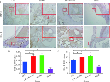
In vivo study of strontium-doped calcium phosphate cement for biological properties
WANG Jing-qi1,2,WANG Xiao1,Δ( )
)
- 1. Department of Stomatology, Peking University Third Hospital, Beijing 100191, China
2. Department of Stomatology, the Second Affiliated Hospital of Hainan Medical University, Haikou 570311, China
CLC Number:
- R783.1

| [1] |
Choudhary S, Halbout P, Alander C, et al. Strontium ranelate promotes osteoblastic differentiation and mineralization of murine bone marrow stromal cells: involvement of prostaglandins[J]. J Bone Miner Res, 2007,22(7):1002-1010.
pmid: 17371157 |
| [2] | Baier M, Staudt P, Klein R. Strontium enhances osseointegration of calcium phosphate cement: a histomorphometric pilot study in ovariectomized rats[J]. J Orthop Surg Res, 2013,8(1):16. |
| [3] | 李曙霞, 王京旗. 掺锶磷酸钙骨水泥材料的生物学性能体外研究[J]. 北京口腔医学, 2017,25(2):81-84. |
| [4] | 王晓娜, 赵静辉. 骨替代材料在口腔种植领域中的成骨效果[J]. 国际口腔医学杂志, 2016,43(1):113-117. |
| [5] |
Meloni SM, Jovanovic SA, Lolli FM, et al. Grafting after sinus lift with anorganic bovine bone alone compared with 50 ∶50 anorganic bovine bone and autologous bone: results of a pilot randomised trial at one year[J]. Br J Oral Maxillofac Surg, 2015,53(5):436-441.
pmid: 25796408 |
| [6] |
Aludden HC, Mordenfeld A, Hallman M, et al. Lateral ridge augmentation with Bio-Oss alone or Bio-Oss mixed with particulate autogenous bone graft: a systematic review[J]. Int J Oral Maxillofac Surg, 2017,46(8):1030-1038.
doi: 10.1016/j.ijom.2017.03.008 pmid: 28366452 |
| [7] | Wang F, Li Q, Wang Z. A comparative study of the effect of Bio-Oss in combination with concentrated growth factors or bone marrow-derived mesenchymal stem cells in canine sinus grafting[J]. J Oral Patho Med, 2017,46(7):528-536. |
| [8] | Orsini G, Scarano A, Degidi M, et al. Histological and ultrastructural evaluation of bone around Bio-Oss particles in sinus augmentation[J]. Oral Dis, 2007,13(13):586-593. |
| [9] | Jensen T, Schou S, Stavropoulos A, et al. Maxillary sinus floor augmentation with Bio-Oss or Bio-Oss mixed with autogenous bone as graft: a systematic review[J]. Int J Oral Maxillofac Surg, 2012,41(1):114-120. |
| [10] |
Urban IA, Nagursky H, Lozada JL, et al. Horizontal ridge augmentation with a collagen membrane and a combination of particulated autogenous bone and anorganic bovine bone-derived mineral: a prospective case series in 25 patients[J]. Int J Periodontics Restorative Dent, 2013,33(3):299-307.
pmid: 23593623 |
| [11] | Tian M, Chen F, Song W. In vivo study of porous strontium-doped calcium poly-phosphate scaffolds for bone substitute applications[J]. J Mater Sci Mater Med, 2009,20(7):1505-1512. |
| [12] | 严广斌. 骨组织形态计量学[J]. 中华关节外科杂志: 电子版, 2016,10(2):100. |
| [13] | 郭冲冲, 杜启翠. 细胞因子与牙槽骨重建关系的研究进展[J]. 口腔医学, 2015,35(8):697-701. |
| [14] | Axelrad TW, Kakar S, Einhorn TA. New technologies for the enhancement of skeletal repair[J]. Injury, 2007,38(l1):49-62. |
| [15] | 苏佳灿. 骨生长因子[M]. 第二军医大学出版社, 2015: 170-182. |
| [16] |
Marelli B, Ghezzi CE, Barralet JE, et al. Three-dimensional mineralization of dense nanofibrillar collagen bioglass hybrid scaffolds[J]. Biomacromolecules, 2010,11(6):1470-1479.
pmid: 20443577 |
| [17] |
Thorwarth M, Rupprecht S, Falk S, et al. Expression of bone matrix proteins during de novo bone formation using a bovine collagen and platelet-rich plasma (prp): an immunohistochemical analysis[J]. Biomaterials, 2005,26(15):2575-2584.
pmid: 15585260 |
| [18] | 郭莉. CPC与人重组骨形成蛋白-2(rhBMP-2)复合修复即刻种植牙骨缺损的效果研究[J]. 现代仪器与医疗, 2015,6(6):104-106. |
| [19] | Dahl SG, Allain P, Marie PJ, et al. Incorporation and distribution of strontium in bone[J]. Bone (New York), 2001,28(4):446-453. |
| [1] | Zheng LI, Longwei LV, Xiao ZHANG, Dandan XIA, Ping ZHANG, Yunsong LIU, Yongsheng ZHOU. Advances in oral and craniofacial bone regeneration modulated by stem cells and biomaterials [J]. Journal of Peking University (Health Sciences), 2026, 58(2): 272-277. |
| [2] | Ziyang YU, Houzuo GUO, Xi JIANG, Weihua HAN, Ye LIN. Imaging study of osteogenesis in maxillary sinus segment of zygomatic implants [J]. Journal of Peking University (Health Sciences), 2025, 57(5): 967-974. |
| [3] | Shuyuan MIN, Yun TIAN. Biocompatibility of 3D printed biodegradable WE43 magnesium alloy scaffolds and treatment of bone defects [J]. Journal of Peking University (Health Sciences), 2025, 57(2): 309-316. |
| [4] | Xinying WANG, Xueyuan CHENG, Yong ZHANG, Fei LI, Jinyu DUAN, Jing QIAO. Therapeutic effect of concentrated growth factors combined with self-curing calcium phosphate cement on periodontal intrabony defects: Clinical and radiographic evaluation [J]. Journal of Peking University (Health Sciences), 2025, 57(1): 42-50. |
| [5] | DU Wen-yu,YANG Jing-wen,JIANG Ting. Early constant observation of the effect of deferoxamine mesylate on improvement of vascularized bone regeneration in SD rat skull critical size defect model [J]. Journal of Peking University (Health Sciences), 2021, 53(6): 1171-1177. |
| [6] | ZHANG Sheng-nan,AN Na,OUYANG Xiang-ying,LIU Ying-jun,WANG Xue-kui. Role of growth arrest-specific protein 6 in migration and osteogenic differentiation of human periodontal ligament cells [J]. Journal of Peking University (Health Sciences), 2021, 53(1): 9-15. |
| [7] | Mei WANG, Bo-wen LI, Si-wen WANG, Yu-hua LIU. Preparation and osteogenic effect study of small intestinal submucosa sponge [J]. Journal of Peking University (Health Sciences), 2020, 52(5): 952-958. |
| [8] | Chang CAO,Fei WANG,En-bo WANG,Yu LIU. Application of β-TCP for bone defect restore after the mandibular third molars extraction: A split-mouth clinical trial [J]. Journal of Peking University(Health Sciences), 2020, 52(1): 97-102. |
| [9] | Ying CHEN,Zhong-ning LIU,Bo LI,Ting JIANG. Preparation of aspirin sustained-release microsphere and its in vitro releasing [J]. Journal of Peking University(Health Sciences), 2019, 51(5): 907-912. |
| [10] | Hao WANG,Yang LIU,Hao-chen LIU,Dong HAN,Hai-lan FENG. Detection and functional analysis of BMP2 gene mutation in patients with tooth agenesis [J]. Journal of Peking University(Health Sciences), 2019, 51(1): 9-15. |
| [11] | SUI Hua-xin, LV Pei-jun, WANG Yong, FENG Yu-chi. Effects of low level laser irradiation on the osteogenic capacity of sodium alginate/gelatin/human adipose-derived stem cells 3D bio-printing construct [J]. Journal of Peking University(Health Sciences), 2018, 50(5): 868-875. |
| [12] | LIU Jing-yin, CHEN Fei, GE Yan-jun, WEI Ling, PAN Shao-xia, FENG Hai-lan. Influence of implants prepared by selective laser melting on early bone healing [J]. Journal of Peking University(Health Sciences), 2018, 50(1): 117-122. |
| [13] | CHEN Fei, PAN Shao-xia, FENG Hai-lan. Distribution and content of transforming growth factor-β1 and vascular endothelial growth factor in each layer of concentrated growth factors [J]. Journal of Peking University(Health Sciences), 2016, 48(5): 860-865. |
| [14] | QIN Xue-yan, ZHAO Hua-xiang, ZHANG Qian, CHEN Feng, LIN Jiu-xiang. NELL-1: a novel highly efficient and specific growth factor [J]. Journal of Peking University(Health Sciences), 2016, 48(2): 380-383. |
| [15] | GE Wen-shu, TANG Yi-man, ZHANG Xiao, LIU Yun-song, ZHOU Yong-sheng. Establishing a luciferase reporter system to evaluate osteogenic differentiation potential of human adipose-derived stem cells [J]. Journal of Peking University(Health Sciences), 2016, 48(1): 170-174. |
|
||