Journal of Peking University (Health Sciences) ›› 2023, Vol. 55 ›› Issue (2): 343-350. doi: 10.19723/j.issn.1671-167X.2023.02.021
Previous Articles Next Articles
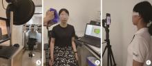
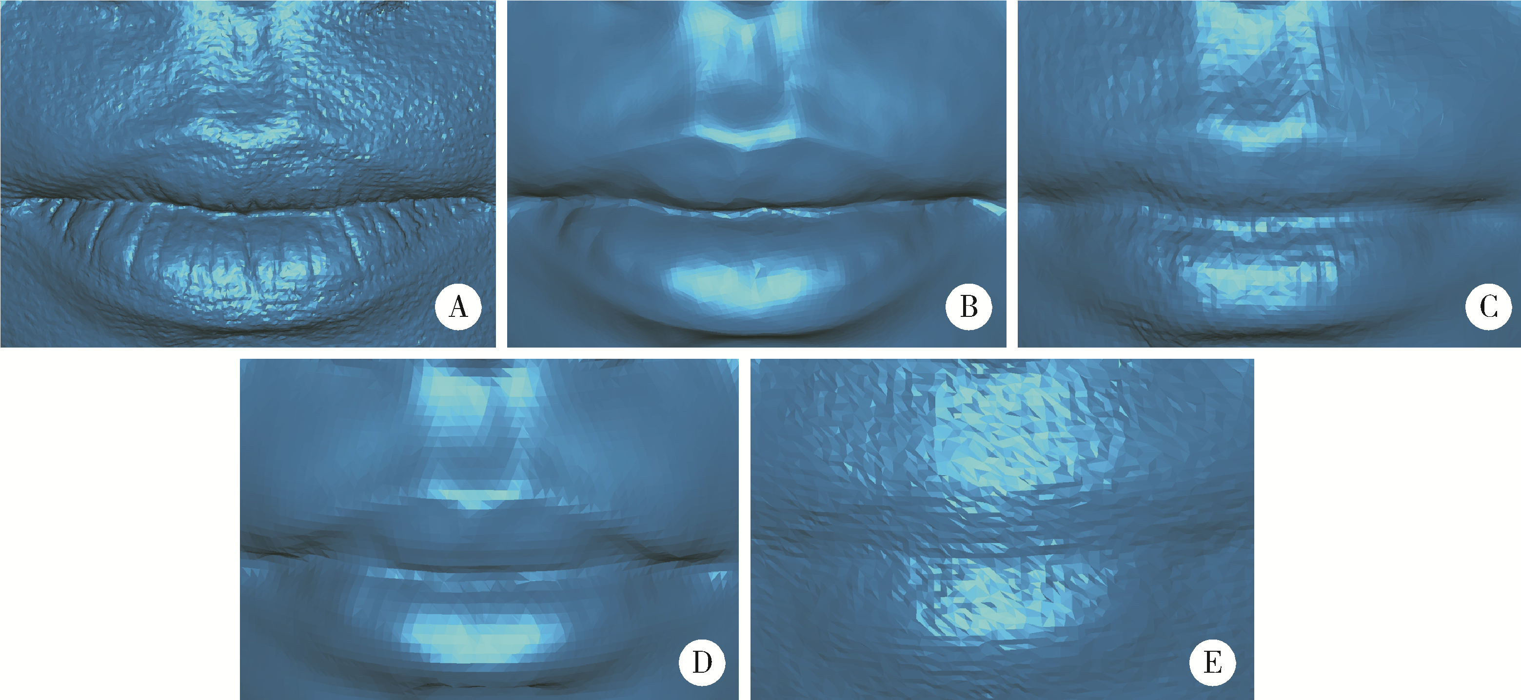
Preliminary evaluation of the trueness of 5 chairside 3D facial scanning techniques
Ao-nan WEN1,2,Wei LIU3,Da-wei LIU4,Yu-jia ZHU2,Ning XIAO2,Yong WANG1,2,*( ),Yi-jiao ZHAO1,2,*()
- 1. Institute of Medical Technology, Peking University Health Science Center, Beijing 100191, China
2. Center of Digital Dentistry, Peking University School and Hospital of Stomatology & National Center for Stomatology & National Clinical Research Center for Oral Diseases & National Engineering Research Center of Oral Biomaterials and Digital Medical Devices & Beijing Key Laboratory of Digital Stomatology & NHC Research Center of Engineering and Technology for Computerized Dentistry & NMPA Key Laboratory for Dental Materials, Beijing 100081, China
3. Yinchuan Stomatology Hospital, Yinchuan 750004, China
4. Department of Orthodontics, Peking University School and Hospital of Stomatology, Beijing 100081, China
CLC Number:
- R780.4


| 1 |
Fink M , Hirschfelder U , Hirschinger V , et al. Assessment of facial soft-tissue profiles based on lateral photographs versus three-dimensional face scans[J]. J Orofac Orthop, 2017, 78 (1): 70- 76.
doi: 10.1007/s00056-016-0055-z |
| 2 |
Topsakal O , Akbas MI , Smith BS , et al. Evaluating the agreement and reliability of a web-based facial analysis tool for rhinoplasty[J]. Int J Comput Assist Radiol Surg, 2021, 16 (8): 1381- 1391.
doi: 10.1007/s11548-021-02423-z |
| 3 | Anas IY , Bamgbose BO , Nuhu S . A comparison between 2D and 3D methods of quantifying facial morphology[J]. Heliyon, 2019, 5 (6): e1880. |
| 4 |
Stebel A , Desmedt D , Bronkhorst E , et al. Rating nasolabial appearance on three-dimensional images in cleft lip and palate: A comparison with standard photographs[J]. Eur J Orthod, 2016, 38 (2): 197- 201.
doi: 10.1093/ejo/cjv024 |
| 5 |
Krneta B , Primo IJ , Zhurov A , et al. Three-dimensional evaluation of facial morphology in children aged 5-6 years with a class Ⅲ malocclusion[J]. Eur J Orthod, 2014, 36 (2): 133- 139.
doi: 10.1093/ejo/cjs018 |
| 6 |
Bockey S , Berssenbrügge P , Dirksen D , et al. Computer-aided design of facial prostheses by means of 3D-data acquisition and following symmetry analysis[J]. J Craniomaxillofac Surg, 2018, 46 (8): 1320- 1328.
doi: 10.1016/j.jcms.2018.05.020 |
| 7 |
Farook TH , Jamayet NB , Abdullah JY , et al. Designing 3D prosthetic templates for maxillofacial defect rehabilitation: A comparative analysis of different virtual workflows[J]. Comput Biol Med, 2020, 118, 103646.
doi: 10.1016/j.compbiomed.2020.103646 |
| 8 |
Duppe K , Becker M , Schonmeyr B . Evaluation of facial anthropometry using three-dimensional photogrammetry and direct mea-suring techniques[J]. J Craniofac Surg, 2018, 29 (5): 1245- 1251.
doi: 10.1097/SCS.0000000000004580 |
| 9 |
van der Meer WJ , Dijkstra PU , Visser A , et al. Reliability and validity of measurements of facial swelling with a stereophotogrammetry optical three-dimensional scanner[J]. Br J Oral Maxillofac Surg, 2014, 52 (10): 922- 927.
doi: 10.1016/j.bjoms.2014.08.019 |
| 10 |
Andrade LM , Rodrigues da Silva AMB , Magri LV , et al. Repeatability study of angular and linear measurements on facial morpho-logy analysis by means of stereophotogrammetry[J]. J Craniofac Surg, 2017, 28 (4): 1107- 1111.
doi: 10.1097/SCS.0000000000003554 |
| 11 | Zhao Y , Xiong Y , Wang Y . Three-dimensional accuracy of facial scan for facial deformities in clinics: A new evaluation method for facial scanner accuracy[J]. PLoS One, 2017, 12 (1): e169402. |
| 12 | Gaber A, Faher MF, Waned MA. Automated grading of facial paralysis using the Kinect v2: A proof of concept study: International Conference on Virtual Rehabilitation (ICVR)[C]. Valencia, Spain: IEEE, 2015: 258-264. |
| 13 |
Sidequersky FV , Verze L , Mapelli A , et al. Quantification of facial movements by optical instruments: Surface laser scanning and optoelectronic three-dimensional motion analyzer[J]. J Craniofac Surg, 2014, 25 (1): e65- e70.
doi: 10.1097/SCS.0000000000000379 |
| 14 | Gaber A , Taher MF , Wahed MA . Quantifying facial paralysis using the Kinect v2[J]. Annu Int Conf IEEE Eng Med Biol Soc, 2015, 2015, 2497- 2501. |
| 15 |
Knoops PG , Beaumont CA , Borghi A , et al. Comparison of three-dimensional scanner systems for craniomaxillofacial imaging[J]. J Plast Reconstr Aesthet Surg, 2017, 70 (4): 441- 449.
doi: 10.1016/j.bjps.2016.12.015 |
| 16 |
Modabber A , Peters F , Kniha K , et al. Evaluation of the accuracy of a mobile and a stationary system for three-dimensional facial scanning[J]. J Craniomaxillofac Surg, 2016, 44 (10): 1719- 1724.
doi: 10.1016/j.jcms.2016.08.008 |
| 17 |
Ahn H , Chang Y , Kim K , et al. Measurement of three-dimensional perioral soft tissue changes in dentoalveolar protrusion patients after orthodontic treatment using a structured light scanner[J]. Angle Orthod, 2014, 84 (5): 795- 802.
doi: 10.2319/112913-877.1 |
| 18 |
Dindaroǧlu F , Kutlu P , Duran GS , et al. Accuracy and reliability of 3D stereophotogrammetry: A comparison to direct anthropometry and 2D photogrammetry[J]. Angle Orthod, 2016, 86 (3): 487- 494.
doi: 10.2319/041415-244.1 |
| 19 | 熊玉雪, 杨慧芳, 赵一姣, 等. 两种评价面部三维表面数据不对称度方法的比较[J]. 北京大学学报(医学版), 2015, 47 (2): 340- 343. |
| 20 |
Zeng W , Chen G , Ju R , et al. The combined application of database and three-dimensional image registration technology in the restoration of total nose defect[J]. J Craniofac Surg, 2018, 29 (5): e484- e487.
doi: 10.1097/SCS.0000000000004500 |
| 21 | 赵一姣, 熊玉雪, 杨慧芳, 等. 2种三维颜面部扫描仪测量精度的定量评价[J]. 实用口腔医学杂志, 2016, 32 (1): 37- 42. |
| 22 |
Artopoulos A , Buytaert JA , Dirckx JJ , et al. Comparison of the accuracy of digital stereophotogrammetry and projection moiré profilometry for three-dimensional imaging of the face[J]. Int J Oral Maxillofac Surg, 2014, 43 (5): 654- 662.
doi: 10.1016/j.ijom.2013.10.005 |
| 23 |
Winder RJ , Darvann TA , McKnight W , et al. Technical validation of the Di3D stereophotogrammetry surface imaging system[J]. Br J Oral Maxillofac Surg, 2008, 46 (1): 33- 37.
doi: 10.1016/j.bjoms.2007.09.005 |
| 24 |
Lo Russo L , Di Gioia C , Salamini A , et al. Integrating intraoral, perioral, and facial scans into the design of digital dentures[J]. J Prosthet Dent, 2020, 123 (4): 584- 588.
doi: 10.1016/j.prosdent.2019.05.030 |
| 25 |
Swennen G , Pottel L , Haers PE . Custom-made 3D-printed face masks in case of pandemic crisis situations with a lack of commercially available FFP2/3 masks[J]. Int J Oral Maxillofac Surg, 2020, 49 (5): 673- 677.
doi: 10.1016/j.ijom.2020.03.015 |
| 26 | Mai H , Lee D . The effect of perioral scan and artificial skin markers on the accuracy of virtual dentofacial integration: stereophotogrammetry versus smartphone three-dimensional face-scanning[J]. Int J Environ Res Public Health, 2021, 18 (1): 229. |
| 27 |
Duran GS , Dindaroglu F , Kutlu P . Hard- and soft-tissue symmetry comparison in patients with Class Ⅲ malocclusion[J]. Am J Orthod Dentofacial Orthop, 2019, 155 (4): 509- 522.
doi: 10.1016/j.ajodo.2018.05.021 |
| 28 |
史雨林, 商洪涛, 田磊, 等. 骨性Ⅲ类错 畸形患者双颌手术前后面部软组织变化的三维研究[J]. 中国修复重建外科杂志, 2018, 32 (5): 612- 616. 畸形患者双颌手术前后面部软组织变化的三维研究[J]. 中国修复重建外科杂志, 2018, 32 (5): 612- 616.
|
| 29 | 刘文静, 史雨林, 许方方, 等. 偏突颌畸形患者手术前后面部软组织的三维测量研究[J]. 现代生物医学进展, 2018, 18 (14): 2669- 2673. |
| 30 | Yamamoto S , Miyachi H , Fujii H , et al. Intuitive facial imaging method for evaluation of postoperative swelling: A combination of 3-dimensional computed tomography and laser surface scanning in orthognathic surgery[J]. J Oral Maxillofac Surg, 2016, 74 (12): 2501- 2506. |
| 31 |
Özsoy U , Uysal H , Hizay A , et al. Three-dimensional objective evaluation of facial palsy and follow-up of recovery with a handheld scanner[J]. J Plast Reconstr Aesthet Surg, 2021, 74 (12): 3404- 3414.
doi: 10.1016/j.bjps.2021.05.003 |
| 32 | 王勇, 赵一姣, 司燕. 与三维测量有关的名词浅析[J]. 中华口腔正畸学杂志, 2009, 16 (2): 111- 113. |
| 33 | 陈俊锴, 孙玉春, 陈虎, 等. 口内三维扫描仪扫描精度的定量评价方法研究[J]. 中华口腔医学杂志, 2021, 56 (9): 920- 925. |
| 34 | 曹悦, 陈俊锴, 邓珂慧, 等. 三款口内三维扫描仪获取无牙颌红膏初印模精度的对比评价[J]. 北京大学学报(医学版), 2020, 52 (1): 129- 137. |
| 35 | Rudy HL , Wake N , Yee J , et al. Three-dimensional facial scanning at the fingertips of patients and surgeons: accuracy and precision testing of iphone Ⅹ three-dimensional scanner[J]. Plast Reconstr Surg, 2020, 146 (6): 1407- 1417. |
| 36 | 赵一姣, 熊玉雪, 杨慧芳, 等. 3种不同原理颜面部扫描仪测量精度的评价[J]. 北京大学学报(医学版), 2014, 46 (1): 76- 80. |
| 37 | Amornvit P , Sanohkan S . The accuracy of digital face scans obtained from 3d scanners: An in vitro study[J]. Int J Environ Res Public Health, 2019, 16 (24): 5061. |
| 38 | Petrides G , Clark JR , Low H , et al. Three-dimensional scanners for soft-tissue facial assessment in clinical practice[J]. J Plast Reconstr Aesthet Surg, 2021, 74 (3): 605- 614. |
| [1] | Aonan WEN, Xiaohui ZHANG, Yongtao YANG, Zixiang GAO, Wenbo LI, Shenyao SHAN, Xiangyi SHANG, Yuwen TIAN, Shuwei GUO, Yizhen WANG, Yong WANG, Yijiao ZHAO. Method of constructing 3D facial smile simulation sequence data based on non-rigid registration [J]. Journal of Peking University (Health Sciences), 2026, 58(1): 139-144. |
| [2] | Wenbo LI, Yufeng SHEN, Yongtao YANG, Shenyao SHAN, Zixiang GAO, Aonan WEN, Xiangyi SHANG, Yuwen TIAN, Shuwei GUO, Yizhen WANG, Yong WANG, Yijiao ZHAO. Development of a surface electromyography index system for orofacial muscles and validation of a discriminant model in unilateral molar occlusal interference [J]. Journal of Peking University (Health Sciences), 2026, 58(1): 89-98. |
| [3] | Fangru LIN, Zhihui TANG. Correlation analysis of peri-implant health after single-tooth dental implant [J]. Journal of Peking University (Health Sciences), 2025, 57(2): 347-353. |
| [4] | Xinkai XU, Jianjiang ZHAO, Sukun TIAN, Zhongning LIU, Xiaoyi ZHAO, Xiaobo ZHAO, Tengfei JIANG, Xiaojun CHEN, Chao MA, Yuchun SUN. Evaluation of the accuracy of three-dimensional data acquisition from liquid- interference surfaces assisted by a scanner head with a compressed airflow system [J]. Journal of Peking University (Health Sciences), 2025, 57(1): 121-127. |
| [5] | Xiaotong LING,Liuyang QU,Danni ZHENG,Jing YANG,Xuebing YAN,Denggao LIU,Yan GAO. Three-dimensional radiographic features of calcifying odontogenic cyst and calcifying epithelial odontogenic tumor [J]. Journal of Peking University (Health Sciences), 2024, 56(1): 131-137. |
| [6] | Da-wei WANG,Hua-dong WANG,Li LI,Xin YIN,Wei HUANG,Ji-dong GUO,Ya-feng YANG,Yi-hao LIU,Yang ZHENG. Efficacy analysis of autologous facet joint bone block in lumbar interbody fusion of osteoporosis patients [J]. Journal of Peking University (Health Sciences), 2023, 55(5): 899-909. |
| [7] | Qian DING,Wen-jin LI,Feng-bo SUN,Jing-hua GU,Yuan-hua LIN,Lei ZHANG. Effects of surface treatment on the phase and fracture strength of yttria- and magnesia-stabilized zirconia implants [J]. Journal of Peking University (Health Sciences), 2023, 55(4): 721-728. |
| [8] | Wen ZHANG,Xiao-jing LIU,Zi-li LI,Yi ZHANG. Effect of alar base cinch suture based on anatomic landmarks on the morphology of nasolabial region in patients after orthognathic surgery [J]. Journal of Peking University (Health Sciences), 2023, 55(4): 736-742. |
| [9] | Wei-wei LI,Hu CHEN,Yong WANG,Yu-chun SUN. Research on friction and wear behaviors of silicon-lithium spray coating on zirconia ceramics [J]. Journal of Peking University (Health Sciences), 2023, 55(1): 94-100. |
| [10] | Hao-zhe YU,Wei-zhen ZENG,Wen-yu WU,Zhong-qiang YAO,Yun FENG. Evaluation of ocular surface status and function in primary Sjögren's syndrome with hypothyroidism [J]. Journal of Peking University (Health Sciences), 2022, 54(4): 705-711. |
| [11] | Hao LUO,Fu-cong TIAN,Xiao-yan WANG. Surface roughness, gloss and sequential polishing times of various chairside computer aided design/manufacturing restorative materials [J]. Journal of Peking University (Health Sciences), 2022, 54(3): 565-571. |
| [12] | WANG Zheng,DING Qian,GAO Yuan,MA Quan-quan,ZHANG Lei,GE Xi-yuan,SUN Yu-chun,XIE Qiu-fei. Effect of porous zirconia ceramics on proliferation and differentiation of osteoblasts [J]. Journal of Peking University (Health Sciences), 2022, 54(1): 31-39. |
| [13] | LI Si-yu,DUAN Xue-fei,CAO Ye. Evaluation of the effect of using ultrasonic instruments to improve the shoulder of the preparations [J]. Journal of Peking University (Health Sciences), 2021, 53(1): 88-94. |
| [14] | Ren-tao TANG,Xin-hai LI,Jiang-li YU,Lin FENG,Xue-jun GAO. Evaluation of microtensile bond strength between resin composite and glass ceramic [J]. Journal of Peking University (Health Sciences), 2020, 52(4): 755-761. |
| [15] | Jing-ying HU,Li LI,Qian-mei ZHOU,Rui-yu DING,Ran SHANG,Wei BAI. Influence of different mixing pads on physical and mechanical properties of glass ionomer cement [J]. Journal of Peking University(Health Sciences), 2019, 51(5): 964-967. |
|
||