北京大学学报(医学版) ›› 2019, Vol. 51 ›› Issue (6): 1078-1084. doi: 10.19723/j.issn.1671-167X.2019.06.018

• 论著 • 上一篇    下一篇

化疗后结直肠癌转移瘤钙化CT影像学表现与化疗反应之间的关系

张菁1,2,周裕文3,邱萌3,杨岚清1,伍兵1,()   

  1. 1. 四川大学华西医院放射科,成都 610041
    2. 重庆大学附属肿瘤医院影像科,重庆 400030
    3. 四川大学华西医院肿瘤中心,成都 610041
  • 收稿日期:2018-01-09 出版日期:2019-12-18 发布日期:2019-12-19
  • 通讯作者: 伍兵 E-mail:bingwu69@163.com
  • 基金资助:
    四川省科技厅项目(2014SZ0150)

Relationship between the CT features of colorectal cancer metastases calcification and tumor response to chemotherapy

Jing ZHANG1,2,Yu-wen ZHOU3,Meng QIU3,Lan-qing YANG1,Bing WU1,()   

  1. 1. Departments of Radiology, West China Hospital,Sichuan University, Chengdu 610041, China
    2. Departments of Radiology, Chongqing University Cancer Hospital, Chongqing 400030, China
    3. Departments of Oncology, West China Hospital,Sichuan University, Chengdu 610041, China
  • Received:2018-01-09 Online:2019-12-18 Published:2019-12-19
  • Contact: Bing WU E-mail:bingwu69@163.com
  • Supported by:
    Supported by the Project of the Science and Technology Department in Sichuan Province(2014SZ0150)

摘要:

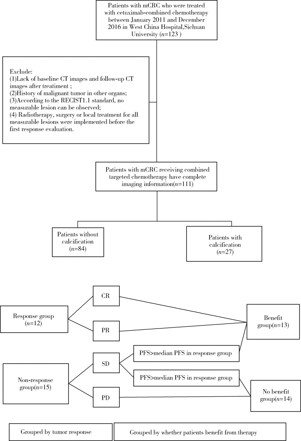
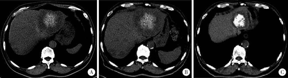
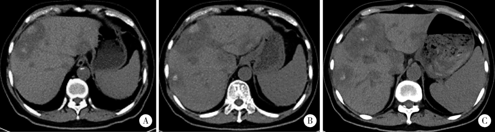
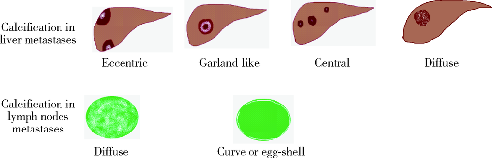
目的 探究结直肠癌转移灶在联合西妥昔单抗靶向化疗后钙化的CT特征与其治疗疗效的关系。方法 回顾性分析2011年1月至2016年12月接受过联合西妥昔单抗靶向化疗且有完整资料的转移性结直肠癌钙化患者。两位影像科医生对患者治疗前后的肿瘤钙化的发生、钙化特征,以及治疗疗效评价进行独立评估。参照《实体肿瘤疗效评价标准(1.1版本)》 对患者的最佳疗效评价进行记录:(1)将完全缓解(complete response,CR)和部分缓解(partial response,PR)归为治疗有反应组,将疾病稳定(stable disease,SD)和疾病进展(progressive disease,PD)归为治疗无反应组;(2)对于疗效评价为SD的患者,由于无进展生存时间(progress free survival,PFS)较长的患者可以认为能从治疗中获益,因此根据PFS长短对其进行进一步分组,将PFS大于治疗有反应组中位PFS的患者与疗效评价为CR或PR的患者归为治疗获益组,余者归为治疗未获益组。对比分析患者转移瘤钙化的不同影像学特征(钙化形态、最大钙化密度、钙化密度-时间增长斜率)的差异。结果 在所有符合要求的111名患者中,出现肿瘤钙化的患者总计27例,共涉及30个部位,其中肝脏转移灶钙化患者19个(63.3%),淋巴结转移8个(26.7%),肺转移2个(6.7%),皮下转移1个(3.3%)。治疗有反应组12例,治疗无反应组15例;治疗获益组13例,治疗无获益组14例。治疗有反应组对比无反应组有较高的钙化密度-时间增长斜率,治疗获益组表现为钙化灶数量增加的比例(61.5%)较治疗无获益组(14.3%)高(P=0.018),最大钙化密度在各分组间差异均无统计学意义。肝转移瘤钙化灶均为无定形钙化,呈中心性钙化(占36.8%)、偏心性钙化(占36.8%)以及花环状钙化(占15.8%)和弥漫性钙化(占10.6%)。淋巴结转移灶可呈弥漫型(占75.0%), 以及曲线或壳状钙化(占25.0%),在各分组间差异无统计学意义。结论 在接受联合西妥昔单抗靶向化疗的晚期结直肠癌发生钙化的患者中,密度增长快、钙化数量的增加可能成为治疗疗效有效的影像学特征,最大钙化密度和钙化形态与疗效无明显关系。

关键词: 结直肠癌, 转移瘤, 钙化

Abstract:

Objective: To investigate the relationship between CT features of metastatic calcification and the response to chemotherapy in colorectal cancer metastases.Methods: A total of 27 patients with 30 sites of calcified metastases who underwent chemotherapy combined with targeted therapy (cetuximab) between January 2011 and December 2016 comprised this retrospective study population. Two radiologists independently evaluated the occurrence of tumor calcification before and after treatment, and evaluated the tumor response after therapy. According to the response evaluation criteria in solid tumors (version 1.1), the best curative effect evaluation of the patients was recorded. The patients were divided into groups as below: (1) Patients who showed complete response (CR) and partial response (PR) were assigned to the response group, and the stable disease (SD) and progressive disease (PD) were assigned to the non-response group. (2) Patients showed CR or PR, or patients showed SD with longer progress free survival (PFS) were assigned to the benefit group, and the remaining patients were assigned to the no benefit group. The difference of different imaging calcification features (morphology, maximum density, and density-time slope) were analyzed.Results: The most common site of metastases calcification was liver (63.3%), followed by lymph nodes (26.7%). There were 12 cases in the response group, 15 cases in the non-response group; and 13 cases in the benefit group, 14 cases in the no benefit group. The density time growth slope was higher in the response group when compared with the non-response group (P=0.025). The proportion of thhe patients with increased number of calcified foci in the benefit group (61.5%) was higher than that in the no benefit group (14.3%), P=0.018. There was no significant difference in the maximum density between the groups. The calcification of liver metastases were all amorphous calcification, with central calcification (36.8%), eccentric calcification (36.8%), garland calcification (15.8%) and diffuse calcification (10.6%). The lymph node metastases could be diffuse (75.0%), and curve or egg-shell calcification (25.0%). There was no statistical difference between the groups.Conclusion: In patients with advanced colorectal cancer metastases treated with cetu-ximab combined chemotherapy, rapid growth of calcification density and increased calcification number may be valuable imaging features of therapeutic efficacy. The maximal calcification density and morphology of calcification are not related to the therapeutic efficacy.

Key words: Colorectal carcinoma, Metastases, Calcification

中图分类号: 

  • R445

图1

患者筛选及分组"

表1

患者基本情况"

Characteristic All patients
(n=111)
Patients with tumor
calcification(n=27)
Age/years, x?±s 59±12 52±12
Gender, n
Male 70 17
Female 41 10
Primary tumor site, n
Rectum 47 12
Left colon 35 9
Right colon 29 6
Degree of differentiation, n
Poorly differentiated 34 4
Moderately differentiated 52 18
Well differentiated 1 0
Unknown 24 5
Combined chemotherapy lines, n
First line 77 21
Second line 31 6
Other 3 0
Combined chemotherapy regimen, n
FOLFOX 36 10
FOLFIRI 69 15
Irinotecan 6 2
Response to therapy, n
Complete response 2 0
Partial response 37 12
Stable disease 57 14
Progressive disease 15 1

表2

不同分组间钙化影像学特征对比"

Groups Maximum density/HU Density-time growth slope/
(HU/month)
Changes of calcification number
Increasing Invariant
Grouped by tumor response
Response group 125(94-191) 11.9±13.8 7 5
Non-response group 136(87-226) 7.9±6.6 3 12
P 0.622 0.025 0.057
Grouped by whether patients benefit from therapy
Benefit group 130(98-202) 12.0±13.2 8 5
No benefit group 133(86-230) 7.7±6.7 2 12
P 0.839 0.059 0.018

图2

典型病例1,转移瘤钙化密度在治疗后明显增加"

图3

典型病例2,钙化数量在治疗后增加"

图4

典型病例3,钙化数量和密度在治疗后未见明显改变"

图5

转移瘤钙化的影像表现模式(依据位置和形态)"

[1] Chen W, Zheng R, Baade PD , et al. Cancer statistics in China, 2015[J]. CA Cancer J Clin, 2016,66(2):115-132.
[2] Siegel RL, Miller KD, Jemal A . Cancer statistics, 2018[J]. CA Cancer J Clin, 2018,68(1):7-30.
[3] Eisenhauer EA, Therasse P, Bogaerts J , et al. New response eva-luation criteria in solid tumours: Revised RECIST guideline (version 1.1)[J]. Eur J Cancer, 2009,45(2):228-247.
[4] Easson AM, Barron PT, Cripps C , et al. Calcification in colorectal hepatic metastases correlates with longer survival[J]. J Surg Oncol, 1996,63(4):221-225.
[5] Hale HL, Husband JE, Gossios K , et al. CT of calcified liver metastases in colorectal carcinoma[J]. Clin Radiol, 1998,53(10):735-741.
[6] 姜昊, 姜慧杰, 潘文彬 , 等. 不同来源肝转移瘤多层螺旋CT影像学特征的分析[J]. 中华结直肠疾病电子杂志, 2017,6(1):41-45.
[7] Roy B, Verma S, Awasthi R , et al. Correlation of phase values with CT Hounsfield and R2*values in calcified neurocysticercosis[J]. J Magn Reson Imaging, 2011,34(5):1060-1064.
[8] Giachelli CM . Ectopic calcification:Gathering hard facts about soft tissue mineralization[J]. Am J Pathol, 1999,154(3):671-675.
[9] Agarwal A, Yeh BM, Breiman RS , et al. Peritoneal calcification: Causes and distinguishing features on CT[J]. AJR Am J Roentgenol, 2004,182(2):441-445.
[10] 邓祥春, 郑波, 童朝阳 , 等. 多层螺旋CT对黏液性与非黏液性结直肠癌的鉴别诊断价值[J]. 中国CT和MRI杂志, 2015,13(8):80-83.
[11] Sweeney DJ, Low VH, Robbins PD , et al. Calcified lymph node metastases in adenocarcinoma of the colon[J]. Australas Radiol, 1994,38(3):233-234.
[12] Caskey CI, Fishman EK . Computed tomography of calcified meta-stases to skeletal muscle from adenocarcinoma of the colon[J]. J Comput Tomogr, 1988,12(3):199-202.
[13] Yoshikawa H, Kameyama M, Ueda T , et al. Ossifying intramuscular metastasis from colon cancer: Report of a case[J]. Dis Colon Rectum, 1999,42(9):1225-1227.
[14] Yu MH, Kim YJ, Park HS , et al. Imaging patterns of intratumoral calcification in the abdominopelvic cavity[J]. Korean J Radiol, 2017,18(2):323-335.
[15] Günhan-Bilgen I, Oktay A . Management of microcalcifications developing at the lumpectomy bed after conservative surgery and radiation therapy[J]. AJR Am J Roentgenol, 2007,188(2):393-398.
[16] Goyer P, BenoistE S, Julie C , et al. Complete calcification of colorectal liver metastases on imaging after chemotherapy does not indicate sterilization of disease[J]. J Visc Surg, 2012,149(4):E271-E274.
[17] Cheng JM, Tirumani SH, Kim KW , et al. Malignant abdominal rocks: where do they come from?[J]. Cancer Imaging, 2013,13(4):527-539.
[1] 穆东亮,薛铖,安彬,王东信. 硬膜外阻滞与结直肠癌患者术后远期生存状态的关系:一项倾向性评分匹配的回顾性研究[J]. 北京大学学报(医学版), 2021, 53(6): 1152-1158.
[2] 高乐,于树青,杨继春,马峻岭,詹思延,孙凤. 全球结直肠癌筛查指南的质量评价[J]. 北京大学学报(医学版), 2019, 51(3): 548-555.
[3] 肖洋,杜瑶瑶,高成,孔炜. microRNA在高磷诱导血管平滑肌细胞钙化早期的动态变化[J]. 北京大学学报(医学版), 2016, 48(5): 756-765.
[4] 李斌斌, 李铁军. 颌骨牙本质生成性影细胞瘤的复发与恶变[J]. 北京大学学报(医学版), 2011, 43(1): 48-51.
[5] 张宝红, 唐朝枢, 杜军保. 钙化血管细胞凋亡基因的变化[J]. 北京大学学报(医学版), 2003, 35(1): 45-49.
Viewed
Full text


Abstract

Cited

  Shared   
  Discussed   
[1] 田增民, 陈涛, Nanbert ZHONG, 李志超, 尹丰, 刘爽. 神经干细胞移植治疗遗传性小脑萎缩的临床研究(英文稿)[J]. 北京大学学报(医学版), 2009, 41(4): 456 -458 .
[2] 郭岩, 谢铮. 用一代人时间弥合差距——健康社会决定因素理论及其国际经验[J]. 北京大学学报(医学版), 2009, 41(2): 125 -128 .
[3] 成刚, 钱振华, 胡军. 艾滋病项目自愿咨询检测的技术效率分析[J]. 北京大学学报(医学版), 2009, 41(2): 135 -140 .
[4] 卢恬, 朱晓辉, 柳世庆, 郑杰, 邱晓彦. 白细胞介素2促进宫颈癌细胞系HeLaS3免疫球蛋白G的表达[J]. 北京大学学报(医学版), 2009, 41(2): 158 -161 .
[5] 袁惠燕, 张苑, 范田园. 离子交换型栓塞微球及其载平阳霉素的制备与性质研究[J]. 北京大学学报(医学版), 2009, 41(2): 217 -220 .
[6] 徐莉, 孟焕新, 张立, 陈智滨, 冯向辉, 释栋. 侵袭性牙周炎患者血清中抗牙龈卟啉单胞菌的IgG抗体水平的研究[J]. 北京大学学报(医学版), 2009, 41(1): 52 -55 .
[7] 董稳, 刘瑞昌, 刘克英, 关明, 杨旭东. 氯诺昔康和舒芬太尼用于颌面外科术后自控静脉镇痛的比较[J]. 北京大学学报(医学版), 2009, 41(1): 109 -111 .
[8] 祁琨, 邓芙蓉, 郭新彪. 纳米二氧化钛颗粒对人肺成纤维细胞缝隙连接通讯的影响[J]. 北京大学学报(医学版), 2009, 41(3): 297 -301 .
[9] 李宏亮*, 安卫红*, 赵扬玉, 朱曦. 妊娠合并高脂血症性胰腺炎行血液净化治疗1例[J]. 北京大学学报(医学版), 2009, 41(5): 599 -601 .
[10] 李伟军, 邢晓芳, 曲立科, 孟麟, 寿成超. PRL-3基因C104S位点突变体和CAAX缺失体的构建及表达[J]. 北京大学学报(医学版), 2009, 41(5): 516 -520 .