北京大学学报(医学版) ›› 2020, Vol. 52 ›› Issue (4): 705-710. doi: 10.19723/j.issn.1671-167X.2020.04.021
术前三维影像重建在治疗肾盂输尿管连接部梗阻中的应用
郑蒙蒙1,2,丁光璞1,朱伟杰1,杨昆霖1,樊书菠1,关豹1,李新飞1,蔡宇坤1,张进生2,李学松1,△( ),周利群1
- 1.北京大学第一医院泌尿外科,北京大学泌尿外科研究所,国家泌尿、男性生殖系肿瘤研究中心,北京 100034
2.首都医科大学附属复兴医院泌尿外科,北京 100038
Application of preoperative three-dimensional image reconstruction in the treatment of ureteropelvic junction obstruction
Meng-meng ZHENG1,2,Guang-pu DING1,Wei-jie ZHU1,Kun-lin YANG1,Shu-bo FAN1,Bao GUAN1,Xin-fei LI1,Yu-kun CAI1,Jin-sheng ZHANG2,Xue-song LI1,△(),Li-qun ZHOU1
- 1. Department of Urology, Peking University First Hospital, Institute of Urology, Peking University, National Urological Cancer Centre,Beijing 100034, China
2. Department of Urology, Fu Xing Hospital, Capital Medical University, Beijing 100038, China
摘要:
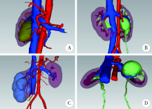
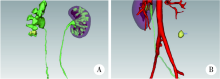
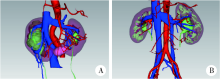
目的: 探讨术前三维影像重建技术在治疗肾盂输尿管连接部梗阻(ureteropelvic junction obstruction,UPJO)中的应用价值。方法: 选择2017年5月至2019年4月北京大学第一医院收治的UPJO患者病例资料进行回顾性分析,共收集病例40例,其中男性22例,女性18例,中位年龄26.5 岁(四分位距23.25~38.75) 岁。40例中11例合并异位血管,14例合并肾结石,3例合并马蹄肾,肾盂成形术后再梗阻6例。40例患者均在术前对泌尿系统增强CT进行三维影像模型重构,观察肾盂输尿管连接部梗阻位置,以及梗阻处与血管及周围组织器官的关系,依据三维重构影像结果制定手术计划。37例患者行腹腔镜下肾盂成形术(其中3例联合软膀胱镜取石,1例联合孙氏镜取石,1例联合输尿管切断再吻合术,3例联合马蹄肾成形术), 2例行腹腔镜下舌黏膜补片法输尿管成形术,1例行机器人辅助腹腔镜下肾盂成形术。结果: 术前泌尿系统增强CT图像经三维重建后,清晰显示了肾盂输尿管连接部梗阻处与血管及周围组织器官的关系,术者依据三维重建模型可于术前清晰地观察异位血管的类型、直径、位置及走形方向,评估肾结石或其他占位的数量、大小、位置和形态,累及的肾盏数,以及在肾盂、肾盏中的解剖分布,综合分析上述信息后,对所有患者施行个体化的手术方案,40例患者手术均顺利完成,无中转开放手术者。平均手术时间 (129.91±37.90) min (75~273 min),平均出血量 (48.1±78.0) mL (10~400 mL),平均住院时间(5.04±1.99) d (2~10 d), 平均术后拔除引流时间 (3.8±1.4) d (2~8 d)。结论: 三维影像重建技术在UPJO治疗方面具有较高的临床应用价值,对辅助术者拟定手术方案具有指导意义,值得进一步临床推广及应用。
中图分类号:
- R691.2


| [1] | 黄强, 杨骥, 林先盛, 等. CT三维重建在肝门部胆管癌的诊疗中的应用价值[J]. 中国普通外科杂志, 2017,26(8):960-967. |
| [2] | 中华医学会数字医学分会, 中国研究型医院学会数字医学临床外科专业委员会. 复杂性肝脏肿瘤三维可视化精准诊治专家共识[J]. 中国实用外科杂志, 2017,37(1):53-59. |
| [3] | 李学松, 杨昆霖, 周利群. IUPU经腹腹腔镜肾盂成型术治疗成人肾盂输尿管连接处梗阻(附视频)[J]. 现代泌尿外科杂志, 2015,20(6):369-372. |
| [4] |
Yang K, Yao L, Li X, et al. A modified suture technique for transperitoneal laparoscopic dismembered pyeloplasty of pelviureteric junction obstruction[J]. Urology, 2015,85(1):263-267.
doi: 10.1016/j.urology.2014.09.031 pmid: 25530399 |
| [5] | Klingler HC, Remzi M, Janetschek G, et al. Comparison of open versus laparoscopic pyeloplasty techniques in treatment of uretero-pelvic junction obstruction[J]. Eur Urol, 2003,44(3):340-345. |
| [6] | 贾晨尧, 陈柯, 刘奇, 等. 基于CT的肾脏可视化三维重建模型在肾蒂血管变异的肾癌根治术中的应用[J]. 广东医学, 2017,38(9):81-84. |
| [7] |
Wang Z, Qi L, Yuan P, et al. Application of three-dimensional visualization technology in laparoscopic partial nephrectomy of renal tumor: a comparative study[J]. J Laparoendosc Adv Surg Tech A, 2017,27(5):516-523.
pmid: 28186431 |
| [8] |
Tolkach Y, Thomann S, Kristiansen G. Three-dimensional reconstruction of prostate cancer architecture with serial immunohistochemical sections: hallmarks of tumour growth, tumour compartmentalisation, and implications for grading and heterogeneity[J]. Histopathology, 2018,72(6):1051-1059.
pmid: 29323728 |
| [9] | Rydberg J, Kopecky KK, Tann M, et al. Evaluation of prospective living renal donors for laparoscopic nephrectomy with multisection CT: the marriage of minimally invasive imaging with minimally invasive surgery[J]. Radiographics, 2001,21(Supp1 1):S223-S236. |
| [10] |
Ukimura O. Image-guided surgery in minimally invasive urology[J]. Curr Opin Urol, 2010,20(2):136-140.
doi: 10.1097/MOU.0b013e3283362610 |
| [11] | 赵海岳, 叶雄俊, 陈伟男, 等. 腹腔镜肾盂成型术中异位血管的处理方法[J]. 北京大学学报(医学版), 2019,51(4):660-664. |
| [12] |
Berkman DS, Landman J, Gupta M. Treatment outcomes after endopyelotomy performed with or without simultaneous nephrolithotomy: 10-year experience[J]. J Endourol, 2009,23(9):1409-1413.
doi: 10.1089/end.2009.0379 pmid: 19694529 |
| [13] | 陈远波, 李虎林, 刘春晓, 等. 数字化肾结石三维模型的建立及虚拟手术仿真[J]. 南方医科大学学报, 2013,33(2):267-270. |
| [14] |
Li HL, Chen YB, Liu C, et al. Construction of a three-dimensional model of renal stones: comprehensive planning for percutaneous nephrolithotomy and assistance in surgery[J]. World J Urol, 2013,31(6):1587-1592.
doi: 10.1007/s00345-012-0998-7 pmid: 23223963 |
| [15] | 杨昆霖, 李学松, 周利群. IUPU经腹腹腔镜联合膀胱软镜肾盂取石及肾盂成型术[J]. 泌尿外科杂志: 电子版, 2015,7(3):4-6. |
| [16] | Simonato A, Gregori A, Lissiani A, et al. The tongue as an alternative donor site for graft urethroplasty: a pilot study[J]. J Urol, 2006,175(2):589-592. |
| [17] |
Singh PB, Das SK, Kumar A, et al. Dorsal onlay lingual mucosal graft urethroplasty: comparison of two techniques[J]. Int J Urol, 2010,15(11):1002-1005.
pmid: 18808427 |
| [18] | Xu YM, Feng C, Sa YL, et al. Outcome of 1-stage urethroplasty using oral mucosal grafts for the treatment of urethral strictures associated with genital lichen sclerosus[J]. Urology, 2014,83(1):232-236. |
| [19] |
Li B, Xu YJ, Hai B, et al. Laparoscopic onlay lingual mucosal graft ureteroplasty for proximal ureteral stricture: initial experience and 9-month follow-up[J]. Int Urol Nephrol, 2016,48(8):1275-1279.
doi: 10.1007/s11255-016-1289-9 pmid: 27115158 |
| [1] | 安立哲,熊六林,陈亮,王焕瑞,陈伟男,黄晓波. 腹腔镜肾盂成形术联合肾盂镜超声碎石取石术治疗肾盂输尿管连接部梗阻合并肾结石[J]. 北京大学学报(医学版), 2022, 54(4): 746-750. |
| [2] | 熊盛炜,王杰,朱伟杰,程嗣达,张雷,李学松,周利群. 二次肾盂成形术在复发性肾盂输尿管连接部梗阻中的研究进展[J]. 北京大学学报(医学版), 2020, 52(4): 794-798. |
| [3] | 陈伟男,叶雄俊,刘士军,熊六林,黄晓波,徐涛,王晓峰. 三种手术方式治疗肾盂输尿管连接部梗阻的疗效及并发症比较[J]. 北京大学学报(医学版), 2016, 48(5): 817-821. |
|
||
