北京大学学报(医学版) ›› 2023, Vol. 55 ›› Issue (1): 22-29. doi: 10.19723/j.issn.1671-167X.2023.01.004
三种方法建立大鼠种植体周炎模型的比较
孟令玮1,李雪2,高胜寒1,李悦1,曹瑞涛1,张毅2,*( ),潘韶霞1,*()
- 1. 北京大学口腔医学院·口腔医院修复科,国家口腔医学中心,国家口腔疾病临床医学研究中心,口腔生物材料和数字诊疗装备国家工程研究中心,口腔数字医学北京市重点实验室,国家卫生健康委员会口腔医学计算机应用工程技术研究中心,国家药品监督管理局口腔生物材料重点实验室,北京 100081
2. 军事科学院军事医学研究院辐射医学研究所,北京 100850
Comparison of three methods for establishing rat peri-implantitis model
Ling-wei MENG1,Xue LI2,Sheng-han GAO1,Yue LI1,Rui-tao CAO1,Yi ZHANG2,*(),Shao-xia PAN1,*()
- 1. Department of Prosthodontics, Peking University School and Hospital of Stomatology & National Center of Stomatology & National Clinical Research Center for Oral Diseases & National Engineering Research Center of Oral Biomaterials and Digi-tal Medical Devices & Beijing Key Laboratory of Digital Stomatology & NHC Research Center of Engineering and Technology for Computerized Dentistry & NMPA Key Laboratory for Dental Materials, Beijing 100081, China
2. Institute of Radiation Medicine, Academy of Military Medical Sciences, Beijing 100850, China
摘要:
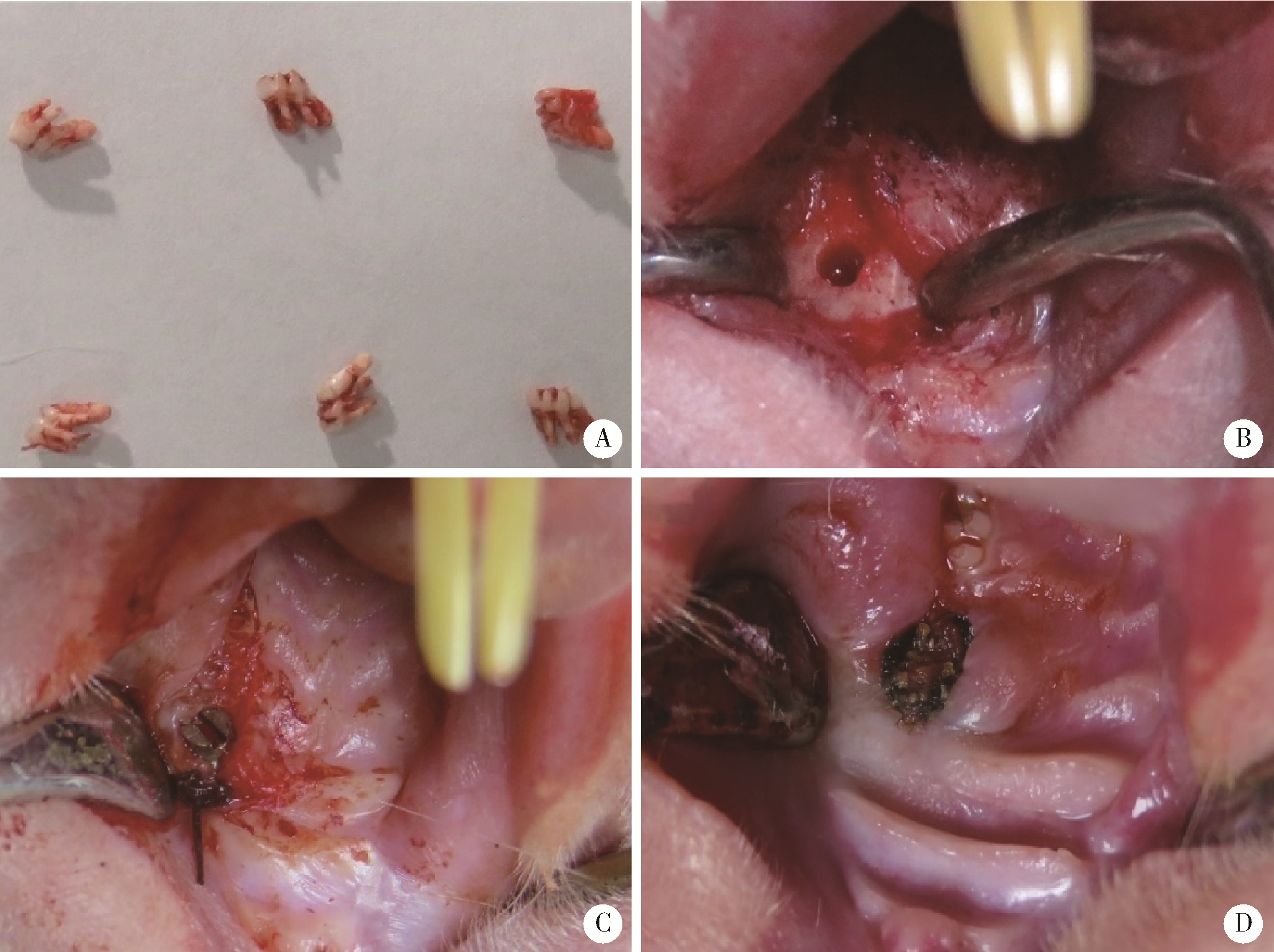
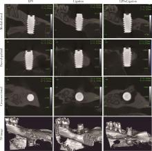
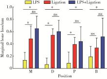
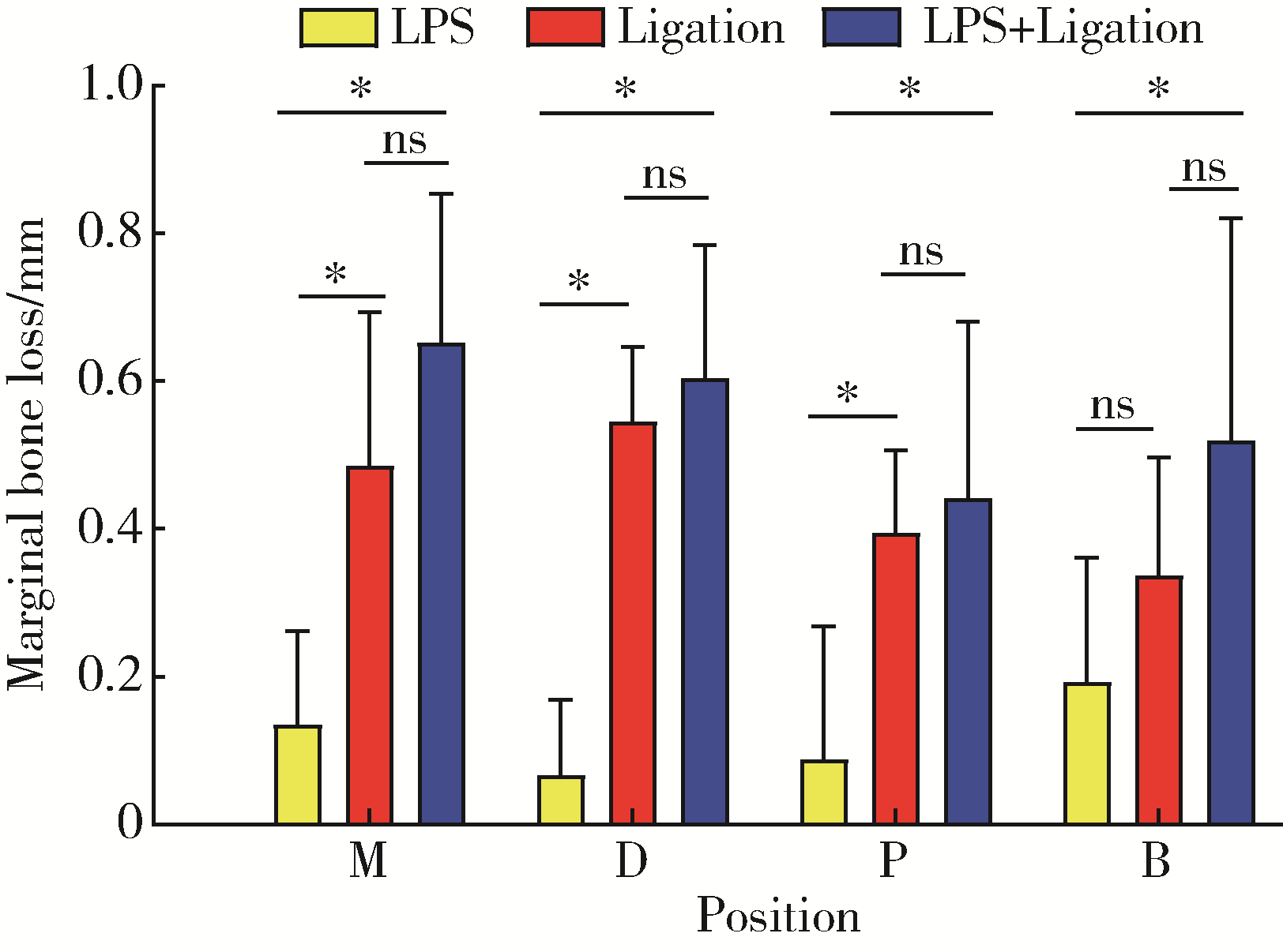
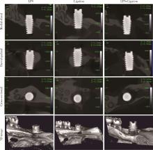
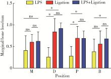
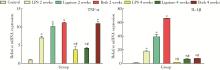
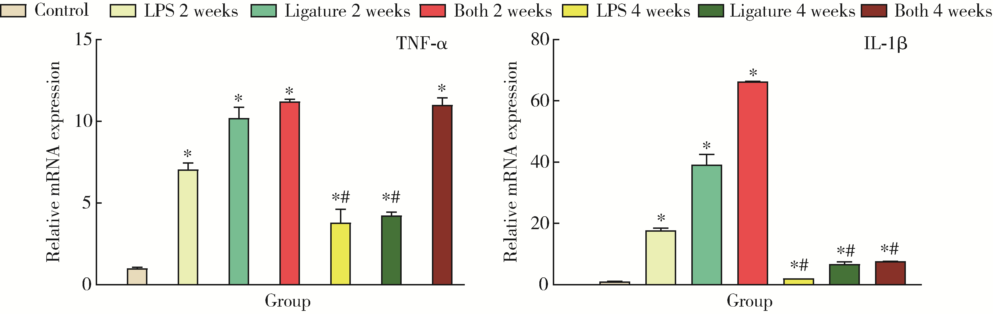
目的: 比较传统棉线结扎与种植体周围局部注射牙龈卟啉单胞菌脂多糖(lipopolysaccharide,LPS)诱导法,以及两种方法联合建立大鼠种植体周炎模型的效率和效果,以探索大鼠种植体周炎模型建模的最佳方法。方法: 纳入39只雄性SD大鼠,拔除上颌左侧第一磨牙,拔牙窝愈合4周后植入钛种植体。种植体植入4周骨结合后,将39只大鼠随机分为4组,分别采取棉线结扎(n=12)、种植体周围局部注射LPS(n=12),以及两种方法联合(n=12)诱导大鼠种植体周炎,同时设无任何处理对照组(n=3)。记录实验组大鼠诱导前及诱导后2周和4周时的探诊深度(probing depth,PD)、探诊出血(bleeding on probing,BOP)和牙龈指数(gingival index,GI)。收集种植体周围牙龈组织,提取RNA,实时荧光定量分析(real time quantitative PCR,RT-qPCR)检测炎症因子白细胞介素-1β(interleukin-1β,IL-1β)和肿瘤坏死因子-α(tumor necrosis factor-alpha,TNF-α)表达。分别于诱导2周和4周时处死实验组动物,收集上颌骨,采用微型CT(micro-CT)观察种植体边缘骨吸收状况。结果: 种植4周后,种植体骨结合良好。3个实验组均可见牙龈红肿,质软,种植体边缘骨吸收。诱导2周和4周时,各实验组PD、GI和BOP均较诱导前增加,但仅BOP在3组间差异有统计学意义(P < 0.05)。炎症诱导2周时,棉线结扎组和联合组在每个位点均可见边缘骨吸收,且联合组每个位点边缘骨吸收程度均大于棉线结扎组,但差异不具有统计学意义(P > 0.05);局部注射组部分种植体的部分位点可见边缘骨吸收。炎症诱导4周时,各实验组各位点均可见边缘骨吸收,其中棉线结扎组和联合组边缘骨吸收大于局部注射组,差异有统计学意义(P < 0.05)。诱导2周和4周时,实验组TNF-α和IL-1β均较对照组表达增加,差异有统计学意义(P < 0.05)。结论: 相比于种植体周围局部注射LPS,棉线结扎及两种方法联合可以更好且更快地诱导大鼠种植体周炎。
中图分类号:
- R781.42


| 1 | Schwarz F , Alcoforado G , Guerrero A , et al. Peri-implantitis: Summary and consensus statements of group 3. The 6th EAO Consensus Conference 2021[J]. Clin Oral Implants Res, 2021, 32 (Suppl 21): 245- 253. |
| 2 | Schliephake H , Sicilia A , Nawas BA , et al. Drugs and diseases: Summary and consensus statements of group 1. The 5 th EAO Consensus Conference 2018[J]. Clin Oral Implants Res, 2018, 29 (Suppl 18): 93- 99. |
| 3 | Roccuzzo M , Layton DM , Roccuzzo A , et al. Clinical outcomes of peri-implantitis treatment and supportive care: A systematic review[J]. Clin Oral Implants Res, 2018, 29 (Suppl 16): 331- 350. |
| 4 |
Fu JH , Wang HL . Breaking the wave of peri-implantitis[J]. Periodontol 2000, 2020, 84 (1): 145- 160.
doi: 10.1111/prd.12335 |
| 5 |
Wancket LM . Animal models for evaluation of bone implants and devices: Comparative bone structure and common model uses[J]. Vet Pathol, 2015, 52 (5): 842- 850.
doi: 10.1177/0300985815593124 |
| 6 |
Schwarz F , Sculean A , Engebretson SP , et al. Animal models for peri-implant mucositis and peri-implantitis[J]. Periodontol 2000, 2015, 68 (1): 168- 181.
doi: 10.1111/prd.12064 |
| 7 |
Kantarci A , Hasturk H , Van Dyke TE . Animal models for periodontal regeneration and peri-implant responses[J]. Periodontol 2000, 2015, 68 (1): 66- 82.
doi: 10.1111/prd.12052 |
| 8 | 朱白雪, 高晓蔚, 戴晓玮. 种植体周围炎动物模型的研究进展[J]. 口腔医学研究, 2018, 38 (8): 747- 751. |
| 9 |
Kensara A , Hefni E , Williams MA , et al. Microbiological profile and human immune response associated with peri-implantitis: A systematic review[J]. J Prosthodont, 2021, 30 (3): 210- 234.
doi: 10.1111/jopr.13270 |
| 10 |
Sun J , Eberhard J , Glage S , et al. Development of a peri-implantitis model in the rat[J]. Clin Oral Implants Res, 2020, 31 (3): 203- 214.
doi: 10.1111/clr.13556 |
| 11 | 杨少强, 廖旭辉, 顾为望, 等. 牙周炎犬种植体周围炎动物模型建立[J]. 口腔医学研究, 2011, 31 (12): 746- 750. |
| 12 | Miyamoto Y , Koretake K , Hirata M , et al. Influence of static overload on the bony interface around implants in dogs[J]. Int J Prosthodont, 2008, 21 (5): 437- 444. |
| 13 | 李星佳, 陈琪欣, 袁长永, 等. 种植体周围炎大鼠模型研究[J]. 口腔医学研究, 2021, 37 (4): 314- 318. |
| 14 |
Reinedahl D , Chrcanovic B , Albrektsson T , et al. Ligature-induced experimental peri-implantitis: A systematic review[J]. J Clin Med, 2018, 7 (12): 492.
doi: 10.3390/jcm7120492 |
| 15 |
Deng S , Hu Y , Zhou J , et al. TLR4 mediates alveolar bone resorption in experimental peri-implantitis through regulation of CD45(+) cell infiltration, RANKL/OPG ratio, and inflammatory cytokine production[J]. J Periodontol, 2020, 91 (5): 671- 682.
doi: 10.1002/JPER.18-0748 |
| 16 |
吴亚菲, 赵筱芩, 陈宇, 等. 不同方法建立大鼠实验性牙周炎模型的比较研究[J]. 四川大学学报(医学版), 2003, 34 (4): 742- 745.
doi: 10.3969/j.issn.1672-173X.2003.04.041 |
| 17 | He Q , Mu Z , Shrestha A , et al. Development of a rat model for type 2 diabetes mellitus peri-implantitis: A preliminary study[J]. Oral Dis, 2021, 28 (7): 1936- 1946. |
| 18 | Orecchioni M , Ghosheh Y , Pramod AB , et al. Macrophage polarization: Different gene signatures in M1(LPS+) vs. classically and M2(LPS-) vs. alternatively activated macrophages[J]. Front Immunol, 2019, 10, 1084. |
| 19 | 柯晓菁, 李厚轩, 闫福华, 等. 牙龈卟啉单胞菌脂多糖对骨髓来源的巨噬细胞和破骨细胞先天免疫反应的影响[J]. 中华口腔医学杂志, 2020, 55 (1): 32- 37. |
| 20 | Park SY , Kim KH , Rhee SH , et al. An immediate peri-implantitis induction model to study regenerative peri-implantitis treatments[J]. Clin Oral Implants Res, 2017, 28 (1): 36- 42. |
| 21 | Hasani-Sadrabadi MM , Sarrion P , Pouraghaei S , et al. An engineered cell-laden adhesive hydrogel promotes craniofacial bone tissue regeneration in rats[J]. Sci Transl Med, 2020, 12 (534): eaay6853. |
| [1] | 孙菲,刘建,李思琪,危伊萍,胡文杰,王翠. 种植体黏膜下微生物在健康种植体和种植体周炎中的构成与差异:一项横断面研究[J]. 北京大学学报(医学版), 2023, 55(1): 30-37. |
| [2] | 朱琳,张维宇,许克新. 环磷酰胺诱导SD大鼠膀胱疼痛综合征模型的有效性[J]. 北京大学学报(医学版), 2022, 54(4): 735-740. |
| [3] | 何伟,杨思雯,陈娟,朱晓俊,陈志忠,马文军. 275 nm和310 nm紫外线对去卵巢骨质疏松大鼠骨代谢的影响[J]. 北京大学学报(医学版), 2022, 54(2): 236-243. |
| [4] | 孙菲,李思琪,危伊萍,钟金晟,王翠,胡文杰. 种植体周病非手术治疗中联合应用甘氨酸粉喷砂的临床效果评价[J]. 北京大学学报(医学版), 2022, 54(1): 119-125. |
| [5] | 陈章健,韩硕,郑湃,贾光. 锐钛矿型纳米二氧化钛经口暴露90天对Sprague-Dawley大鼠血常规指标的影响[J]. 北京大学学报(医学版), 2021, 53(6): 1205-1208. |
| [6] | 王贵红,左婷,李然,左正才. 瑞巴派特在大鼠痛风性关节炎急性发作中的作用[J]. 北京大学学报(医学版), 2021, 53(4): 716-720. |
| [7] | 尹雪倩, 张晓玄, 文婧, 刘思奇, 刘欣然, 周若宇, 王军波. 荞麦、燕麦、豌豆复配对糖尿病大鼠血糖的影响[J]. 北京大学学报(医学版), 2021, 53(3): 447-452. |
| [8] | 白枫,何倚帆,牛亚楠,杨若娟,曹静. 超细颗粒物对大鼠离体灌注心脏功能的影响[J]. 北京大学学报(医学版), 2021, 53(2): 240-245. |
| [9] | 周迪,陈章健,胡贵平,阎腾龙,龙昌茂,冯慧敏,贾光. 纳米二氧化钛亚急性经口暴露对大鼠氧化/抗氧化生物标志和炎性因子的影响[J]. 北京大学学报(医学版), 2020, 52(5): 821-827. |
| [10] | 陈章健,韩硕,郑湃,周淑佩,贾光. 纳米二氧化钛与葡萄糖亚慢性联合经口暴露对幼年大鼠血清叶酸和维生素B12水平的影响[J]. 北京大学学报(医学版), 2020, 52(3): 451-456. |
| [11] | 韩硕,陈章健,周迪,郑湃,张家赫,贾光. 纳米二氧化钛经口暴露90天对大鼠粪便代谢组的影响[J]. 北京大学学报(医学版), 2020, 52(3): 457-463. |
| [12] | 白珊珊,莫思怡,徐啸翔,刘云,谢秋菲,曹烨. 大鼠咬合干扰致口颌面痛敏的自我赏罚实验行为学特点[J]. 北京大学学报(医学版), 2020, 52(1): 51-57. |
| [13] | 何姣,袁戈恒,张俊清,郭晓蕙. 早期糖尿病周围神经病变大鼠模型的建立[J]. 北京大学学报(医学版), 2019, 51(6): 1150-1154. |
| [14] | 王伟,侯进,黄文强. 运动导致兴奋脑区组织液流动一过性加速[J]. 北京大学学报(医学版), 2019, 51(2): 206-209. |
| [15] | 王玉洁,郭向阳,王军. 重复异丙酚麻醉对新生大鼠海马细胞凋亡及远期学习记忆能力的影响[J]. 北京大学学报(医学版), 2017, 49(2): 310-314. |
|
