北京大学学报(医学版) ›› 2026, Vol. 58 ›› Issue (1): 214-219. doi: 10.19723/j.issn.1671-167X.2026.01.029

• 病例报告 • 上一篇    下一篇

上颌第二磨牙慢性根尖周炎合并器械分离的根管再治疗1例

臧海玲1, 梁宇红2,*()   

  1. 1. 北京大学国际医院口腔科, 北京 102206
    2. 北京大学口腔医学院·口腔医院急诊科, 国家口腔医学中心, 国家口腔疾病临床医学研究中心, 口腔生物材料和数字诊疗装备国家工程研究中心, 口腔数字医学北京市重点实验室, 国家卫生健康委员会口腔医学计算机应用工程技术研究中心, 国家药品监督管理局口腔生物材料重点实验室, 北京 100081
  • 收稿日期:2025-10-13 出版日期:2026-02-18 发布日期:2026-01-05
  • 通讯作者: 梁宇红

Endodontic retreatment of a maxillary second molar with chronic apical periodontitis and separated instrument: A case report

Hailing ZANG1, Yuhong LIANG2,*()   

  1. 1. Department of Stomatology, Peking University International Hospital, Beijing 102206, China
    2. Department of Oral Emergency, Peking University School and Hospital of Stomatology & National Center for Stomatology & National Clinical Research Center for Oral Diseases & National Engineering Research Center of Oral Biomaterials and Digital Medical Devices & Beijing Key Laboratory of Digital Stomatology & NHC Research Center of Engineering and Technology for Computerized Dentistry & NMPA Key Laboratory for Dental Materials, Beijing 100081, China
  • Received:2025-10-13 Online:2026-02-18 Published:2026-01-05
  • Contact: Yuhong LIANG

RICH HTML


摘要:

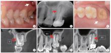
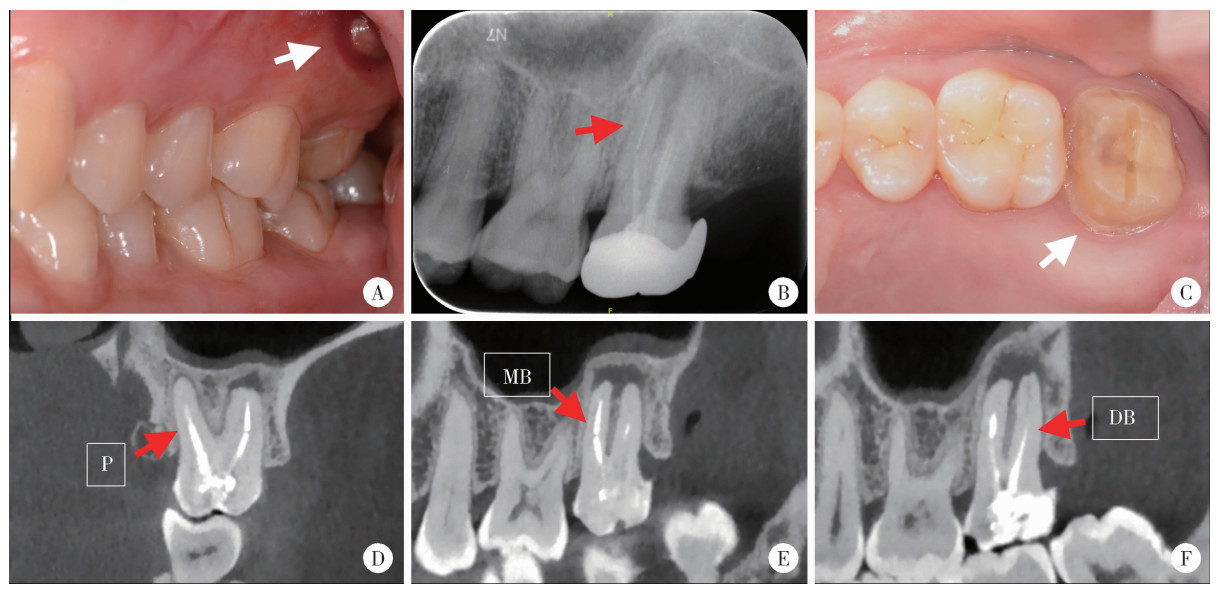
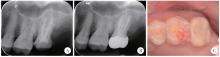
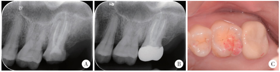
本文报告1例成功处理涉及器械分离、纤维桩取出、医源性根管穿孔及慢性根尖周炎等复杂情况的左上第二磨牙(27)显微根管再治疗病例。患者主诉左上后牙反复牙龈肿包半年, 该患牙曾于两年前行根管治疗及桩核冠修复。临床检查发现颊侧根尖区牙龈存在窦道; 锥形束CT(cone-beam CT, CBCT)检查发现近中颊根根尖区存在一长度约5 mm的金属分离器械, 腭根内有一纤维桩延伸至根管中段, 近中颊根、远中颊根和腭根根管内充填物均存在明显欠填, 且根尖周均可见低密度影像。诊断为27慢性根尖周炎(牙髓治疗后), 治疗计划为27显微根管再治疗。橡皮障隔离, 在牙科显微镜下, 利用超声技术取出了腭根内的纤维桩及近中颊根分离器械的上段(2.5 mm)。再次探查根管时发现近中颊根根管冠1/3处的医源性穿孔。鉴于分离器械下段位置深在, 较难取出, 评估风险和获益后, 治疗策略调整为旁路通过, 最终成功绕过器械并疏通根管至工作长度。经过彻底的化学-机械预备后, 采用热牙胶垂直加压技术进行三维充填, 并使用iRoot BP生物陶瓷材料对穿孔部位进行严密修补, 完成患牙全冠修复。术后10个月随访时, 患者无临床症状, 影像学检查示根尖周病变愈合良好。本病例表明, 综合运用CBCT、牙科显微镜、超声技术和生物陶瓷材料, 并采取保守而灵活的治疗策略, 能够提高多重并发症根管再治疗病例的成功率。

关键词: 根管再治疗, 器械分离, 根管穿孔, 旁路通过, 生物陶瓷

Abstract:

This case report describes the micro-endodontic retreatment and comprehensive management of a complex left maxillary second molar presenting with multiple complications. The patient, a 41-year-old female, presented with recurrent gingival swelling associated with the tooth for more than six months. The tooth had a history of root canal treatment, fiber post placement, and full crown restoration two years earlier. Cone-beam CT (CBCT) revealed a separated metallic instrument approximately 5 mm in length in the apical region of the mesiobuccal root, a fiber post in the palatal root, underfilled root canal obturation, and periapical radiolucency involving the mesiobuccal, distobuccal and palatal roots. A diagnosis of chronic periapical periodontitis (post-endodontic treatment) was established. The primary challenges included the limited access to the left maxillary second molar, the deeply embedded apical instrument segment, and the high risk for complications, such as root fracture or secondary perforation during retrieval attempts. The treatment strategy involved removal of the separated instrument and retreatment under a dental operating microscope. With ultrasonic assistance, the palatal fiber post and the coronal segment (2.5 mm) of the separated instrument from the mesiobuccal canal were successfully removed. An iatrogenic lateral perforation occurred in the mesiobuccal canal near the furcation area during the instrument retrieval procedure. Given the depth and high risk of further damage from retrieving the remaining apical segment (2 mm), a conservative and flexible approach was adopted using the bypass technique. After successful bypassing, thorough chemo-mechanical debridement was performed, followed by obturation using the warm vertical compaction technique. The iatrogenic perforation on the distal wall of the mesiobuccal canal was immediately repaired with a bioceramic material (iRoot BP) for its superior sealing and bioactivity. The tooth was then restored with a full crown. A 10-month follow-up showed that the patient was asymptomatic with complete fistula healing and radiographic examination revealed favorable healing of the periapical lesion. This case demonstrates that, in complex root canal retreatment cases involving retained instruments and iatrogenic perforations, the integration of advanced diagnostic tools (e. g. CBCT), microsurgical techniques (e. g. microscope and ultrasound), and modern restorative materials (e. g. bioceramics), along with a dynamic strategy combining instrument retrieval and bypassing, is essential for achieving predictable long-term success.

Key words: Root canal retreatment, Instrument separation, Perforation, Bypass, Bioceramics

中图分类号: 

  • R781.3

图1

左侧上颌第二磨牙术前口内照及影像学检查"

图2

左侧上颌第二磨牙器械取出、侧穿修补、根管充填图像及根尖片"

图3

左侧上颌第二磨牙复查根尖片及口内照"

1
岳林, 王晓燕. 牙体牙髓病学[M]. 3版 北京: 北京大学医学出版社, 2022: 407.
2
Ng YL , Mann V , Gulabivala K . A prospective study of the factors affecting outcomes of nonsurgical root canal treatment: Part 1:Periapical health[J]. Int Endod J, 2011, 44 (7): 583- 609.

doi: 10.1111/j.1365-2591.2011.01872.x
3
Gulabivala K , Ng YL . Factors that affect the outcomes of root canal treatment and retreatment: A reframing of the principles[J]. Int Endod J, 2023, 56 (Suppl 2): 82- 115.
4
Terauchi Y , Sexton C , Bakland LK , et al. Factors affecting the removal time of separated instruments[J]. J Endod, 2021, 47 (8): 1245- 1252.

doi: 10.1016/j.joen.2021.05.003
5
Terauchi Y , Ali WT , Abielhassan MM . Present status and future directions: Removal of fractured instruments[J]. Int Endod J, 2022, 55 (Suppl 3): 685- 709.
6
Fan Y , Gao Y , Wang X , et al. Expert consensus on management of instrument separation in root canal therapy[J]. Int J Oral Sci, 2025, 17 (1): 46.

doi: 10.1038/s41368-025-00372-w
7
Patel S , Brown J , Semper M , et al. European Society of Endodontology position statement: Use of cone beam computed tomography in Endodontics: European Society of Endodontology (ESE) deve-loped by[J]. Int Endod J, 2019, 52 (12): 1675- 1678.

doi: 10.1111/iej.13187
8
Baratto-Filho F , Vavassori de Freitas J , Fagundes Tomazinho FS , et al. Cone-beam computed tomography detection of separated endodontic instruments[J]. J Endod, 2020, 46 (11): 1776- 1781.

doi: 10.1016/j.joen.2020.08.011
9
Cujé J , Bargholz C , Hülsmann M . The outcome of retained instrument removal in a specialist practice[J]. Int Endod J, 2010, 43 (7): 545- 554.

doi: 10.1111/j.1365-2591.2009.01652.x
10
Gorni FG , Ionescu AC , Ambrogi F , et al. Prognostic factors and primary healing on root perforation repaired with MTA: A 14-year longitudinal study[J]. J Endod, 2022, 48 (9): 1092- 1099.

doi: 10.1016/j.joen.2022.06.005
11
Camilleri J , Atmeh A , Li X , et al. Present status and future directions: Hydraulic materials for endodontic use[J]. Int Endod J, 2022, 55 (Suppl 3): 710- 777.
12
Liang YH , Jiang L , Gao XJ , et al. Detection and measurement of artificial periapical lesions by cone-beam computed tomography[J]. Int Endod J, 2014, 47 (4): 332- 338.

doi: 10.1111/iej.12148
13
梁宇红, 岳林. 锥形束CT在牙髓根尖周病诊治中的合理应用与思考[J]. 中华口腔医学杂志, 2019, 54 (9): 591- 597.
14
Madarati AA , Hunter MJ , Dummer PM . Management of intracanal separated instruments[J]. J Endod, 2013, 39 (5): 569- 581.

doi: 10.1016/j.joen.2012.12.033
15
McGuigan MB , Louca C , Duncan HF . Endodontic instrument fracture: Causes and prevention[J]. Br Dent J, 2013, 214 (7): 341- 348.

doi: 10.1038/sj.bdj.2013.324
16
Spili P , Parashos P , Messer HH . The impact of instrument fracture on outcome of endodontic treatment[J]. J Endod, 2005, 31 (12): 845- 850.

doi: 10.1097/01.don.0000164127.62864.7c
17
Suter B , Lussi A , Sequeira P . Probability of removing fractured instruments from root canals[J]. Int Endod J, 2005, 38 (2): 112- 123.

doi: 10.1111/j.1365-2591.2004.00916.x
18
Al-Fouzan KS . Incidence of rotary ProFile instrument fracture and the potential for bypassing in vivo[J]. Int Endod J, 2003, 36 (12): 864- 867.

doi: 10.1111/j.1365-2591.2003.00733.x
19
Ordinola-Zapata R , Martins JNR , Versiani MA , et al. Micro-CT analysis of danger zone thickness in the mesiobuccal roots of maxillary first molars[J]. Int Endod J, 2019, 52 (4): 524- 529.

doi: 10.1111/iej.13025
20
Clauder T . Present status and future directions: Managing perforations[J]. Int Endod J, 2022, 55 (Suppl 4): 872- 891.
21
Yang N , Yang W , Shen R , et al. In vitro and in vivo evaluation of iRoot BP Plus as a coronal sealing material for regenerative endodontic procedures[J]. Clin Oral Investig, 2024, 28 (1): 70.

doi: 10.1007/s00784-023-05468-3
22
Dong X , Xu X . Bioceramics in endodontics: Updates and future perspectives[J]. Bioengineering, 2023, 10 (3): 354.

doi: 10.3390/bioengineering10030354
23
Abdelmotelb MA , Gomaa YF , Khattab NMA , et al. Premixed bioceramics versus mineral trioxide aggregate in furcal perforation repair of primary molars: In vitro and in vivo study[J]. Clin Oral Investig, 2021, 25 (8): 4915- 4925.

doi: 10.1007/s00784-021-03800-3
[1] 雍颹,钱锟,朱文昊,赵晓一,刘畅,潘洁. 成年恒牙牙髓切断后牙髓钙化的X线片评价[J]. 北京大学学报(医学版), 2023, 55(1): 88-93.
[2] 王爽,彭楚芳,刘鹤. 新型生物陶瓷材料用于乳磨牙牙髓切断术的临床疗效[J]. 北京大学学报(医学版), 2022, 54(6): 1196-1201.
[3] 钱锟,潘洁,朱文昊,赵晓一,刘畅,雍颹. 两种硅酸钙类材料用于成熟恒牙牙髓切断术的临床效果[J]. 北京大学学报(医学版), 2022, 54(1): 113-118.
[4] 雷玥,杨颖婷,战园. 生物陶瓷材料在乳牙牙髓切断术中的应用[J]. 北京大学学报(医学版), 2019, 51(1): 70-74.
Viewed
Full text


Abstract

Cited

  Shared   
  Discussed   
No Suggested Reading articles found!