北京大学学报(医学版) ›› 2023, Vol. 55 ›› Issue (1): 156-159. doi: 10.19723/j.issn.1671-167X.2023.01.024
腓骨远端撕脱骨折的影像学诊断:踝关节X线与CT三维重建的比较
- 北京大学第三医院运动医学科,北京大学运动医学研究所,运动医学关节伤病北京市重点实验室,北京 100191
Radiographic diagnosis of distal fibula avulsion fractures: Comparison of ankle X-ray and three-dimensional reconstruction of CT
Shi-kai XIONG,Wei-li SHI,An-hong WANG,Xing XIE,Qin-wei GUO*( )
)
- Department of Sports Medicine, Peking University Third Hospital; Institute of Sports Medicine, Peking University; Beijing Key Laboratory of Joint Injuries in Sports Medicine; Beijing 100191, China
摘要:
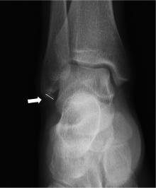
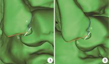
目的: 探讨X线检查、CT三维重建(three dimensional reconstruction of computed tomography, 3D-CT)诊断腓骨远端撕脱骨折的敏感度差异,分析撕脱骨折块的影像学表现。方法: 收集2018年1—10月就诊于北京大学第三医院运动医学科行外踝韧带止点重建术的92例腓骨远端撕脱骨折的患者,根据纳入和排除标准,最终入组60例。将术中诊断作为金标准,统计术前踝关节正侧位X线检查以及3D-CT对腓骨远端撕脱骨折的诊断敏感度,并测量骨块最大径以及移位程度。在3D-CT上,用骨块中心点至腓骨前结节的距离(a)和至腓骨尖的距离(b)的比值(a/b值)来表示骨块位移程度。结果: 60例患者中,术前踝关节正侧位X线检查和3D-CT的阳性诊断例数分别为36例和52例,敏感度分别为60.0%和86.7%(P=0.004)。X线检查和3D-CT上撕脱骨块的平均直径分别为(9.2±3.9) mm和(10.5±3.2) mm。撕脱骨块中心点至腓骨前结节的平均距离(a)为(17.5±3.6) mm,至腓骨尖的平均距离(b)为(17.4±4.8) mm,a/b值平均为1.1±0.7。各项测量结果的组内相关系数(intraclass correlation efficient, ICC)范围为0.891~0.998,具有高度一致性。结论: 与X线检查相比,3D-CT诊断腓骨远端撕脱骨折具有更高的敏感度,可准确定位,便于手术方案的制定,建议对于临床上腓骨远端可疑撕脱骨折的病例应该进行3D-CT检查。
中图分类号:
- R683.42
| 1 |
Waterman BR , Owens BD , Davey S , et al. The epidemiology of ankle sprains in the United States[J]. J Bone Joint Surg Am, 2010, 92 (13): 2279- 2284.
doi: 10.2106/JBJS.I.01537 |
| 2 |
Milgrom C , Shlamkovitch N , Finestone A , et al. Risk factors for lateral ankle sprain: A prospective study among military recruits[J]. Foot Ankle, 1991, 12 (1): 26- 30.
doi: 10.1177/107110079101200105 |
| 3 |
Bridgman SA , Clement D , Downing A , et al. Population based epidemiology of ankle sprains attending accident and emergency units in the West Midlands of England, and a survey of UK practice for severe ankle sprains[J]. Emerg Med J, 2003, 20 (6): 508- 510.
doi: 10.1136/emj.20.6.508 |
| 4 |
Cordova ML , Sefton JM , Hubbard TJ . Mechanical joint laxity associated with chronic ankle instability: A systematic review[J]. Sports Health, 2010, 2 (6): 452- 459.
doi: 10.1177/1941738110382392 |
| 5 |
Reiner MM , Sharpe JJ . The role of the accessory malleolar ossicles and malleolar avulsion fractures in lateral ankle ligament reconstruction[J]. Foot Ankle Spec, 2018, 11 (4): 308- 314.
doi: 10.1177/1938640017729498 |
| 6 |
Kim BS , Choi WJ , Kim YS , et al. The effect of an ossicle of the lateral malleolus on ligament reconstruction of chronic lateral ankle instability[J]. Foot Ankle Int, 2010, 31 (3): 191- 196.
doi: 10.3113/FAI.2010.0191 |
| 7 |
Choi WJ , Lee JW , Han SH , et al. Chronic lateral ankle instability: The effect of intra-articular lesions on clinical outcome[J]. Am J Sports Med, 2008, 36 (11): 2167- 2172.
doi: 10.1177/0363546508319050 |
| 8 |
Chun TH , Park YS , Sung KS . The effect of ossicle resection in the lateral ligament repair for treatment of chronic lateral ankle instability[J]. Foot Ankle Int, 2013, 34 (8): 1128- 1133.
doi: 10.1177/1071100713481457 |
| 9 |
Haraguchi N , Kato F , Hayashi H . New radiographic projections for avulsion fractures of the lateral malleolus[J]. J Bone Joint Surg Br, 1998, 80 (4): 684- 688.
doi: 10.1302/0301-620X.80B4.0800684 |
| 10 |
Boutis K , Narayanan UG , Dong FF , et al. Magnetic resonance imaging of clinically suspected Salter-Harris I fracture of the distal fibula[J]. Injury, 2010, 41 (8): 852- 856.
doi: 10.1016/j.injury.2010.04.015 |
| 11 |
Nakasa T , Fukuhara K , Adachi N , et al. Evaluation of anterior talofibular ligament lesion using 3-dimensional computed tomography[J]. J Comput Assist Tomogr, 2006, 30 (3): 543- 547.
doi: 10.1097/00004728-200605000-00032 |
| 12 |
Allen GM , Wilson DJ , Bullock SA , et al. Extremity CT and ultrasound in the assessment of ankle injuries: Occult fractures and ligament injuries[J]. Br J Radiol, 2020, 93 (1105): 20180989.
doi: 10.1259/bjr.20180989 |
| 13 |
Boszczyk A , Fudalej M , Kwapisz S , et al. X-ray features to predict ankle fracture mechanism[J]. Forensic Sci Int, 2018, 291, 185- 192.
doi: 10.1016/j.forsciint.2018.08.042 |
| 14 | Han SH , Choi WJ , Kim S , et al. Ossicles associated with chronic pain around the malleoli of the ankle[J]. J Bone Joint Surg Br, 2008, 90 (8): 1049- 1054. |
| 15 |
Vahvanen V , Westerlund M , Nikku R . Lateral ligament injury of the ankle in children. Follow-up results of primary surgical treatment[J]. Acta Orthop Scand, 1984, 55 (1): 21- 25.
doi: 10.3109/17453678408992305 |
| 16 |
Takakura Y , Yamaguchi S , Akagi R , et al. Diagnosis of avulsion fractures of the distal fibula after lateral ankle sprain in children: A diagnostic accuracy study comparing ultrasonography with radiography[J]. BMC Musculoskelet Disord, 2020, 21 (1): 276.
doi: 10.1186/s12891-020-03287-1 |
| 17 |
Davidson RS , Mistovich RJ . Operative indications and treatment for chronic symptomatic os subfibulare in children[J]. JBJS Essent Surg Tech, 2014, 4 (3): e18.
doi: 10.2106/JBJS.ST.M.00065 |
| 18 | Ahn HW , Lee KB . Comparison of the modified Broström procedure for chronic lateral ankle instability with and without subfibular ossicle[J]. Am J Sports Med, 2016, 44 (12): 3158- 3164. |
| 19 |
Kim BS , Woo S , Kim JY , et al. Radiologic findings for prediction of rehabilitation outcomes in patients with chronic symptomatic os subfibulare[J]. Radiol Med, 2017, 122 (10): 766- 773.
doi: 10.1007/s11547-017-0786-y |
| 20 |
Kubo M , Yasui Y , Sasahara J , et al. Simultaneous ossicle resection and lateral ligament repair give excellent clinical results with an early return to physical activity in pediatric and adolescent patients with chronic lateral ankle instability and os subfibulare[J]. Knee Surg Sports Traumatol Arthrosc, 2020, 28 (1): 298- 304.
doi: 10.1007/s00167-019-05718-6 |
| [1] | 李文菁,张保宙,李恒,赖良鹏,杜辉,孙宁,龚晓峰,李莹,王岩,武勇. 胫距跟融合治疗终末期踝和后足病变的中短期临床结果[J]. 北京大学学报(医学版), 2024, 56(2): 299-306. |
| [2] | 李新飞, 彭意吉, 余霄腾, 熊盛炜, 程嗣达, 丁光璞, 杨昆霖, 唐琦, 米悦, 吴静云, 张鹏, 谢家馨, 郝瀚, 王鹤, 邱建星, 杨建, 李学松, 周利群. 肾部分切除术前CT三维可视化评估标准的初步探究[J]. 北京大学学报(医学版), 2021, 53(3): 613-622. |
| [3] | 侯宗辰,敖英芳,胡跃林,焦晨,郭秦炜,黄红拾,任爽,张思,谢兴,陈临新,赵峰,皮彦斌,李楠,江东. 慢性踝关节不稳患者足底压力特征及相关因素分析[J]. 北京大学学报(医学版), 2021, 53(2): 279-285. |
| [4] | 江东,胡跃林,焦晨,郭秦炜,谢兴,陈临新,赵峰,皮彦斌. 慢性踝关节不稳合并后踝撞击同期手术中长期疗效及影响因素分析[J]. 北京大学学报(医学版), 2019, 51(3): 505-509. |
| [5] | 王荣丽,周志浩,席宇诚,王启宁,王宁华,黄真. 机器人辅助脑瘫儿童踝关节康复临床初步研究[J]. 北京大学学报(医学版), 2018, 50(2): 207-212. |
| [6] | 戴帆帆,刘怡,许天民,陈贵. 探索成人正畸前后下颌三维数字化模型的重叠方法[J]. 北京大学学报(医学版), 2018, 50(2): 271-278. |
| [7] | 程明轩,姜婷,孙玉春,张皓羽. 比较口内扫描和模型扫描对数字化牙列模型咬合定量分析的影响[J]. 北京大学学报(医学版), 2018, 50(1): 136-140. |
| [8] | 龚晓峰,吕艳伟,王金辉,王岩,武勇,王满宜. 踝关节CT与踝关节骨折分型的相关性研究[J]. 北京大学学报(医学版), 2017, 49(2): 281-285. |
| [9] | 姬洪全,周方,田耘,张志山,郭琰,吕扬,杨钟玮,侯国进. 手术治疗Maisonneuve骨折失误1例报告[J]. 北京大学学报(医学版), 2017, 49(2): 354-356. |
| [10] | 魏菱,陈虎,周永胜,孙玉春,潘韶霞. 数字化全口义齿个别托盘制作与临床应用时间[J]. 北京大学学报(医学版), 2017, 49(1): 86-091. |
| [11] | 王子昀,吴新宝. 手术复位不良导致的腓骨前脱位1例[J]. 北京大学学报(医学版), 2016, 48(2): 361-365. |
| [12] | 叶红强,柳玉树,刘云松,宁静,赵一姣,周永胜. 口内数码摄影辅助构建三维彩色数字牙列模型[J]. 北京大学学报(医学版), 2016, 48(1): 138-142. |
| [13] | 张子安,吴新宝,王满宜. 踝关节骨折合并脱位急诊手术与择期手术的结果对比[J]. 北京大学学报(医学版), 2015, 47(5): 791-795. |
| [14] | 何颖, 郭传瑸, 邓旭亮, 王兴, 王晓霞. 北方正常人群颅颌面三维比例测量及面部对称性分析[J]. 北京大学学报(医学版), 2015, 47(4): 708-713. |
| [15] | 陈硕, 刘筱菁, 李自力, 梁成, 王晓霞, 傅开元, 伊彪. 下颌后缩畸形患者正颌外科术后髁突改建的三维影像评价[J]. 北京大学学报(医学版), 2015, 47(4): 703-707. |
|
||


