北京大学学报(医学版) ›› 2019, Vol. 51 ›› Issue (2): 206-209. doi: 10.19723/j.issn.1671-167X.2019.02.002
运动导致兴奋脑区组织液流动一过性加速
王伟1,侯进2,∆( ),黄文强3
- 1. 佛山市第一人民医院影像科, 广东佛山 528000
2. 广州医科大学附属第二医院放射科, 广州 510260
3. 首都师范大学心理学院, 北京 100048
Temporary acceleration of interstitial fluid drainage in excited brain region induced by movement
Wei WANG1,Jin HOU2,∆(),Wen-qiang HUANG3
- 1. Department of Radiology, The First People’s Hospital of Foshan, Foshan 528000, Guangdong, China;
2. Department of Radiology, The Second Affiliated Hospital of Guangzhou Medical University, Guangzhou 510260, China
3. School of Psychology, Capital Normal University, Beijing 100048, China
摘要:
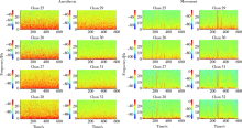
目的: 研究运动后兴奋脑区组织液(interstitial fluid, ISF)流动的变化。方法: 将20只雄性Sprague-Dawley大鼠随机分为对照组(12只)和运动组(8只), 两组大鼠均采用异氟烷进行麻醉,并动态监测尾状核区ISF流动情况,其中运动组在实验前期被放入到特制的转轮中进行运动20 min,对照组则给予持续麻醉,所有大鼠无偏瘫,运动能力良好。另外将5只大鼠采用异氟烷气体麻醉后向尾状核内植入微电极,记录运动态和麻醉态下尾状核神经元电活动。采用立体定位技术注射磁性示踪剂至尾状核ISF内,在注射前和注射后不同时间点进行一系列的磁共振扫描,直到示踪剂所致的高信号消失,采用细胞间隙定量分析系统(1.2版)对图像进行后处理和分析,监测ISF流动过程和测量相关参数,得到可反映示踪剂总量的加权信号强度(加权ΔSI)和其在ISF内的半衰期,分别计算运动组的运动前,运动后10、40、70、130和190 min各时间点以及对照组相同时间点的加权ΔSI和半衰期,采用独立样本t检验对两组测量值进行比较。结果: 微电极检测显示麻醉态和运动态下尾状核区的场电位显著不同;各时间点运动组和对照组的加权ΔSI(单位:信号强度×mm 3)分别为:运动前(60 257.1±23 069.2 vs. 61 072.0±19 547.3), 运动后10 min(83 624.3±21 475.7 vs. 71 218.1±12 586.5), 运动后40 min (57 336.0±36 243.4 vs. 69 756.1±13 306.0), 运动后70 min (43 705.9±10 246.3 vs. 55 443.2±20 733.3)、运动后130 min(7 734.9±2 645.2 vs. 8 967.6±2 007.3)和运动后190 min(2 497.3±987.5 vs. 3 013.2±1 760.8)。相对于对照组,运动组加权ΔSI在运动后40 min出现一过性的降低(P<0.05), 其余各时间点两组的加权ΔSI差异无统计学意义,两组示踪剂半衰期差异无统计学意义[(104.3±54.1) min vs. (113.4±47.3) min, P>0.05]。 结论: 运动可以导致兴奋脑区ISF流动一过性加速。
中图分类号:
- R445


| [1] |
He C, Chen F, Li B , et al. Neurophysiology of HCN channels: from cellular functions to multiple regulations[J]. Prog Neuro-biol, 2014,112(1):1-23.
doi: 10.1016/j.pneurobio.2013.10.001 |
| [2] |
Frischknecht R, Gundelfinger ED . The brain’s extracellular matrix and its role in synaptic plasticity[J]. Adv Exp Med Biol, 2012,970(1):153-171.
doi: 10.1007/978-3-7091-0932-8 |
| [3] |
Deco G, Rolls ET, Albantakis L , et al. Brain mechanisms for perceptual and reward-related decision-making[J]. Prog Neuro-biol, 2013,103(4):194-213.
doi: 10.1016/j.pneurobio.2012.01.010 |
| [4] | 杨双风, 韩鸿宾, 彭芸 . 大鼠生后发育过程中脑细胞外间隙的解剖及生理特性的变化[J]. 中国比较医学杂志, 2015,25(3):73-79. |
| [5] | Shi C, Lei Y, Han H , et al. Transportation inthe interstitial space of the brain can be regulated by neuronal excitation[J]. Sci Rep, 2015,5(12):17673. |
| [6] |
Liu B, Bai X, Zhou F , et al. Mutual information based three-dimensional registration of rat brain magnetic resonance imaging time-series[J]. Comput Electr Eng, 2013,39(5):1473-1484.
doi: 10.1016/j.compeleceng.2012.11.026 |
| [7] |
Han H, Shi C, Fu Y , et al. A novel MRI tracer-based method for measuring waterdiffusion in the extracellular space of the rat brain[J]. IEEE J Biomed Health Inform, 2014,18(3):978-983.
doi: 10.1109/JBHI.2014.2308279 |
| [8] |
Lei Y, Han H, Yuan F , et al. The brain interstitial system: Ana-tomy, modeling, in vivo measurement, and applications[J]. Prog Neurobiol, 2017,157(10):230-246.
doi: 10.1016/j.pneurobio.2015.12.007 |
| [9] | 刘娥, 张艺璇, 孙琳琳 , 等. 水通道蛋白4在阿尔兹海默病脑组织间液引流改变中的作用[J]. 北京大学学报(医学版), 2018,50(2):203-206. |
| [10] |
Xie L, Kang H, Xu Q , et al. Sleep drives metabolite clearance from the adult brain[J]. Science, 2013,342(6156):373-377.
doi: 10.1126/science.1241224 |
| [11] | 李学义, 王伟, 韩鸿宾 , 等. 采用磁共振示踪法探讨大鼠脑细胞间隙内物质转运清除规律[J]. 中国医学影像技术, 2018,34(1):1-4. |
| [12] |
Lv D, Li J, Li H , et al. Imaging and quantitative analysis of the interstitial space in the caudate nucleus in a rotenone-induced rat model of Parkinson’s disease using tracer-based MRI[J]. Aging Dis, 2017,8(1):1-6.
doi: 10.14336/AD.2016.0625 |
| [13] |
Watanabe M, Munoz DP . Saccade suppression by electrical microstimulation in monkey caudate nucleus[J]. J Neurosci, 2010,30(7):2700-2709.
doi: 10.1523/JNEUROSCI.5011-09.2010 |
| [14] |
Kravitz AV, Kreitzer AC . Striatal mechanisms underlying movement, reinforcement, and punishment[J]. Physiology, 2012,27(3):167-177.
doi: 10.1152/physiol.00004.2012 |
| [15] |
Hikosaka O, Kim HF, Yasuda M , et al. Basal ganglia circuits for reward value-guided behavior[J]. Annu Rev Neurosci, 2014,37(8):289-306.
doi: 10.1146/annurev-neuro-071013-013924 |
| [16] |
Lee SH, Koh JS, Ryu CW , et al. Changes of motor deactivation regions in patients with intracranial lesions[J]. J Korean Neurosurg Soc, 2013,54(6):453-460.
doi: 10.3340/jkns.2013.54.6.453 |
| [17] |
Hughes LE, Altena E, Barker RA , et al. Perseveration and choice in Parkinson’s disease: the impact of progressive frontostriatal dysfunction on action decisions[J]. Cerebral Cortex, 2013,23(7):1572-1581.
doi: 10.1093/cercor/bhs144 |
| [18] |
Hou J, Wang W, Quan X , et al. Quantitativevisualization of dynamic tracer transportation in the extracellular space of deep brain regions using tracer-based magnetic resonance imaging[J]. Med Sci Monit, 2017,23(9):4260-4268.
doi: 10.12659/MSM.903010 |
| [19] | 滕泽, 王伟, 关湘萍 , 等. 大脑类淋巴系统的研究进展[J]. 中华老年心脑血管病杂志, 2017,19(9):1001-1003. |
| [1] | 孟庆伟, 范梦, 郭煌达, 章涵宇, 王梦莹, 王斯悦, 彭和香, 王雪珩, 侯天姣, 秦雪英, 陈大方, 李劲, 武轶群, 吴涛, 陈洪波, 胡永华. 老年人心源性卒中抗凝治疗的预后[J]. 北京大学学报(医学版), 2026, 58(3): 536-542. |
| [2] | 王欧洋, 朱鹏磊, 林杰, 吴昊. LncRNA DANCR调节miR-656/BMPR1A轴对脑胶质瘤细胞恶性行为的影响[J]. 北京大学学报(医学版), 2026, 58(3): 616-623. |
| [3] | 王泽远, 于栓宝, 郑浩轲, 陶金, 范雅峰, 张雪培. 基于临床特征和多参数MRI的前列腺癌盆腔淋巴结转移的术前预测模型[J]. 北京大学学报(医学版), 2025, 57(4): 684-691. |
| [4] | 邹晶, 高天姿, 汪洋, 任蒙蒙, 刘东阳, 龙仁, 成雨萌, 刘萌, 徐正仁, 谢肇恒, 吕鹏宇, 袁兰, 韩鸿宾. 99mTc-DTPA经脑细胞外间隙给药后的动态分布及消除规律[J]. 北京大学学报(医学版), 2025, 57(3): 562-568. |
| [5] | 钟鹏, 胡晓丹, 王振洲. 大鼠脑创伤半暗带光学相干断层血管造影及微血管密度定量[J]. 北京大学学报(医学版), 2025, 57(2): 262-266. |
| [6] | 孙建军, 马千权, 尹晓亮, 杨辰龙, 张嘉, 陈素华, 吴超, 谢京城, 韩芸峰, 林国中, 司雨, 杨军, 邬海博, 赵强. 任意维度重建磁共振对骶管囊肿进行精准分型对于指导微创手术和康复的意义[J]. 北京大学学报(医学版), 2025, 57(2): 303-308. |
| [7] | 邢念增,王明帅,杨飞亚,尹路,韩苏军. 前列腺免活检创新理念的临床实践及其应用前景[J]. 北京大学学报(医学版), 2024, 56(4): 565-566. |
| [8] | 田宇轩,阮明健,刘毅,李德润,吴静云,沈棋,范宇,金杰. 双参数MRI改良PI-RADS评分4分和5分病灶的最大径对临床有意义前列腺癌的预测效果[J]. 北京大学学报(医学版), 2024, 56(4): 567-574. |
| [9] | 李晋娜,许丽娜,李敏,宋怡,张静,贾龙斌. 急性脑梗死患者血清BDNF、IL-18、hs-CRP水平与血管性认知障碍的相关性[J]. 北京大学学报(医学版), 2024, 56(4): 708-714. |
| [10] | 龙仁, 毛鑫, 高天姿, 解倩, 谈瀚博, 李子寅, 韩鸿宾, 袁兰. 熊果酸改善精神分裂症小鼠脱髓鞘和脑组织间液引流紊乱[J]. 北京大学学报(医学版), 2024, 56(3): 487-494. |
| [11] | 刘毅,袁昌巍,吴静云,沈棋,肖江喜,赵峥,王霄英,李学松,何志嵩,周利群. 靶向穿刺+6针系统穿刺对PI-RADS 5分患者的前列腺癌诊断效能[J]. 北京大学学报(医学版), 2023, 55(5): 812-817. |
| [12] | 袁昌巍,李德润,李志华,刘毅,山刚志,李学松,周利群. 多参数磁共振成像中动态对比增强状态在诊断PI-RADS 4分前列腺癌中的应用[J]. 北京大学学报(医学版), 2023, 55(5): 838-842. |
| [13] | 张展奕,张帆,颜野,曹财广,李长剑,邓绍晖,孙悦皓,黄天亮,管允鹤,李楠,陆敏,胡振华,张树栋. 近红外荧光靶向探针用于前列腺神经血管束术中成像[J]. 北京大学学报(医学版), 2023, 55(5): 843-850. |
| [14] | 刘颖,霍然,徐慧敏,王筝,王涛,袁慧书. 磁共振血管壁成像评估颈动脉中重度狭窄患者斑块特征与脑血流灌注的相关性[J]. 北京大学学报(医学版), 2023, 55(4): 646-651. |
| [15] | 傅强,高冠英,徐雁,林卓华,孙由静,崔立刚. 无症状髋关节前上盂唇撕裂超声与磁共振检查的对比研究[J]. 北京大学学报(医学版), 2023, 55(4): 665-669. |
|
||
