北京大学学报(医学版) ›› 2019, Vol. 51 ›› Issue (5): 944-948. doi: 10.19723/j.issn.1671-167X.2019.05.025
上颌前突患者鼻唇区软组织三维形态测量方法的建立
张添文,王晓霞( ),李自力,伊彪,梁成,王兴
- 北京大学口腔医学院·口腔医院,口腔颌面外科 国家口腔疾病临床医学研究中心 口腔数字化医疗技术和材料国家工程实验室 口腔数字医学北京市重点实验室,北京 100081
Establishment of three-dimensional measurement methods of nasolabial soft tissue for patients with maxillary protrusion
Tian-wen ZHANG,Xiao-xia WANG(),Zi-li LI,Biao YI,Cheng LIANG,Xing WANG
- Department of Oral and Maxillofacial Surgery, Peking University School and Hospital of Stomatology & National Clinical Research Center for Oral Diseases & National Engineering Laboratory for Digital and Material Technology of Stomatology & Beijing Key Laboratory of Digital Stomatology, Beijing 100081, China
摘要:
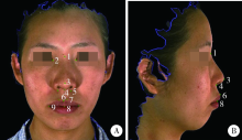
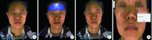
目的:利用3dMD照相机建立上颌前突患者鼻唇区软组织形态及其正颌手术前后变化的三维测量方法。方法:30例女性上颌前突患者,术前及术后均采用3dMD照相机拍摄面部三维照片,利用3dMD patient软件在三维照片上定位鼻唇区标志点,并进行软组织线距、角度、曲线距离、术前术后三维体积变化等10个指标的测量。3位测量者,每位测量者间隔1周各测量2次数据,分别对每位测量者2次测量结果及3位测量者之间做标准一致性检验,验证其可重复性。结果:每位测量者2次测量的10个测量指标组内相关系数(correlation coefficients,ICC)均大于0.8;3位测量者之间做标准一致性检验,其中内眦宽度、鼻高度、鼻尖突度、鼻唇角、人中长度、上唇红曲线高度、上唇高的组内相关系数大于0.8,鼻翼基底宽度、鼻背长与上唇三维体积变化的组内相关系数分别为0.680、0.627与0.528。结论:利用3dMD照相机及3dMD patient软件可较全面准确地对术前术后患者鼻唇区软组织三维形态进行测量和分析,但鼻尖点与鼻翼基底点的三维定位以及上唇区手术前后三维体积变化的重复性稍差。
中图分类号:
- R782.2


| [1] | 李东, 白宇明, 段银钟 , 等. 双颌前突患者正颌手术及拔牙矫治的软组织变化分析[J]. 第四军医大学学报, 2004,25(18):1704-1706. |
| [2] | 张君孝, 乔鸣芳 . 60例广东省双颌前突患者头影测量分析研究[J]. 华西口腔医学杂志, 2001,19(1):32-34. |
| [3] | Ioi H, Nakata S, Nakasima A ,et a1.Effect of facial convexity on antero-posterior lip positions of the most favored Japanese facial profiles[J]. Angle Orthod, 2005,75(3):326-332. |
| [4] |
何颖, 郭传瑸, 邓旭亮 , 等. 北方正常牙合人群颅颌面三维比例测量及面部对称性分析[J]. 北京大学学报(医学版), 2015,47(4):708-713.
doi: 10.3969/j.issn.1671-167X.2015.04.031 |
| [5] | 白玉兴, 郭宏铭, 刘凤德 , 等. 面部软组织三维重建及测量系统的研制与应用[J]. 中华口腔医学杂志, 2001,36(4):61-63. |
| [6] | Ort R, Metzler P, Kruse AL , et al. The reliability of a three-Dimensional photo system- (3dMDface-) based evaluation of the face in cleft lip infants[J]. Plastic Surg Int, 2012,2012(6):1-8. |
| [7] | Lübbers H, Medinger L, Kruse A , et al. Precision and Accuracy of the 3dMD photogrammetric system in craniomaxillofacial application[J]. J Craniofac Surg, 2010,21(3):763-767. |
| [8] | Aldridge K, Boyadjiev SA, Capone GT , et al. Precision and error of three-dimensional phenotypic measures acquired from 3dMD photogrammetric images[J]. Am J Med Genet Part A, 2005,138A(3):247-253. |
| [9] | Tzou CJ, Artner NM, Pona I , et al. Comparison of three-dimensional surface-imaging systems[J]. J Plast Reconstr Aesthet Surg, 2014,67(4):489-497. |
| [10] | 袁玲君, 沈国芳, 吴勇 , 等. 骨性Ⅲ类错牙合正颌术后颊部宽度变化的三维研究[J]. 中华口腔正畸学杂志, 2012,19(2):70-74. |
| [11] | Hellak AF, Kirsten B, Schauseil M , et al. Influence of maxillary advancement surgery on skeletal and soft-tissue changes in the nose: a retrospective cone-beam computed tomography study[J]. Head Face Med, 2015,11(1):12-23. |
| [12] | Kau CH, Cronin AJ, Richmond S . A three-dimensional evaluation of postoperative swelling following orthognathic surgery at 6 months[J]. Plast Reconstr Surg, 2007,119(7):2192-2199. |
| [13] | Tian K, Li Q, Wang X , et al. Reproducibility of natural head position in normal Chinese people[J]. Am J Orthodo Dentofacial Orthop, 2015,148(3):503-510. |
| [14] | Metzger TE, Kula KS, Eckert GJ , et al. Orthodontic soft-tissue parameters: a comparison of cone-beam computed tomography and the 3dMD imaging system[J]. Am J Orthod Dentofacial Orthop, 2013,144(5):672-681. |
| [15] | Fourie Z, Damstra J, Gerrits PO , et al. Evaluation of anthropometric accuracy and reliability using different three-dimensional scanning systems[J]. Forensic Sci Int, 2011,207(1/2/3):127-134. |
| [16] | 王宗琦, 王晓霞, 李自力 , 等. 3种缝合方法控制上颌Le Fort Ⅰ型截骨术后鼻翼宽度的效果比较[J]. 北京大学学报(医学版), 2015,47(1):104-108. |
| [17] | Oh K, Seo S, Park J , et al. Post-operative soft tissue changes in patients with mandibular prognathism after bimaxillary surgery[J]. J Craniomaxillofac Surg, 2013,41(3):204-211. |
| [18] | Dantas WRM, Da Silveira MMF, Do Egito Vasconcelos BC, et al. Evaluation of the nasal shape after orthognathic surgery[J]. Braz J Otorhinolaryngol, 2015,81(1):19-23. |
| [19] | Metzler P, Geiger EJ, Chang CC , et al. Surgically assisted maxillary expansion imparts three-dimensional nasal change[J]. J Oral Maxillofac Surg, 2014,72(10):2005-2014. |
| [20] | Patel A, Islam SMS, Murray K , et al. Facial asymmetry assessment in adults using three-dimensional surface imaging[J]. Prog Orthod, 2015,16(1):36. |
| [21] | Rustemeyer J, Martin A . Soft tissue response in orthognathic surgery patients treated by bimaxillary osteotomy: cephalometry compared with 2-D photogrammetry[J]. Oral Maxillofac Surg, 2013,17(1):33-41. |
| [22] | Joss CU, Triaca A, Antonini M , et al. Soft tissue stability in segmental distraction of the anterior mandibular alveolar process. A 2-year follow-up[J]. Int J Oral Maxillofac Surg, 2012,41(5):560-565. |
| [23] | 倪媛媛, 马俊青, 李强 , 等. 江苏地区正常人群上唇三维形态相关指标的研究[J]. 口腔生物医学, 2013,4(3):125-129. |
| [1] | 王旭,章晶晶,原福松,王郁,李成皓,Juha Eerik Varrela,岳江,葛立宏. 萌出诱导矫治器治疗儿童个别前牙反牙合的三维数字化分析[J]. 北京大学学报(医学版), 2018, 50(3): 532-537. |
|
||
