北京大学学报(医学版) ›› 2019, Vol. 51 ›› Issue (5): 937-943. doi: 10.19723/j.issn.1671-167X.2019.05.024

• 论著 • 上一篇    下一篇

三维颅面水平参考平面的确定方法

金珉廷,刘怡()   

  1. 北京大学口腔医学院·口腔医院,正畸科 国家口腔疾病临床研究中心 口腔数字化医疗技术和材料国家工程试验室 口腔数字医学北京市重点实验室,北京 100081
  • 收稿日期:2017-09-07 出版日期:2019-10-18 发布日期:2019-10-23
  • 通讯作者: 刘怡 E-mail:lyortho@163.com

Using three-dimensional craniofacial images to construct horizontal reference plane

Min-jung KIM,Yi LIU()   

  1. Department of Orthodontics, Peking University School and Hospital of Stomatology & National Clinical Research Center for Oral Diseases & National Engineering Laboratory for Digital and Material Technology of Stomatology & Beijing Key Laboratory of Digital Stomatology, Beijing 100081, China
  • Received:2017-09-07 Online:2019-10-18 Published:2019-10-23
  • Contact: Yi LIU E-mail:lyortho@163.com

摘要:

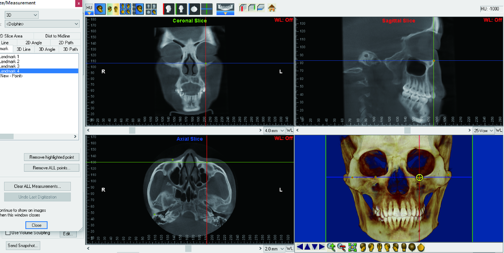
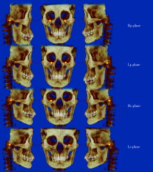
目的:比较在三维影像选取双侧耳点和双侧眶下点构建的不同水平面之间差异,分析不同水平面对双侧耳点和双侧眶下点位置的影响,为颅面部三维头影测量水平参考平面的确定提供依据。方法:选取32名正畸治疗前面部基本对称患者(颏下点离正中矢状面≤2 mm), 获取DICOM格式的大视野锥形束CT数据并导入到Dolphin软件,将鼻根点、蝶鞍点和枢椎齿突最高点构建正中矢状面,测量双侧耳点和双侧眶下点中随机三点构建的4种水平参考平面。分别定义为,平面1:水平面由右侧耳点和双侧眶下点构成;平面2:水平面由左侧耳点和双侧眶下点构成;平面3:水平面由双侧耳点与右侧眶下点构成;平面4:水平面由双侧耳点与左侧眶点构成。记录4个平面在三维空间当中的俯仰角、侧偏角和横滚角。间隔两周,一位研究者进行两次测量。计算组间相关系数(interclass correlation coefficient,ICC)比较两次测量结果的一致性,检验测量者自身的可靠性,进行单因素重复测量方差分析检验组内4个平面之间的差异,按年龄分为13~17岁组和≥18岁组。以枢椎齿突最高点为原点计算双侧耳点和双侧眶下点位置,应用圆周长公式分析头部转动对双侧耳点和双侧眶下点的影响。结果:单因素重复测量方差分析结果显示,不同三点构建的4种平面之间俯仰角、侧偏角和横滚角差异均无统计学意义(P=0.196、0.314、0.341)。头位转动对双侧耳点和双侧眶下点的影响分析结果为:1°俯仰角变化产生耳点约0.5 mm、眶下点约1.6 mm的变化;1°侧偏角变化产生耳点约1.1 mm、眶下点约1.5 mm的变化;1°横滚角变化产生耳点约1.2 mm、眶下点约0.7 mm的变化。结论:对于面部基本对称个体,应用三维头颅影像对双侧耳点和双侧眶下点中随机选取三个点构建的4种水平面之间差异无统计学意义;以双侧眶下点和右侧耳点构建的水平面可能最适合临床使用;头部不同方向的转动使双侧耳点和双侧眶下点产生不同位置的变化。

关键词: 头位, 眶耳平面, 锥形束计算机断层扫描, 三维头影测量

Abstract:

Objective: To compare four different three-dimensional horizontal planes and detect anatomical landmarks so as to provide theoretical reference for horizontal reference plane constructed by three-dimensional cephalometry. Methods: The subjects of this study were 32 facial symmetry patients (menton from mid-sagittal plane ≤2 mm). Cone-bead computed tomography (CBCT) was obtained before orthodontic treatment, and the data were imported into Dolphin imaging soft in DICOM format. The sagittal plane was passing through the Nasion, Sella and Dent. Four horizontal reference planes were constructed by three points of bilateral porion and bilateral orbitale. Plane 1: horizontal reference plane constructed by right porion and bilateral orbitale. Plane 2: horizontal reference plane constructed by left porion and bilateral orbitale. Plane 3: horizontal reference plane constructed by bilateral porion and right orbitale. Plane 4: horizontal reference plane constructed by bilateral porion and left orbitale. Pitch, yaw, roll for four planes were measured three dimensionally. All the samples were measured two times by one judge at an interval of two weeks. The two times measuring results were evaluated with Intraclass correlation coefficient (ICC) for verifying reliability. The multiple sets of repeated measurement analysis were used to compare the four different planes. Based on ages, the samples were divided into two groups (group 1: ages 13 to 17, group 2: over 18 years), the mean and standard deviation of landmark coordinates measured with Dent as the origin point, the circumference formula was applied to calculate the change of landmark position generated by head rotation. Results: No significant differences of pitch, yaw and roll among the four planes (P=0.196, 0.314, and 0.341). One degree of pitch rotation made changes of porion and orbitale approximate 0.5 mm, and 1.6 mm, respectively. One degree of yaw rotation made changes of porion and orbitale approximate 1.1 mm, and 1.5 mm, respectively. One degree of roll rotation made changes of porion and orbitale approximate 1.2 mm, and 0.7 mm, respectively. Conclusion: There was no significant difference among the four horizontal planes constructed by any three points of bilateral orbitales and bilateral porions. It has the highest concordance using bilateral orbitales and one porion to construct horizontal plane in this study, probably the best option in clinical practice. Different head rotation generated different distance changes of anatomical landmarks.

Key words: Head position, Frankfort plane, Cone beam computed tomography, Three-dimensional cephalometry

中图分类号: 

  • R783.5

表1

解剖标志点的定义"

Landmark Definition
Menton The lowest point on the symphyseal shadow of the mandible
Sella The geometric center of the pituitary fossa
Dentes epistrophei The most posterior point of dentes epistrophei
Nasion The most anterior point on frontonasal suture in the midsagittal plane
Orbitale The lowest point on the inferior rim of the orbit
Porion The most superiorly positioned point of the external auditory meatus

表2

旋转角正负的确定"

Items + -
Pitch Downward Upward
Yaw Right posterior Left after
Roll Lower right Left lower

表3

三维旋转角度的定义"

Rotarion Definition
Pitch Rotates around the X-axis, intersection between horizontal reference plane and rotated horizontal plane projected to MSP
Yaw Rotates around the Y-axis, intersection between MSP reference plane and rotated MSP projected to horizontal reference plane
Roll Rotates around the Z-axis, intersection between horizontal reference plane and rotated horizontal plane projected to coronal plane

表4

以Rp平面为基准不同平面之间旋转角度的差异/(°)"

Items Rp-Lp Rp-Ro Rp-Lo
Pitch -0.078±0.892 0.023±0.505 -0.121±0.850
Yaw 0.032±0.177 -0.002±0.010 0.002±0.008
Roll -0.013±0.091 -0.048±0.753 0.117±0.636

图1

双侧眶下点和双侧耳点的定点"

图2

4种水平平面的确定: Rp平面由双侧眶下点与右侧耳点构成;Lp平面由双侧眶下点与左侧耳点构成;Ro平面由双侧耳点与右侧眶下点构成;Lo平面由双侧耳点与左侧眶点构成"

图3

三维虚拟空间旋转角度的定义"

表5

不同年龄段以Dent点为原点的标志点的位置/mm"

Age groups Coordinate Right orbitale Left orbitale Right porion Left porion
X 31.017±2.651 31.067±2.180 59.344±3.323 61.578±5.017
13-17 years old Y 26.967±3.998 26.800±3.944 26.700±3.803 26.739±3.826
Z 80.622±3.944 80.467±4.149 4.811±3.790 4.033±3.524
X 32.364±2.623 32.507±3.546 59.064±6.088 60.964±4.532
≥18 years old Y 28.393±4.482 28.193±4.452 28.179±4.519 27.943±4.715
Z 83.300±2.986 83.179±3.417 6.207±2.806 7.421±2.252

表6

不同年龄段以Dent点为原点计算头部不同方向1°转动对双侧耳点和双侧眶下点产生的距离变化/mm"

Age groups Rotation Right orbitale Left orbitale Right porion Left porion
Pitch 1.484 1.480 0.474 0.472
13-17 years old Yaw 1.508 1.505 1.039 1.077
Roll 0.717 0.716 1.135 1.172
Pitch 1.536 1.533 0.504 0.505
≥18 years old Yaw 1.560 1.559 1.037 1.072
Roll 0.751 0.751 1.142 1.170
[1] Proffit WR . Contemporary orthodontics[M]. 5th ed. St.Louis: Elsevier, 2013: 134-137.
[2] Jacobson A . Radiographic cephalometry: from basics to 3-D imaging[M]. 2nd ed. Chicago: Quintessence Pub, 2006: 153-160.
[3] Moorrees CFA, Kean MR . Natural head position, a basic consi-deration in the interpretation of cephalometric radiographs[J]. Am J Phys Anthropol, 1958,16(2):213-234.
[4] Finlay LM . Craniometry and cephalometry: a history prior to the advent of radiography[J]. Angle Orthod, 1980,50(4):312-321.
[5] Downs WB . Analysis of the dentofacial profile[J]. Angle Orthod, 1956,26(4):191-212.
[6] Zebeib AM, Naini FB . Variability of the inclination of anatomic horizontal reference planes of the craniofacial complex in relation to the true horizontal line in orthognathic patients[J]. Am J Orthod Dentofacial Orthop, 2014,146(6):740-747.
[7] Barbera AL, Sampson WJ, Townsend GC . Variation in natural head position and establishing corrected head position[J]. Homo, 2014,65(3):187-200.
[8] Hsung T, Lo J, Li T , et al. Automatic detection and reproduction of natural head position in stereo-photogrammetry[J]. PLoS One, 2015,10(6):e130877.
[9] Kovacs L, Zimmermann A, Brockmann G , et al. Three-dimensional recording of the human face with a 3D laser scanner[J]. J Plast Reconstr Aesthet Surg, 2006,59(11):1193-1202.
[10] Xia JJ, McGrory JK, Gateno J, et al. A new method to orient 3-dimensional computed tomography models to the natural head position: a clinical feasibility study[J]. J Oral Maxillofac Surg, 2011,69(3):584-591.
[11] Tian K, Li Q, Wang X , et al. Reproducibility of natural head position in normal Chinese people[J]. Am J Orthod Dentofacial Orthop, 2015,148(3):503-510.
[12] Damstra J, Fourie Z, DeWit M, et al. A three-dimensional comparison of a morphometric and conventional cephalometric mid-sagittal planes for craniofacial asymmetry[J]. Clin Oral Investig, 2012,16(1):285-294.
[13] Lee JK, Jung PK, Moon CH . Three-dimensional cone beam computed tomographic image reorientation using soft tissues as reference for facial asymmetry diagnosis[J]. Angle Orthod, 2014,84(1):38-47.
[14] Oh S, Kim CY, Hong J . A comparative study between data obtained from conventional lateral cephalometry and reconstructed three-dimensional computed tomography images[J]. J Korean Assoc Oral Maxillofac Surg, 2014,40(3):123-129.
[15] Severt TR, Proffit WR . The prevalence of facial asymmetry in the dentofacial deformities population at the University of North Carolina[J]. Int J Adult Orthodon Orthognath Surg, 1997,12(3):171-176.
[16] Steiner C . Cephalometrics for you and me[J]. Am J Orthod, 1953,39(10):729-755.
[17] Steiner C . Cephalometrics in clinical practice[J]. Angle Orthod, 1959,29(1):8-29.
[18] Steiner C . The use of cephalometrics as an aid to planning and assessing orthodontic treatment[J]. Am J Orthod, 1960,46(10):721-735.
[19] Kim MS, Lee EJ, Song IJ , et al. The location of midfacial landmarks according to the method of establishing the midsagittal reference plane in three-dimensional computed tomography analysis of facial asymmetry[J]. Imaging Sci Dent, 2015,45(4):227.
[20] Kim HJ, Kim BC, Kim JG , et al. Construction and validation of the midsagittal reference plane based on the skull base symmetry for three-dimensional cephalometric craniofacial analysis[J]. J Craniofac Surg, 2014,25(2):338-342.
[21] Xiong Y, Zhao Y, Yang H , et al. Comparison between interactive closest point and procrustes analysis for determining the median sagittal plane of three-dimensional facial data[J]. J Craniofac Surg, 2016,27(2):441-444.
[22] 王斯维, 黎敏, 杨慧芳 , 等. 3种生成大视野锥形束CT数据正中矢状面方法的比较[J]. 北京大学学报(医学版), 2016,48(2):330-335.
doi: 10.3969/j.issn.1671-167X.2016.02.028
[23] Lim YK, Chu EH, Lee DY , et al. Three-dimensional evaluation of soft tissue change gradients after mandibular setback surgery in skeletal class Ⅲ malocclusion[J]. Angle Orthod, 2010,80(5):896-903.
[24] Kim MG, Lee JW, Cha KS , et al. Three-dimensional symmetry and parallelism of the skeletal and soft-tissue poria in patients with facial asymmetry[J]. Korean J Orthod, 2014,44(2):62-68.
[1] 杨雨卉,黄一平,李巍然. 骨皮质切开加速正畸牙齿移动对牙根吸收的影响[J]. 北京大学学报(医学版), 2021, 53(2): 434-437.
[2] 高璐,谷岩. 中国人群腭中缝形态特点分期与Demirjian牙龄的相关性[J]. 北京大学学报(医学版), 2021, 53(1): 133-138.
[3] 周境,刘怡. 不同垂直骨面型骨性Ⅱ类青少年女性颞下颌关节锥形束CT测量分析[J]. 北京大学学报(医学版), 2021, 53(1): 109-119.
[4] 杜仁杰,焦剑,周彦恒,施捷. 侵袭性牙周炎患者正畸前后的咬合变化[J]. 北京大学学报(医学版), 2019, 51(5): 919-924.
[5] 王高南,焦剑,周彦恒,施捷. 正畸牙齿位置的移动对角化龈宽度的影响[J]. 北京大学学报(医学版), 2019, 51(5): 931-936.
[6] 王秀婧,张怡美,周彦恒. 骨性Ⅲ类错牙合畸形患者正畸-正颌联合治疗的稳定性[J]. 北京大学学报(医学版), 2019, 51(1): 86-92.
[7] 王怡然,周彦恒,王雪东,魏松,刘伟涛. 上颌反复扩缩前方牵引三维变化的锥形束CT分析[J]. 北京大学学报(医学版), 2018, 50(4): 685-693.
[8] 戴帆帆,刘怡,许天民,陈贵. 探索成人正畸前后下颌三维数字化模型的重叠方法[J]. 北京大学学报(医学版), 2018, 50(2): 271-278.
[9] 张又文,辛天艺,焦剑,周彦恒,施捷. 慢性牙周炎的减数正畸治疗[J]. 北京大学学报(医学版), 2018, 50(2): 308-313.
[10] 马静,江久汇. 骨性Ⅱ类和Ⅲ类高角错牙合患者下切牙区的牙槽骨形态分析[J]. 北京大学学报(医学版), 2018, 50(1): 98-103.
[11] 柳玉树,李峥,赵一姣,叶红强,周彦秋,胡文杰,刘云松,寻春雷,周永胜. 数字化正畸修复联合治疗设计在前牙美学重建中的应用[J]. 北京大学学报(医学版), 2018, 50(1): 78-84.
[12] 徐筱,徐莉,江久汇,吴佳琪,李小彤,靖无迪. 锥形束CT评判安氏Ⅲ类错牙合上前牙骨开裂与骨开窗的准确性分析[J]. 北京大学学报(医学版), 2018, 50(1): 104-109.
[13] 柳大为,李晶,郭亮,荣起国,周彦恒. 舌侧矫治器关闭间隙上前牙牙周膜应力变化的三维有限元分析[J]. 北京大学学报(医学版), 2018, 50(1): 141-147.
[14] 常大桐,周彦恒,刘伟涛. 上颌反复快速扩缩对上气道影响的锥束CT研究[J]. 北京大学学报(医学版), 2017, 49(4): 685-690.
[15] 张达,王林川,周彦恒,刘晓默,李晶. 3D打印间接粘接托槽精度[J]. 北京大学学报(医学版), 2017, 49(4): 704-708.
Viewed
Full text


Abstract

Cited

  Shared   
  Discussed   
[1] 张三. 中文标题测试[J]. 北京大学学报(医学版), 2010, 42(1): 1 -10 .
[2] 赵磊, 王天龙 . 右心室舒张末期容量监测用于肝移植术中容量管理的临床研究[J]. 北京大学学报(医学版), 2009, 41(2): 188 -191 .
[3] 万有, , 韩济生, John E. Pintar. 孤啡肽基因敲除小鼠电针镇痛作用增强[J]. 北京大学学报(医学版), 2009, 41(3): 376 -379 .
[4] 张燕, 韩志慧, 钟延丰, 王盛兰, 李玲玲, 郑丹枫. 骨骼肌活组织检查病理诊断技术的改进及应用[J]. 北京大学学报(医学版), 2009, 41(4): 459 -462 .
[5] 赵奇, 薛世华, 刘志勇, 吴凌云. 同向施压测定自酸蚀与全酸蚀粘接系统粘接强度[J]. 北京大学学报(医学版), 2010, 42(1): 82 -84 .
[6] 林红, 王玉凤, 吴野平. 学校生活技能教育对小学三年级学生行为问题影响的对照研究[J]. 北京大学学报(医学版), 2007, 39(3): 319 -322 .
[7] 丰雷, 程嘉, 王玉凤. 注意缺陷多动障碍儿童的运动协调功能[J]. 北京大学学报(医学版), 2007, 39(3): 333 -336 .
[8] 李岳玲, 钱秋瑾, 王玉凤. 儿童注意缺陷多动障碍成人期预后及其预测因素[J]. 北京大学学报(医学版), 2007, 39(3): 337 -340 .
[9] . 书讯[J]. 北京大学学报(医学版), 2007, 39(3): 225 -328 .
[10] 牟向东, 王广发, 刁小莉, 阙呈立. 肺黏膜相关淋巴组织型边缘区B细胞淋巴瘤一例[J]. 北京大学学报(医学版), 2007, 39(4): 346 -350 .