,正畸正颌治疗,锥形束CT,牙根吸收," /> ,正畸正颌治疗,锥形束CT,牙根吸收,"/> 锥形束CT三维体积测量评估骨性Ⅲ类错<inline-formula id="bjdxxbyxb-54-4-719-M1"><img src="/fileup/inline_graphic/2022/1660206314480_bjdxxbyxb-54-4-719-M1.jpg"/></inline-formula>正畸正颌治疗后的牙根吸收

北京大学学报(医学版) ›› 2022, Vol. 54 ›› Issue (4): 719-726. doi: 10.19723/j.issn.1671-167X.2022.04.022

• 论著 • 上一篇    下一篇

锥形束CT三维体积测量评估骨性Ⅲ类错正畸正颌治疗后的牙根吸收

高娟1,2,吕航苗1,马慧敏1,赵一姣3,李小彤1,*()   

  1. 1. 北京大学口腔医学院·口腔医院正畸科,国家口腔医学中心,国家口腔疾病临床医学研究中心,口腔生物材料和数字诊疗装备国家工程研究中心,口腔数字医学北京市重点实验室,国家卫生健康委员会口腔医学计算机应用工程技术研究中心,北京 100081
    2. 贵阳市口腔医院正畸科,贵阳 550002
    3. 北京大学口腔医学院·口腔医院口腔医学数字化研究中心,北京 100081
  • 收稿日期:2021-05-12 出版日期:2022-08-18 发布日期:2022-08-11
  • 通讯作者: 李小彤 E-mail:xiaotonglee@hotmail.com
  • 基金资助:
    北京大学口腔医院新技术新疗法基金(PKUSSNCT-19B04);国家重大疾病多学科合作诊疗能力建设项目(PKUSSNMP-201902);贵阳市科技计划项目:筑科合同([2019]9-7-9)

Evaluation of root resorption after surgical orthodontic treatment of skeletal Class Ⅲ malocclusion by three-dimensional volumetric measurement with cone-beam CT

Juan GAO1,2,Hang-miao LV1,Hui-min MA1,Yi-jiao ZHAO3,Xiao-tong LI1,*()   

  1. 1. Department of Orthodontics, Peking University School and Hospital of Stomatology & National Center of Stomatology & National Clinical Research Center for Oral Diseases & National Engineering Research Center of Oral Biomaterials and Digital Medical Devices & Beijing Key Laboratory of Digital Stomatology & NHC Research Center of Engineering and Technology for Computerized Dentistry, Beijing 100081, China
    2. Department of Orthodontics, Guiyang Stomatological Hospital, Guiyang 550002, China
    3. Digital Technology of Stomatology, Peking University School and Hospital of Stomatology, Beijing 100081, China
  • Received:2021-05-12 Online:2022-08-18 Published:2022-08-11
  • Contact: Xiao-tong LI E-mail:xiaotonglee@hotmail.com
  • Supported by:
    the New Technology and New Therapy Fund of Peking University Stomatology Hospital(PKUSSNCT-19B04);the National Multi-disciplinary Collaborative Diagnosis and Treatment Capacity Building Project for Major Diseases(PKUSSNMP-201902);the Guiyang Science and Technology Project([2019]9-7-9)

摘要:

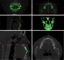
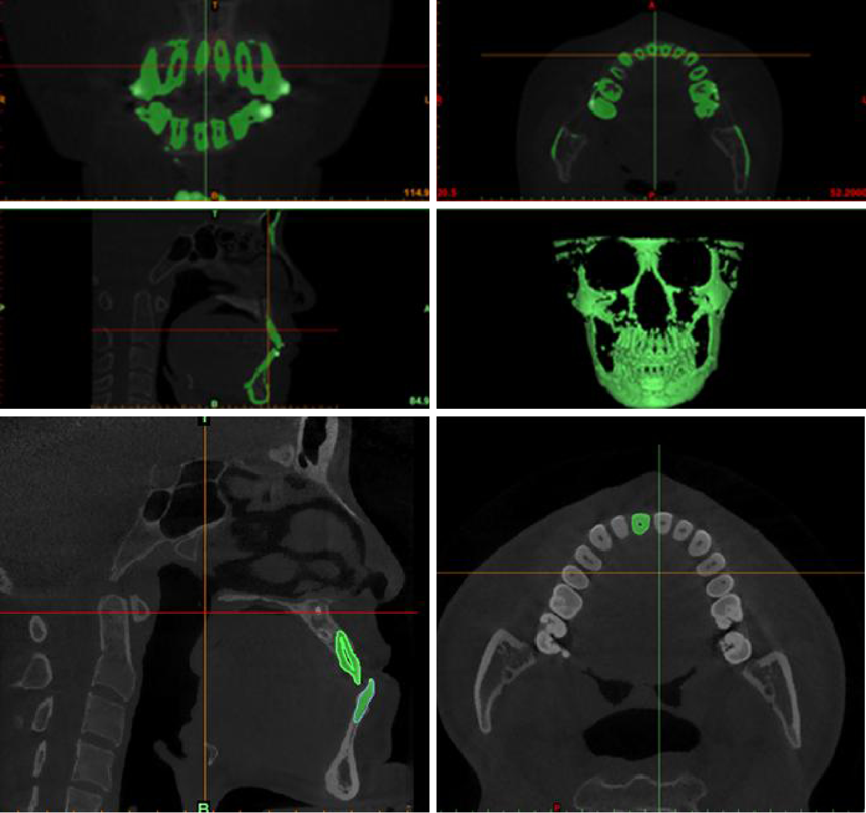
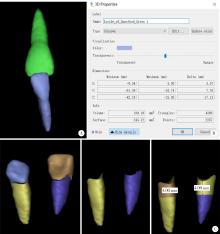
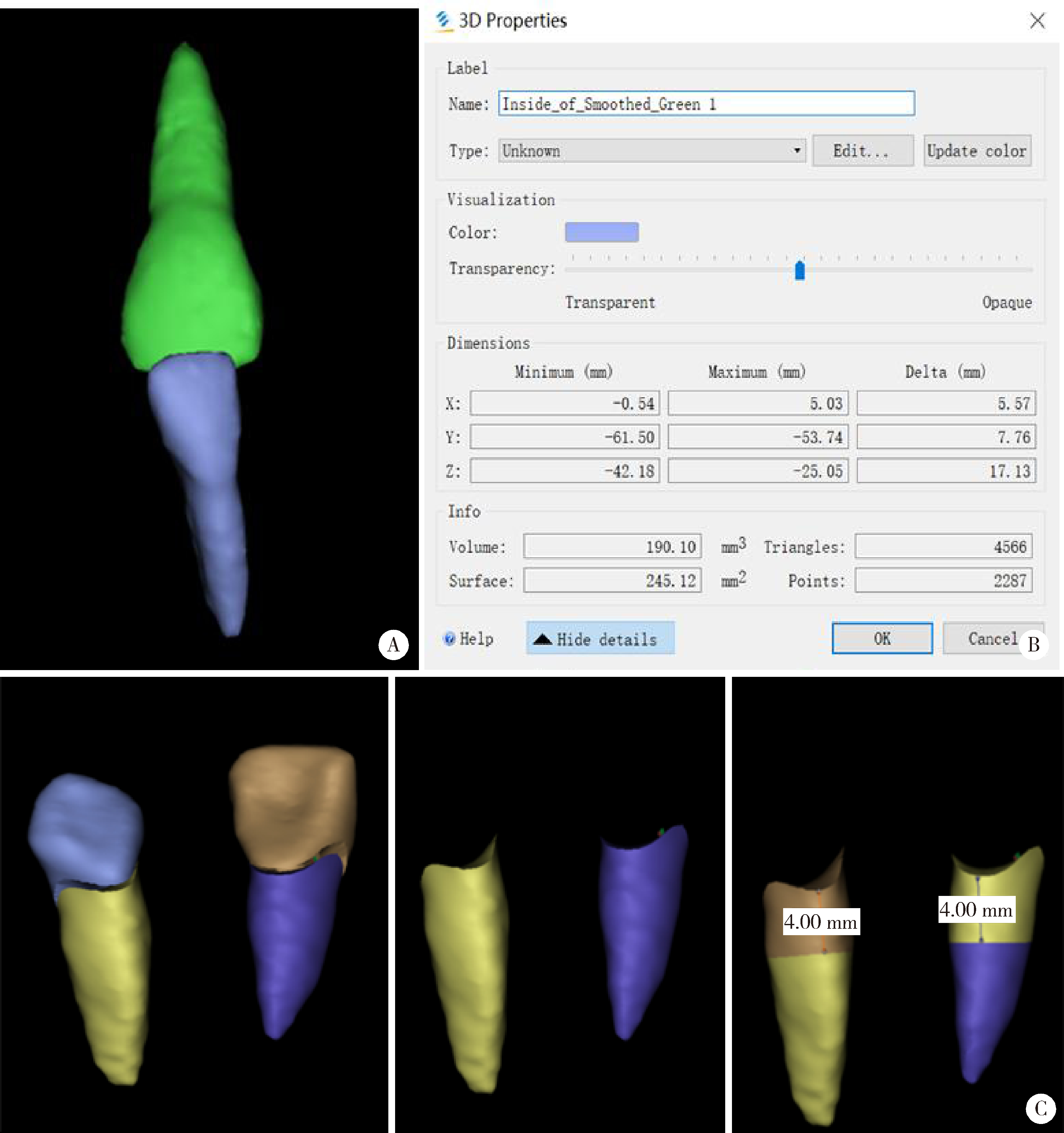
目的: 探索运用锥形束CT(cone-beam computed tomography,CBCT)三维重建技术测量牙根体积的方法,研究骨性Ⅲ类错患者在正畸正颌联合治疗过程中,上下颌中切牙的牙根长度、牙根体积变化规律和牙根吸收的特点。方法: 选取20例因骨性Ⅲ类错行正畸正颌联合治疗的患者,于正畸去代偿治疗前1周内(T0)、正畸去代偿治疗后正颌手术前1周内(T1)及术后正畸治疗结束后1周内(T2)三个时间点拍摄CBCT,应用三维重建技术测量上下颌中切牙的牙根长度、牙根体积(包括牙根总体积、根颈部及根体部体积),计算长度减小百分比和体积减小百分比,并测量正畸治疗前后牙齿移动的距离。数据用SPSS 20.0软件进行统计分析,服从正态分布的组间两两比较采用最小显著性差异法(least significant difference,LSD),不服从正态分布的组间比较采用非参数检验。比较上下颌中切牙牙根长度和牙根体积变化的差异,分析其变化规律和牙根吸收的特点。结果: 骨性Ⅲ类错患者在正畸正颌联合治疗过程中,上下颌中切牙的牙根长度和牙根体积均明显减小(P < 0.05),根颈部和根体部的牙根体积均明显减小(P < 0.05),根体部牙根体积的减小量更加显著,上中切牙根体部牙根体积减小量百分比为(30.51±23.23)%,下中切牙为(23.24±11.96)%。与上中切牙相比,下中切牙牙根体积的减小量和减小量百分比均较小,且上中切牙根体部的体积减小量大于下中切牙,差异有统计学意义(P < 0.05)。术前正畸阶段去代偿治疗上颌中切牙以控根移动方式腭向移动,下颌中切牙移动方式为唇向倾斜移动。结论: 骨性Ⅲ类错患者在正畸正颌联合治疗过程中,上下颌中切牙的牙根长度和牙根总体积均减小,变化规律相似。牙根体积测量提示牙根根颈部也存在牙根吸收。上下颌中切牙牙根吸收量的差异可能与牙齿移动的距离和方式相关。利用CBCT三维重建体积测量评估牙根吸收可以弥补牙根长度测量的局限性。

关键词: ')">骨性Ⅲ类错, 正畸正颌治疗, 锥形束CT, 牙根吸收

Abstract:

Objective: To explore the method of measuring root volume with cone-beam computed tomography (CBCT) three-dimensional reconstruction technology, and to study root length and root volume of upper and lower central incisors in patients with skeletal Class Ⅲ malocclusion treated by surgical orthodontic treatment. Methods: Twenty patients with skeletal Class Ⅲ malocclusion undergoing surgical orthodontic treatment were selected. CBCT data at three time points, before decompensation treatment (T0), after decompensation treatment (before orthognathic surgery, T1), and the end of post-operative orthodontic treatment (T2) were collected. Three-dimensional reconstruction technology was used to measure the root length and root volume of the upper and lower central incisors (including total root volume, cervical root and apical root), calculate the percentage of reduction volume, and measure the distance of tooth movement after orthodontic treatment. Data were statistically analyzed by SPSS 20.0 software. Least significant difference (LSD) method was used for pair comparison between the groups subject to normal distribution, and non-parametric test was used for comparison between the groups not subject to normal distribution. The differences of root length and root volume of upper and lower incisors were compared, and the characteristics of root absorption were analyzed. Results: Root length and root volume of the upper and lower central incisors were reduced during the surgical orthodontic treatment (P < 0.05) in cases. Both the root volume of cervical root and apical root were significantly reduced (P < 0.05), the reduction of apical root was more significant. The percentage of root volume reduction of the upper central incisor was (30.51±23.23)%, and lower central incisor (23.24±11.96)%. Compared with the upper central incisor, the root volume reduction amount and percentage of the lower central incisor were smaller, and apical root volume reduction of the upper central incisor was greater than that of the lower central incisor, which was statistically significant (P < 0.05). During pre-surgical orthodontic treatment, maxillary central incisor palatal moving was in a controlled tipping manner, and the mandibular central incisor tipped labially. Conclusion: In patients with skeletal Class Ⅲ malocclusion, root length and total root volume of upper and lower central incisors decreased during surgical orthodontic treatment. Root volume measurement indicated that the cervical root also had root resorption. The difference in root resorption of the upper and lower central incisors might be related to the distance and direction of teeth movement. CBCT three-dimensional reconstruction will compensate for the limitation of root length measurement in evaluating root resorption.

Key words: Skeletal Class Ⅲ malocclusion, Surgical orthodontic treatment, Cone-beam computed tomography, Tooth root resorption

中图分类号: 

  • R783.5

图1

上下颌中切牙三维重建示意图"

图2

Mimics软件建模、虚拟分割及测量"

图3

二维牙根长度的测量"

表1

骨性Ⅲ类错患者正畸正颌联合治疗过程中上下颌中切牙牙根体积和长度的变化"

Change parts T0, $\bar x \pm s$ T1, $\bar x \pm s$ T2, $\bar x \pm s$ P Comparison among groups
Upper central incisor
    Root length/mm 10.47±2.13 9.41±2.36 8.95±2.37 < 0.001 T0>T1>T2
    Total root volume/mm3 156.90±31.77 145.29±31.49 135.16±31.46 < 0.001 T0>T1>T2
    Root apical volume/mm3 62.55±21.74 53.13±22.47 46.88±21.96 < 0.001 T0>T1>T2
    Root cervical volume/mm3 94.34±15.16 92.15±15.83 88.27±15.41 0.001 (T0, T1)>T2
Under central incisor
    Root length/mm 10.97±0.91 10.16±0.93 9.72±0.86 < 0.001 T0>T1>T2
    Total root volume/mm3 98.07±15.18 89.04±17.10 82.69±16.37 < 0.001 T0>T1>T2
    Root apical volume/mm3 43.71±8.95 37.72±9.28 33.72±9.25 < 0.001 T0>T1>T2
    Root cervical volume/mm3 54.35±8.28 51.32±10.27 48.96±9.05 < 0.001 (T0, T1)>(T1, T2)

表2

骨性Ⅲ类错患者正畸正颌联合治疗后(T2~T0)上下颌中切牙牙根体积和根长减小量及减少百分比"

Items Upper central incisor, $\bar x \pm s$ Under central incisor, $\bar x \pm s$ P
Root length/mm 1.51±1.34 1.24±0.70 0.41
Total root volume/mm3 21.73±13.81 15.38±7.78 0.08
Root apical volume/mm3 15.66±9.63 9.99±5.54 0.04
Root cervical volume/mm3 6.07±7.06 5.39±4.54 0.69
Percentage of root length/% 14.86±12.39 11.21±5.94 0.24
Volume percentage of total root/% 13.96±8.44 15.93±8.15 0.39
Volume percentage of root apical/% 30.51±23.23 23.24±11.96 0.15
Volume percentage of root neck/% 6.37±7.55 10.03±8.52 0.11

表3

骨性Ⅲ类错患者正畸正颌联合治疗后(T2~T0)上下颌中切牙的根颈部与根体部体积减小量及减少百分比的比较"

Items Root neck, $\bar x \pm s$ Root body, $\bar x \pm s$ P
Upper central incisor
    Volume change/mm3 6.07±7.06 15.66±9.63 < 0.001
    Volume reduction percentage/% 6.37±7.54 30.51±23.23 < 0.001
Under central incisor
    Volume change/mm3 5.39±4.53 9.99±5.54 0.005
    Volume reduction percentage/% 10.02±8.51 23.23±11.96 < 0.001

表4

骨性Ⅲ类错患者正畸正颌联合治疗的术前、术后正畸阶段上下颌中切牙牙齿移动量的比较"

Items T0, $\bar x \pm s$ T1, $\bar x \pm s$ T1′, $\bar x \pm s$ T2, $\bar x \pm s$ ΔT1-T0, $\bar x \pm s$ ΔT2-T1′, $\bar x \pm s$ P1 (T1-T0) P2 (T2-T1)
Upper central incisor
    S-edge/mm 70.43±5.72 66.51±5.63 64.24±6.09 65.66±5.58 -3.92±1.78 1.42±2.73 < 0.001 0.048
    V-edge/mm 76.14±3.72 77.26±3.54 75.24±3.67 74.11±3.21 1.12±1.63 -1.13±2.25 0.012 0.055
    S-root/mm 57.75±5.54 56.31±6.00 57.55±4.79 56.82±4.77 -1.44±1.63 -0.73±1.42 0.002 0.050
    V-root/mm 57.17±4.04 57.84±3.79 56.12±4.13 56.36±3.78 0.67±1.14 0.24±1.57 0.027 0.537
Under central incisor
    S-edge/mm 71.44±7.72 74.61±8.45 61.81±6.17 62.49±5.67 3.16±1.97 1.06±2.79 < 0.001 0.151
    V-edge/mm 77.16±5.07 78.06±4.49 74.36±3.84 72.25±3.54 0.89±1.82 -2.30±2.63 0.070 0.003
    S-root/mm 64.79±8.09 64.42±9.03 53.18±6.33 54.57±5.81 -0.37±2.05 1.91±3.68 0.485 0.056
    V-root/mm 96.76±4.02 95.55±3.95 91.32±3.70 89.55±3.82 -1.22±1.60 -1.84±2.31 0.011 0.006
1 Weltman B , Vig KW , Fields HW , et al. Root resorption associated with orthodontic tooth movement: A systematic review[J]. Am J Orthod Dentofacial Orthop, 2010, 137 (4): 462- 476.
doi: 10.1016/j.ajodo.2009.06.021
2 赵志河. 口腔正畸学[M]. 7版 北京: 人民卫生出版社, 2020.
3 Sawicka M , Bedini R , Pecci R , et al. The application of X-ray microtomography for the assessement of root resorption caused by the orthodontic treatment of premolars[J]. Ann Ist Super Sanita, 2012, 48 (1): 71- 74.
4 Zahed Zahedani S , Oshagh M , Momeni Danaei S , et al. Roeinpeikar SMM: A comparison of apical root resorption in incisors after fixed orthodontic treatment with standard edgewise and straight wire (MBT) method[J]. J Dent Shiraz Univ Med Sci, 2013, 14 (3): 103- 110.
5 Hodges RJ , Atchison KA , White SC . Impact of cone-beam computed tomography on orthodontic diagnosis and treatment planning[J]. Am J Orthod Dentofacial Orthop, 2013, 143 (5): 665- 674.
doi: 10.1016/j.ajodo.2012.12.011
6 张婕, 李小彤. 骨性安氏Ⅲ类手术患者前牙区的牙槽骨厚度[J]. 北京大学学报(医学版), 2016, 48 (1): 111- 115.
doi: 10.3969/j.issn.1671-167X.2016.01.020
7 Giudice AL , Quinzi V , Ronsivalle V , et al. Evaluation of imaging software accuracy for 3-dimensional analysis of the mandibular condyle. A comparative study using a surface-to-surface matching technique[J]. Int J Environ Res Public Health, 2020, 17 (13): 4789.
doi: 10.3390/ijerph17134789
8 Deng Y , Sun Y , Xu T . Evaluation of root resorption after comprehensive orthodontic treatment using cone beam computed tomography (CBCT): A meta-analysis[J]. BMC Oral Health, 2018, 18 (1): 116.
doi: 10.1186/s12903-018-0579-2
9 熊再道, 赵桂芝, 柯杰, 等. 上颌快速扩弓后牙根吸收变化的三维测量研究[J]. 实用口腔医学杂志, 2018, 34 (1): 65- 68.
doi: 10.3969/j.issn.1001-3733.2018.01.014
10 Yildirim M , Akin M . Comparison of root resorption after bone-borne and tooth-borne rapid maxillary expansion evaluated with the use of microtomography[J]. Am J Orthod Dentofacial Orthop, 2019, 155 (2): 182- 190.
doi: 10.1016/j.ajodo.2018.03.021
11 Lo Giudice A , Galletti C , Gay-Escoda C , et al. CBCT assessment of radicular volume loss after rapid maxillary expansion: A syste-matic review[J]. J Clin Exp Dent, 2018, 10 (5): e484- e494.
12 Puttaravuttiporn P , Wongsuwanlert M , Charoemratrote C , et al. Volumetric evaluation of root resorption on the upper incisors using cone beam computed tomography after 1 year of orthodontic treatment in adult patients with marginal bone loss[J]. Angle Orthod, 2018, 88 (6): 710- 718.
doi: 10.2319/121717-868.1
13 Aras I , Unal I , Huniler G , et al. Root resorption due to orthodontic treatment using self-ligating and conventional brackets: A cone-beam computed tomography study[J]. J Orofac Orthop, 2018, 79 (3): 181- 190.
doi: 10.1007/s00056-018-0133-5
14 Ma H , Li W , Xu L , et al. Morphometric evaluation of the alveolar bone around central incisors during surgical orthodontic treatment of high-angle skeletal Class Ⅲ malocclusions[J]. Orthod Craniofac Res, 2021, 24 (1): 87- 95.
doi: 10.1111/ocr.12408
15 Kook YA , Kim G , Kim Y . Comparison of alveolar bone loss around incisors in normal occlusion samples and surgical skeletal Class Ⅲ patients[J]. Angle Orthod, 2012, 82 (4): 645- 652.
doi: 10.2319/070111-424.1
16 吕航苗, 高娟, 马慧敏, 等. 锥束CT三维重建技术应用于骨性Ⅲ类错患者上前牙牙根吸收的研究[J]. 中华口腔正畸学杂志, 2020, 27 (3): 129- 133.
doi: 10.3760/cma.j.cn115797-20200407-20303
17 王芳, 王建国, 张锡忠. 成人骨性Ⅲ类错术前正畸后切牙牙根吸收的CBCT研究[J]. 天津医药, 2015, 43 (4): 390- 392.
18 Suteerapongpun P , Sirabanchongkran S , Wattanachai T , et al. Root surface areas of maxillary permanent teeth in anterior normal overbite and anterior open bite assessed using cone-beam computed tomography[J]. Imaging Sci Dent, 2017, 47 (4): 241- 246.
doi: 10.5624/isd.2017.47.4.241
19 Bartley N , Türk T , Colak C , et al. Physical properties of root cementum: Part 17. Root resorption after the application of 2. 5° and 15° of buccal root torque for 4 weeks: A microcomputed tomography study[J]. Am J Orthod Dentofacial Orthop, 2011, 139 (4): e353- e360.
doi: 10.1016/j.ajodo.2010.01.033
20 Hohmann A , Wolfram U , Geiger M , et al. Periodontal ligament hydrostatic pressure with areas of root resorption after application of a continuous torque moment[J]. Angle Orthod, 2007, 77 (4): 653- 659.
doi: 10.2319/060806-234
21 Segal GR , Schiffman PH , Tuncay OC . Meta analysis of the treatment-related factors of external apical root resorption[J]. Orthod Craniofac Res, 2004, 7 (2): 71- 78.
doi: 10.1111/j.1601-6343.2004.00286.x
22 Martins DR , Tibola D , Janson G , et al. Effects of intrusion combined with anterior retraction on apical root resorption[J]. Eur J Orthod, 2012, 34 (2): 170- 175.
doi: 10.1093/ejo/cjq178
23 Li X , Xu J , Yin Y , et al. Association between root resorption and tooth development: A quantitative clinical study[J]. Am J Orthod Dentofacial Orthop, 2020, 157 (5): 602- 610.
doi: 10.1016/j.ajodo.2019.11.011
24 马宁, 李巍然, 陈晓红, 等. 上切牙内收前后的牙根吸收研究[J]. 现代口腔医学杂志, 2015, 29 (6): 330- 334.
[1] 章锦花,潘洁,孙志鹏,王霄. 不同根管内容物对口腔颌面锥形束CT诊断牙根纵裂准确性的影响[J]. 北京大学学报(医学版), 2023, 55(2): 333-338.
[2] 潘孟乔,刘建,徐莉,徐筱,侯建霞,李小彤,王晓霞. 牙周-正畸-正颌联合治疗骨性安氏Ⅲ类错畸形患者下前牙牙周表型的长期观察[J]. 北京大学学报(医学版), 2023, 55(1): 52-61.
[3] 叶佳学,梁宇红. 牙髓专科医师应用锥形束CT的现况调查[J]. 北京大学学报(医学版), 2023, 55(1): 114-119.
[4] 刘伟涛,王怡然,王雪东,周彦恒. 锥形束CT研究上颌反复扩缩前方牵引后上颌骨缝的三维变化[J]. 北京大学学报(医学版), 2022, 54(2): 346-355.
[5] 杨雨卉,黄一平,李巍然. 骨皮质切开加速正畸牙齿移动对牙根吸收的影响[J]. 北京大学学报(医学版), 2021, 53(2): 434-437.
[6] 孟圆,张丽琪,赵雅宁,柳登高,张祖燕,高岩. 67例上颌根尖周囊肿的三维影像特点分析[J]. 北京大学学报(医学版), 2021, 53(2): 396-401.
[7] 曹畅,王菲,王恩博,刘宇. β-磷酸三钙用于下颌第三磨牙拔除术后骨缺损修复的自身对照研究[J]. 北京大学学报(医学版), 2020, 52(1): 97-102.
[8] 谢晓艳,贾淑梅,孙志辉,张祖燕. 分辨率设置与锥形束CT检测牙根外吸收的可靠性[J]. 北京大学学报(医学版), 2019, 51(1): 75-79.
[9] 赵一姣,刘怡,孙玉春,王勇. 一种基于曲率连续算法的冠、根三维数据融合方法[J]. 北京大学学报(医学版), 2017, 49(4): 719-723.
[10] 沈潇,施捷,徐莉,焦剑,路瑞芳,孟焕新. 伴错牙合畸形的侵袭性牙周炎患者牙周-正畸联合治疗的临床评价[J]. 北京大学学报(医学版), 2017, 49(1): 60-066.
[11] 陈小贤, 林碧琛, 钟洁, 葛立宏. 改良乳牙根管充填材料体内降解匹配性与临床效果[J]. 北京大学学报(医学版), 2015, 47(3): 529-535.
[12] 白洁, 赵玉鸣, 秦满. 儿童恒牙全脱出牙周组织预后的回顾性研究[J]. 北京大学学报(医学版), 2015, 47(2): 312-316.
[13] 杨祥, 张趁英, 郑树国. 颅骨锁骨发育不良患者乳牙牙根吸收特点及牙齿结构分析[J]. 北京大学学报(医学版), 2011, 43(1): 98-101.
[14] 钟金晟, 欧阳翔英, 柳登高, 曹采方. 锥形束CT测量离体下颌磨牙Ⅱ°根分叉病变效果的评价[J]. 北京大学学报(医学版), 2010, 42(1): 41-45.
[15] 刘怡, James MAH, 许天民. 锥形束计算机断层扫描中牙齿的分割精度[J]. 北京大学学报(医学版), 2010, 42(1): 98-102.
Viewed
Full text


Abstract

Cited

  Shared   
  Discussed   
[1] 王学庆, 万有, 于英心, 韩济生. 关节炎大鼠背根神经节细胞的膜电生理学特征[J]. 北京大学学报(医学版), 2001, 33(1): 50 -53 .
[2] 唐志慧, 曾祥龙. 恒牙早期正常骨面型青少年上气道形态和舌骨位置的X线头影测量研究[J]. 北京大学学报(医学版), 2002, 34(2): 140 -143 .
[3] 关志忱, 魏本林, 孟作为. 远程无线排尿日记开发及20例年轻人客观排尿情况报告[J]. 北京大学学报(医学版), 2010, 42(4): 476 -479 .
[4] 张春丽, 王荣福, 李太华, 付占立. 新型有机锗倍半氧化物的抗肿瘤活性及其在荷瘤裸鼠的体内分布[J]. 北京大学学报(医学版), 2008, 40(2): 208 -210 .
[5] 胡维亨, 任军. 人乙型肝炎病毒DNA阳性血清对人骨髓间充质干细胞向肝细胞分化的影响[J]. 北京大学学报(医学版), 2008, 40(5): 459 -464 .
[6] 王军, 肖水芳, 秦永, 王全桂, 陈丽. 以面神经麻痹为首诊表现的Wegener肉芽肿病一例[J]. 北京大学学报(医学版), 2007, 39(4): 434 -436 .
[7] 柳晓辉, 那加, 刘玲玲, 罗斌. 头颈部血管肉瘤3例[J]. 北京大学学报(医学版), 2001, 33(3): 288 -289 .
[8] 张震康. 口腔医学科学研究的重要进展和方向[J]. 北京大学学报(医学版), 2002, 34(2): 97 -98 .
[9] 梁成, 王兴, 伊彪, 李自力, 王晓霞. 骨性颞下颌关节强直伴小颌畸形及阻塞性睡眠呼吸暂停综合征的牵引成骨治疗[J]. 北京大学学报(医学版), 2002, 34(2): 112 -116 .
[10] 张勇, 栾庆先. 牙周维护治疗在保持牙周长期疗效中的作用[J]. 北京大学学报(医学版), 2011, 43(1): 29 -33 .