北京大学学报(医学版) ›› 2023, Vol. 55 ›› Issue (1): 114-119. doi: 10.19723/j.issn.1671-167X.2023.01.017
牙髓专科医师应用锥形束CT的现况调查
叶佳学,梁宇红*( )
)
- 北京大学口腔医学院·口腔医院牙体牙髓科, 国家口腔医学中心, 国家口腔疾病临床医学研究中心, 口腔生物材料和数字诊疗装备国家工程研究中心, 口腔数字医学北京市重点实验室, 国家卫生健康委员会口腔医学计算机应用工程技术研究中心, 国家药品监督管理局口腔生物材料重点实验室, 北京 100081
A prevalence survey of cone-beam computed tomography use among endodontic practitioners
Jia-xue YE,Yu-hong LIANG*()
- Department of Cariology and Endodontology, Peking University School and Hospital of Stomatology & National Center of Stomatology & National Clinical Research Center for Oral Diseases & National Engineering Research Center of Oral Biomaterials and Digital Medical Devices & Beijing Key Laboratory of Digital Stomatology & NHC Research Center of Engineering and Technology for Computerized Dentistry & NMPA Key Laboratory for Dental Materials, Beijing 100081, China
摘要:
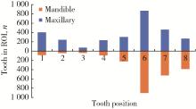
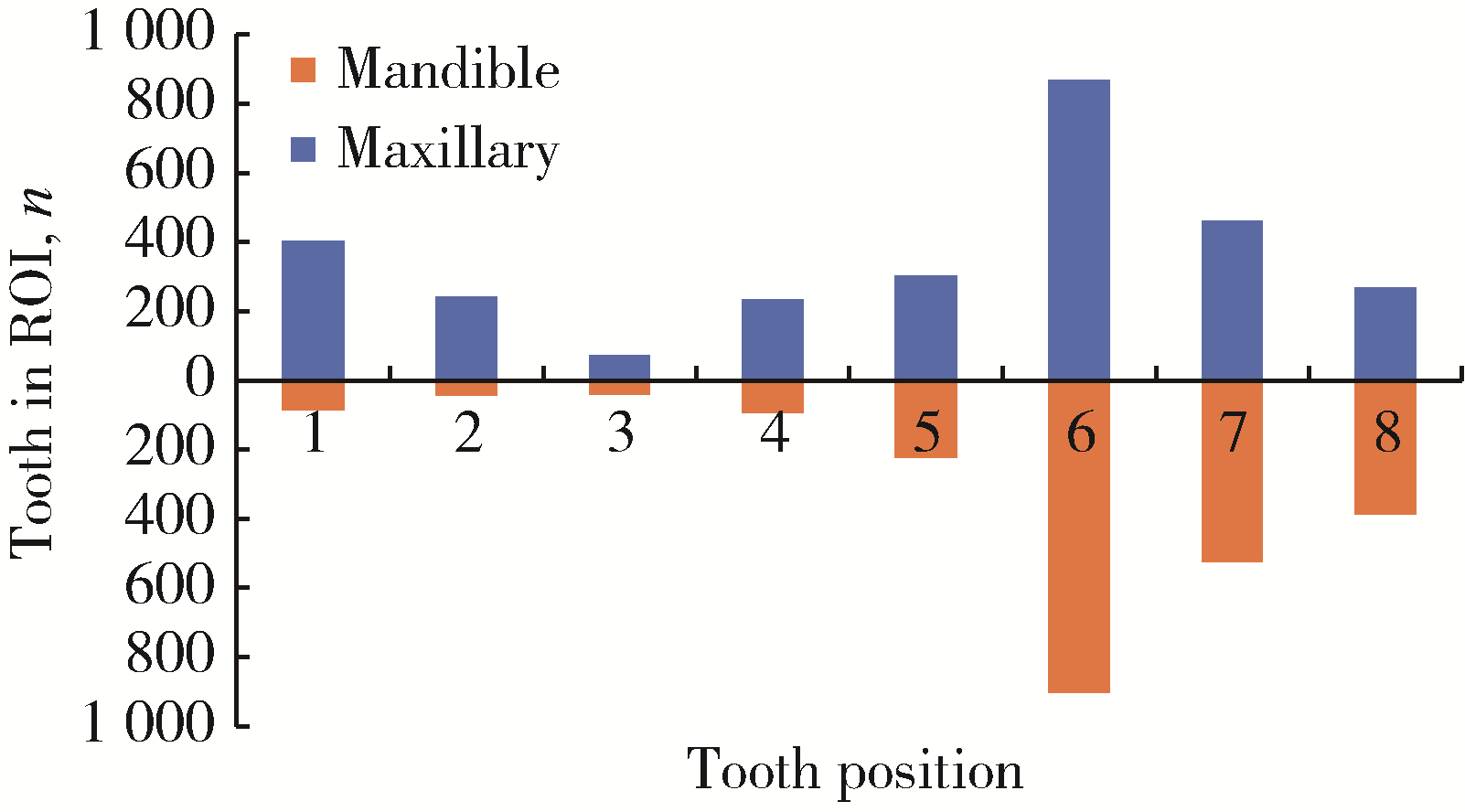
目的: 调查锥形束CT(cone-beam computed tomography, CBCT)在牙髓专科医师的临床使用情况, 分析CBCT在牙髓根尖周病诊治中作用, 为CBCT的合理应用提供参考。方法: 收集2021年1至12月北京大学口腔医院牙体牙髓科就诊并拍摄CBCT的患者临床资料, 纳入患者临床资料完整, 包括临床病历、放射申请单/报告及二维和三维影像学资料, 除外因正畸或修复等原因而拍摄者。分析临床资料, 调查应用CBCT的牙髓医师经验及培训背景、全年接诊患者数量, 同时检索CBCT检查目的和兴趣区、CBCT拍摄的技术参数(如机型和视野)、拍摄后的报告, 评估对诊断的影响。应用Wilcoxon和Mann-Whitney检验比较CBCT兴趣区的分布, 应用卡方检验及两两比较分析不同临床经验的牙髓专科医师(高、中、低年资)应用CBCT的情况。结果: 共61名临床医师全年接诊34 952人, 申请CBCT扫描共3 308份, 其中受检者3 218例(男∶女约为1 ∶2), 年龄中位数35岁(28, 49), 占全年接诊患者数量10%;其中98%的CBCT检查在拍摄二维根尖片后, 96%的CBCT扫描投照视野小于10 cm×10 cm。3 308份CBCT中83%的扫描兴趣区为上下颌后牙区, 拍摄数高于前牙17%(Z=-2.278, P < 0.05), 受检最多的上下颌第一磨牙占受检患牙的35%。统计CBCT扫描目的主要包括三方面: 明确临床诊断、指导手术和非手术牙髓治疗(包括牙髓治疗并发症处理)及疗效评估, 分别为1 111份(34%)、1 745份(54%)、311份(10%), 其他占2%。CBCT用于明确临床诊断, 主要应用于慢性根尖周炎、牙根折裂、牙根吸收和牙外伤, 其中353份CBCT检查用于牙根折裂的诊断和鉴别诊断, 阳性诊断率为35%(125/353)。为揭示根管系统解剖而拍摄的846份CBCT中, 297份为治疗失败后明确是否有遗漏根管, 其中58%(171/297)病例确认存在遗漏根管。在治疗并发症的处理中, CBCT主要用于辅助侧穿的诊断和分离器械的定位。311份CBCT检查应用于疗效评价, 包括根管治疗后240份和根尖手术后71份, 拍摄原因为复查或治疗后有临床症状、二维根尖片显示持续存在病损。使用CBCT的61名医师包括高年资医师23名, 占开具CBCT检查总数的45%, 中、低年资医师分别为15、23名, 开具CBCT检查占比分别为30%和25%。高年资与低年资医师申请CBCT检查各占接诊患者的10%, 高于中年资医师的8%(χ12=39.4, χ22=29.1, P < 0.001)。主任医师申请比例为18%, 高于副主任医师的9%(χ12=139.4, P < 0.001)。CBCT拍摄后医师改变诊断或调整计划者约31% (1 009/3 308)。结论: 牙髓专科医师应用CBCT获得更加丰富的临床信息, 有助于诊断和精准治疗及预后判断。
中图分类号:
- R781.3
| 1 |
Uraba S , Ebihara A , Komatsu K , et al. Ability of cone-beam computed tomography to detect periapical lesions that were not detected by periapical radiography: A retrospective assessment according to tooth group[J]. J Endod, 2016, 42 (8): 1186- 1190.
doi: 10.1016/j.joen.2016.04.026 |
| 2 | 姜岚, 陈晨, 高学军, 等. 锥形束CT与根尖X线片诊断根尖病变的准确性对比[J]. 中华口腔医学杂志, 2013, 48 (z1): 1- 5. |
| 3 |
Patel S , Dawood A , Whaites E , et al. New dimensions in endodontic imaging: Part 1. Conventional and alternative radiographic systems[J]. Int Endod J, 2009, 42 (6): 447- 462.
doi: 10.1111/j.1365-2591.2008.01530.x |
| 4 |
Patel S , Dawood A , Wilson R , et al. The detection and management of root resorption lesions using intraoral radiography and cone beam computed tomography: An in vivo investigation[J]. Int Endod J, 2009, 42 (9): 831- 838.
doi: 10.1111/j.1365-2591.2009.01592.x |
| 5 |
Ahlowalia M , Patel S , Anwar H , et al. Accuracy of CBCT for volumetric measurement of simulated periapical lesions[J]. Int Endod J, 2013, 46 (6): 538- 546.
doi: 10.1111/iej.12023 |
| 6 |
Metska ME , Aartman IH , Wesselink PR , et al. Detection of vertical root fractures in vivo in endodontically treated teeth by cone-beam computed tomography scans[J]. J Endod, 2012, 38 (10): 1344- 1347.
doi: 10.1016/j.joen.2012.05.003 |
| 7 |
Özer SY . Detection of vertical root fractures by using cone beam computed tomography with variable voxel sizes in an in vitro model[J]. J Endod, 2011, 37 (1): 75- 79.
doi: 10.1016/j.joen.2010.04.021 |
| 8 |
Patel S , Brady E , Wilson R , et al. The detection of vertical root fractures in root filled teeth with periapical radiographs and CBCT scans[J]. Int Endod J, 2013, 46 (12): 1140- 1152.
doi: 10.1111/iej.12109 |
| 9 |
Zou X , Liu D , Yue L , et al. The ability of cone-beam compute-rized tomography to detect vertical root fractures in endodontically treated and nonendodontically treated teeth: A report of 3 cases[J]. Oral Surg Oral Med Oral Pathol Oral Radiol Endod, 2011, 111 (6): 797- 801.
doi: 10.1016/j.tripleo.2010.12.015 |
| 10 |
Chavda R , Mannocci F , Andiappan M , et al. Comparing the in vivo diagnostic accuracy of digital periapical radiography with cone-beam computed tomography for the detection of vertical root fracture[J]. J Endod, 2014, 40 (10): 1524- 1529.
doi: 10.1016/j.joen.2014.05.011 |
| 11 |
Bernardes RA , de Paulo RS , Pereira LO , et al. Comparative study of cone beam computed tomography and intraoral periapical radiographs in diagnosis of lingual-simulated external root resorptions[J]. Dent Traumatol, 2012, 28 (4): 268- 272.
doi: 10.1111/j.1600-9657.2011.01113.x |
| 12 |
Estrela C , Bueno MR , Porto OC , et al. Influence of intracanal post on apical periodontitis identified by cone-beam computed tomography[J]. Braz Dent J, 2009, 20 (5): 370- 375.
doi: 10.1590/S0103-64402009000500003 |
| 13 |
American Association of Endodontists , American Academy of Oral and Maxillofacial Radiology . Use of cone-beam computed tomography in endodontics joint position statement of the American Association of Endodontists and the American Academy of Oral and Maxillofacial Radiology[J]. Oral Surg Oral Med Oral Pathol Oral Radiol Endod, 2011, 111 (2): 234- 237.
doi: 10.1016/j.tripleo.2010.11.012 |
| 14 |
Setzer FC , Hinckley N , Kohli MR , et al. A survey of cone-beam computed tomographic use among endodontic practitioners in the United States[J]. J Endod, 2017, 43 (5): 699- 704.
doi: 10.1016/j.joen.2016.12.021 |
| 15 |
Alzamazmi ZT , Abulhamael AM , Talim DJ , et al. Cone-beam computed tomographic usage: Survey of american endodontists[J]. J Contemp Dent Pract, 2019, 20 (10): 1132- 1137.
doi: 10.5005/jp-journals-10024-2661 |
| 16 |
梁宇红, 岳林. 锥形束CT在牙髓根尖周病诊治中的合理应用与思考[J]. 中华口腔医学杂志, 2019, 54 (9): 591- 597.
doi: 10.3760/cma.j.issn.1002-0098.2019.09.003 |
| 17 |
Ludlow JB , Timothy R , Walker C , et al. Correction to effective dose of dental CBCT: A meta analysis of published data and additional data for nine CBCT units[J]. Dentomaxillofac Radiol, 2015, 44 (7): 20159003.
doi: 10.1259/dmfr.20159003 |
| 18 |
Ludlow JB , Ivanovic M . Comparative dosimetry of dental CBCT devices and 64-slice CT for oral and maxillofacial radiology[J]. Oral Surg Oral Med Oral Pathol Oral Radiol Endod, 2008, 106 (1): 106- 114.
doi: 10.1016/j.tripleo.2008.03.018 |
| 19 |
Patel S , Brown J , Semper M , et al. European Society of Endodontology position statement: Use of cone beam computed tomography in endodontics European Society of Endodontology (ESE) deve-loped by[J]. Int Endod J, 2019, 52 (12): 1675- 1378.
doi: 10.1111/iej.13187 |
| 20 |
中华口腔医学会牙体牙髓病学专业委员会. 牙体牙髓病诊疗中口腔放射学的应用指南[J]. 中华口腔医学杂志, 2021, 56 (4): 311- 317.
doi: 10.3760/cma.j.cn112144-20210125-00039 |
| 21 | Mathew AI, Lee SC, Ha WN, et al. Cone-beam computed tomography-predictors and characteristics of usage in Australia and New Zealand: A multifactorial analysis[J/OL]. Aust Endod J, 2022, 7 (2022-07-13)[2022-09-13]. https://pubmed.ncbi.nlm.nih.gov/35830370. |
| 22 | Rajeevan M , Chandler NP , Makdissi J , et al. A survey of cone beam computed tomography (CBCT) use among endodontic practitioners in the UK[J]. Endo-Endod Pract Tod, 2018, 12 (1): 29- 33. |
| 23 |
Bhatt M , Coil J , Chehroudi B , et al. Clinical decision-making and importance of the AAE/AAOMR position statement for CBCT examination in endodontic cases[J]. Int Endod J, 2021, 54 (1): 26- 37.
doi: 10.1111/iej.13397 |
| 24 |
Mota de Almeida FJ , Knutsson K , Flygare L . The effect of cone beam CT (CBCT) on therapeutic decision-making in endodontics[J]. Dentomaxillofac Radiol, 2014, 43 (4): 20130137.
doi: 10.1259/dmfr.20130137 |
| 25 |
Rodriguez G , Patel S , Duran-Sindreu F , et al. Influence of cone-beam computed tomography on endodontic retreatment strategies among general dental practitioners and endodontists[J]. J Endod, 2017, 43 (9): 1433- 1437.
doi: 10.1016/j.joen.2017.04.004 |
| 26 |
Ee J , Fayad MI , Johnson BR . Comparison of endodontic diagnosis and treatment planning decisions using cone-beam volumetric tomography versus periapical radiography[J]. J Endodont, 2014, 40 (7): 910- 916.
doi: 10.1016/j.joen.2014.03.002 |
| 27 | Aljuhani A , Dutta S , Mandorah A . Evaluation of knowledge and perspective of endodontic residents and general dentist towards the endodontic application of CBCT in Saudi Arabia[J]. J Res Med Dent Sci, 2020, 8 (7): 459- 464. |
| [1] | 薄士仕,高承志. 基于卷积神经网络实现锥形束CT牙齿分割及牙位标定[J]. 北京大学学报(医学版), 2024, 56(4): 735-740. |
| [2] | 章锦花,潘洁,孙志鹏,王霄. 不同根管内容物对口腔颌面锥形束CT诊断牙根纵裂准确性的影响[J]. 北京大学学报(医学版), 2023, 55(2): 333-338. |
| [3] | 潘孟乔,刘建,徐莉,徐筱,侯建霞,李小彤,王晓霞. 牙周-正畸-正颌联合治疗骨性安氏Ⅲ类错 |
| [4] | 高娟,吕航苗,马慧敏,赵一姣,李小彤. 锥形束CT三维体积测量评估骨性Ⅲ类错 |
| [5] | 刘伟涛,王怡然,王雪东,周彦恒. 锥形束CT研究上颌反复扩缩前方牵引后上颌骨缝的三维变化[J]. 北京大学学报(医学版), 2022, 54(2): 346-355. |
| [6] | 孟圆,张丽琪,赵雅宁,柳登高,张祖燕,高岩. 67例上颌根尖周囊肿的三维影像特点分析[J]. 北京大学学报(医学版), 2021, 53(2): 396-401. |
| [7] | 曹畅,王菲,王恩博,刘宇. β-磷酸三钙用于下颌第三磨牙拔除术后骨缺损修复的自身对照研究[J]. 北京大学学报(医学版), 2020, 52(1): 97-102. |
| [8] | 谢晓艳,贾淑梅,孙志辉,张祖燕. 分辨率设置与锥形束CT检测牙根外吸收的可靠性[J]. 北京大学学报(医学版), 2019, 51(1): 75-79. |
| [9] | 赵一姣,刘怡,孙玉春,王勇. 一种基于曲率连续算法的冠、根三维数据融合方法[J]. 北京大学学报(医学版), 2017, 49(4): 719-723. |
| [10] | 钟金晟, 欧阳翔英, 柳登高, 曹采方. 锥形束CT测量离体下颌磨牙Ⅱ°根分叉病变效果的评价[J]. 北京大学学报(医学版), 2010, 42(1): 41-45. |
| [11] | 刘怡, James MAH, 许天民. 锥形束计算机断层扫描中牙齿的分割精度[J]. 北京大学学报(医学版), 2010, 42(1): 98-102. |
| [12] | 王瑞永, 马绪臣, 张万林, 柳登高. 健康成年人颞下颌关节间隙锥形束计算机体层摄影术测量分析[J]. 北京大学学报(医学版), 2007, 39(5): 503-506. |
|
||
