北京大学学报(医学版) ›› 2023, Vol. 55 ›› Issue (1): 120-123. doi: 10.19723/j.issn.1671-167X.2023.01.018
口腔内窥镜在口腔解剖标志识别中的应用
- 北京大学口腔医学院·口腔医院修复科, 国家口腔医学中心, 国家口腔疾病临床医学研究中心, 口腔生物材料和数字诊疗装备国家工程研究中心, 口腔数字医学北京市重点实验室, 国家卫生健康委员会口腔医学计算机应用工程技术研究中心, 国家药品监督管理局口腔生物材料重点实验室, 北京 100081
A study on the application of intraoral camera in the identification of oral anatomical landmarks
Shu-ting CHIU,Ebrahimi FARAZ,Xiao ZHANG,Hong-qiang YE*( ),Yun-song LIU*(
 )
)
- Department of Prosthodontics, Peking University School and Hospital of Stomatology & National Center of Stomatology & National Clinical Research Center for Oral Diseases & National Engineering Research Center of Oral Biomaterials and Digi-tal Medical Devices & Beijing Key Laboratory of Digital Stomatology & NHC Research Center of Engineering and Technology for Computerized Dentistry & NMPA Key Laboratory for Dental Materials, Beijing 100081, China
摘要:
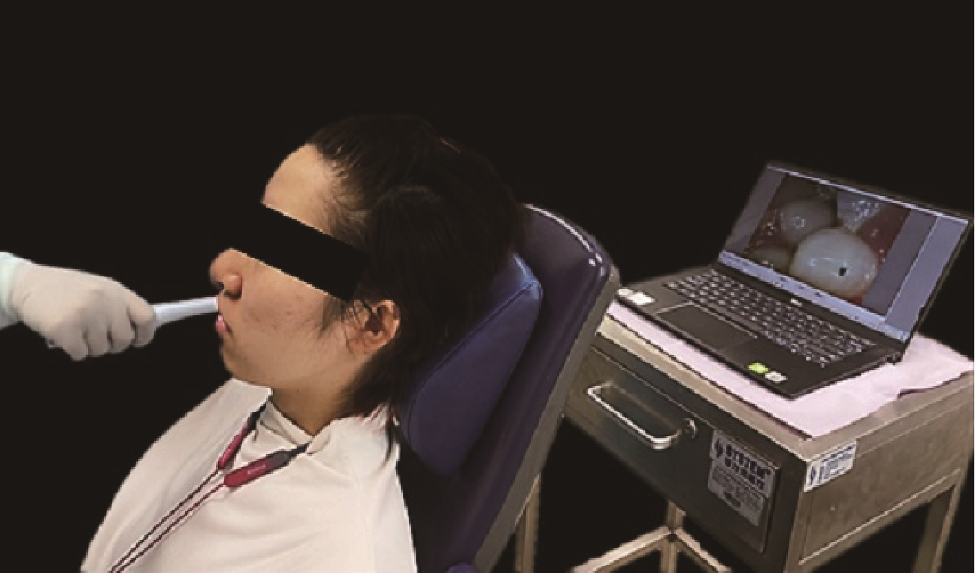
目的: 初步探索口腔内窥镜辅助口腔解剖标志识别的应用场景, 以改进临床诊疗模式, 培养爱伤观念, 加强医患沟通, 辅助专家示教, 提高临床诊疗效率。方法: 在口腔解剖标志识别中应用新型口腔内窥镜, 并开发4种应用场景, (1)临床诊疗场景, 医生使用口腔内窥镜对患者口内进行全面检查并拍摄录像及照片; (2)医患沟通场景, 医生向患者讲述治疗计划时, 将口腔内窥镜拍摄的录像或照片展示给患者; (3)专家示教场景, 在专家操作时, 专家在患者口内使用口腔内窥镜进行示教, 年轻医生在投影屏上学习口腔解剖标志, 配合理论知识的学习; (4)疑难病例记录场景, 临床诊疗的过程中, 遇到疑难病例时可以使用口腔内窥镜进行录像和拍照, 供年轻医生讨论, 专家进行点评和指导。结果: 口腔内窥镜的应用, (1)改进临床诊疗模式, 提高临床诊疗效率; (2)激发年轻医生学习兴趣, 巧妙地将解剖标志的理论知识与临床实践相结合, 从而提升学习效果; (3)培养爱伤观念, 强化轻柔操作的重要性; (4)加强医患沟通, 医生可以更形象化地与患者交流, 使患者对自身情况更加了解, 增加患者对医生的信任。结论: 口腔内窥镜可以辅助口腔临床诊疗, 如口内解剖标志识别等, 对于改进临床诊疗模式、激发学习兴趣、培养爱伤观念、提升医患沟通效果有一定的促进作用。
中图分类号:
- R781.0
| 1 | 周永胜. 口腔修复学[M]. 3版 北京: 北京大学医学出版社, 2020: 289- 292. |
| 2 | 孙玉春, 王勇, 邓珂慧, 等. 功能易适数字化全口义齿的自主创新研发[J]. 北京大学学报(医学版), 2020, 52 (2): 390- 394. |
| 3 | 张志愿. 口腔颌面外科学[M]. 8版 北京: 人民卫生出版社, 2020: 45- 49. |
| 4 |
刘济远, 唐休发, 王了, 等. 口腔内窥镜在牙槽外科临床实习教学中的应用初探[J]. 中国继续医学教育, 2020, 12 (35): 54- 57.
doi: 10.3969/j.issn.1674-9308.2020.35.015 |
| 5 |
Wander P , Ireland RS . Dental photography in record keeping and litigation[J]. Br Dent J, 2014, 217 (3): 133- 137.
doi: 10.1038/sj.bdj.2014.649 |
| 6 |
Christensen GJ . Important clinical uses for digital photography[J]. J Am Dent Assoc, 2005, 136 (1): 77- 79.
doi: 10.14219/jada.archive.2005.0030 |
| 7 | Ferrazzano GF , Orlando S , Cantile T , et al. An experimental in vivo procedure for the standardised assessment of sealants retention over time[J]. Eur J Paediatr Dent, 2016, 17 (3): 176- 180. |
| 8 |
Alzayyat NA , Hafez RM , Yassen AA , et al. Accuracy of the light-induced fluorescent intraoral camera in occlusal caries detection[J]. J Contemp Dent Pract, 2021, 22 (4): 365- 372.
doi: 10.5005/jp-journals-10024-3082 |
| 9 |
Signori C , Collares K , Cumerlato CBF , et al. Validation of assessment of intraoral digital photography for evaluation of dental restorations in clinical research[J]. J Dent, 2018, 71, 54- 60.
doi: 10.1016/j.jdent.2018.02.001 |
| 10 | Vetchaporn S , Rangsri W , Ittichaicharoen J , et al. Validity and reliability of intraoral camera with fluorescent aids for oral potentially malignant disorders screening in teledentistry[J]. Int J Dent, 2021, 2021, 6814027. |
| 11 | 马莎, 朱文卓, 赵豫梅, 等. 基于当代医患关系对医学生医德教育的思考与探讨[J]. 中国继续医学教育, 2022, 14 (13): 162- 165. |
| 12 | 王姝侠, 彭雪寒, 孟忆贫, 等. 论医学生爱伤意识的培养[J]. 重庆医学, 2013, 42 (25): 3068- 3069. |
| 13 | 董向辉, 靳占奎. PBL方法联合SEGUE量表提升骨科规培医师医患沟通能力的应用效果[J]. 中国当代医药, 2022, 29 (27): 157- 161. |
| [1] | 陈晨,梁宇红. 复杂根管上颌磨牙的根管治疗3例[J]. 北京大学学报(医学版), 2024, 56(1): 190-195. |
| [2] | 赵菡,卫彦,张学慧,杨小平,蔡晴,宁成云,徐明明,刘雯雯,黄颖,何颖,郭亚茹,江圣杰,白云洋,吴宇佳,郭雨思,郑晓娜,李文静,邓旭亮. 口腔硬组织修复材料仿生设计制备和临床转化[J]. 北京大学学报(医学版), 2024, 56(1): 4-8. |
| [3] | 周团锋,杨雪,王睿捷,程明轩,张华,韦金奇. 数字化制作简易口内哥特式弓在全口义齿修复正中关系确定中的应用[J]. 北京大学学报(医学版), 2023, 55(1): 101-107. |
| [4] | 雍颹,钱锟,朱文昊,赵晓一,刘畅,潘洁. 成年恒牙牙髓切断后牙髓钙化的X线片评价[J]. 北京大学学报(医学版), 2023, 55(1): 88-93. |
| [5] | 李伟伟,陈虎,王勇,孙玉春. 氧化锆陶瓷表面硅锂喷涂层的摩擦磨损性能[J]. 北京大学学报(医学版), 2023, 55(1): 94-100. |
| [6] | 彭俐,王祖华,孙玉春,渠薇,韩扬,梁宇红. 根尖切除手术导板的计算机辅助设计及三维打印[J]. 北京大学学报(医学版), 2018, 50(5): 905-910. |
| [7] | 谭京,魏秀霞,张庆辉,周永胜. 使用改良半固定桥修复单个缺失后牙的3年临床观察[J]. 北京大学学报(医学版), 2018, 50(2): 314-317. |
| [8] | 焦洋, 王继德, 邓久鹏. 不同表面处理对氧化锆晶相结构及性能的影响[J]. 北京大学学报(医学版), 2018, 50(1): 49-52. |
| [9] | 廖宇,刘晓强,陈立,周建锋,谭建国. 不同表面处理方法对氧化锆与树脂水门汀粘接强度的影响[J]. 北京大学学报(医学版), 2018, 50(1): 53-57. |
| [10] | 张皓羽,姜婷,程明轩,张玉玮. 类瓷树脂及玻璃陶瓷牙合贴面疲劳实验前后的磨耗及表面粗糙度的变化[J]. 北京大学学报(医学版), 2018, 50(1): 73-77. |
| [11] | 唐琳,张一,李皓,刘玉华,周永胜,李博文,吴唯伊,王思雯. 不同浓度的碳二亚胺乙醇溶液处理对牙本质自酸蚀粘接剂粘接强度的影响[J]. 北京大学学报(医学版), 2017, 49(6): 1055-1059. |
| [12] | 李皓,刘玉华,罗志强. 生物活性玻璃用于缓解活髓牙全冠预备后敏感的效果评价[J]. 北京大学学报(医学版), 2017, 49(4): 709-713. |
| [13] | 赵一姣,刘怡,孙玉春,王勇. 一种基于曲率连续算法的冠、根三维数据融合方法[J]. 北京大学学报(医学版), 2017, 49(4): 719-723. |
|
||


