北京大学学报(医学版) ›› 2023, Vol. 55 ›› Issue (1): 124-132. doi: 10.19723/j.issn.1671-167X.2023.01.019

• 论著 • 上一篇    下一篇

初诊IgA肾病患者的肠道菌群及其与疾病进展因素的相关分析

包文晗,唐雯*()   

  1. 北京大学第三医院肾内科,北京 100191
  • 收稿日期:2021-02-05 出版日期:2023-02-18 发布日期:2023-01-31
  • 通讯作者: 唐雯 E-mail:tanggwen@126.com
  • 基金资助:
    中华医学会临床医学科研专项资金项目(14050460583);中华国际医学交流基金会(Z-2017-24-203)

Changes of gut microflora in newly diagnosed IgA nephropathy patients and its correlation with clinical risk factors

Wen-han BAO,Wen TANG*()   

  1. Department of Nephrology, Peking University Third Hospital, Beijing 100191, China
  • Received:2021-02-05 Online:2023-02-18 Published:2023-01-31
  • Contact: Wen TANG E-mail:tanggwen@126.com
  • Supported by:
    the Clinical Medical Research Special Fund Project of Chinese Medical Association(14050460583);the China International Medical Foundation(Z-2017-24-203)

RICH HTML

16   

摘要:

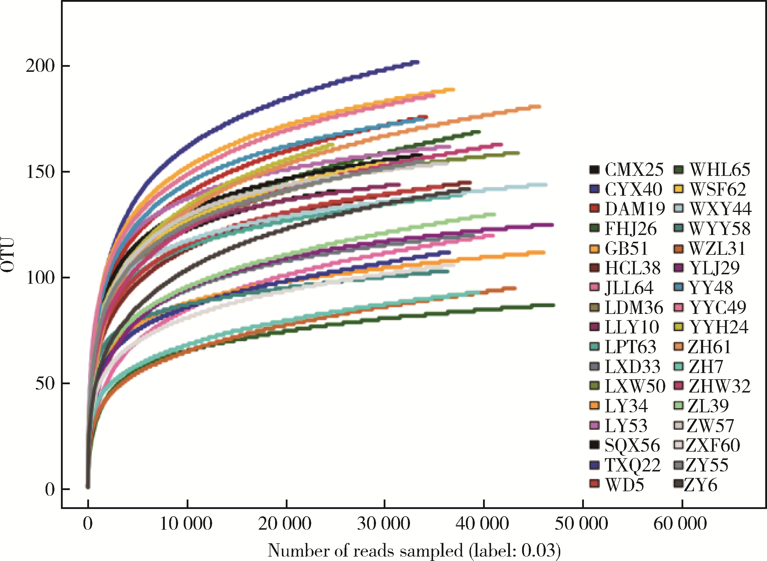
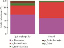
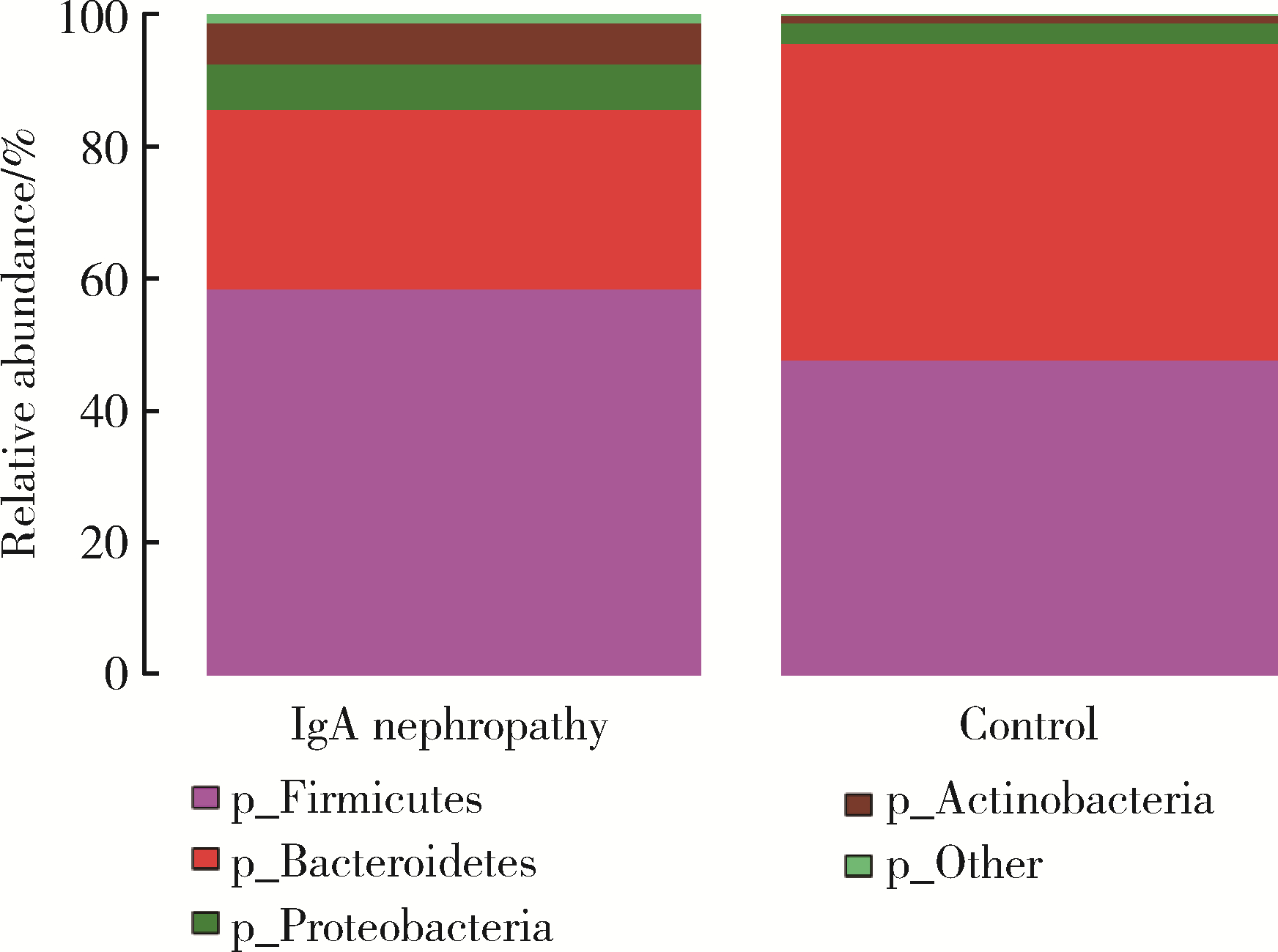
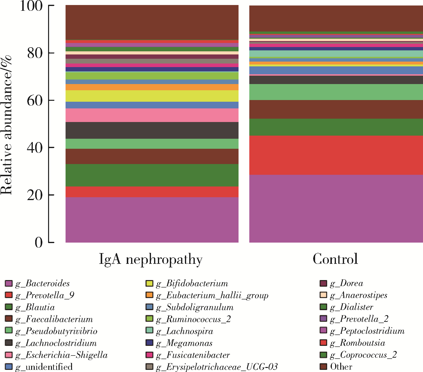
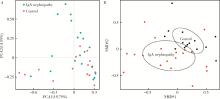
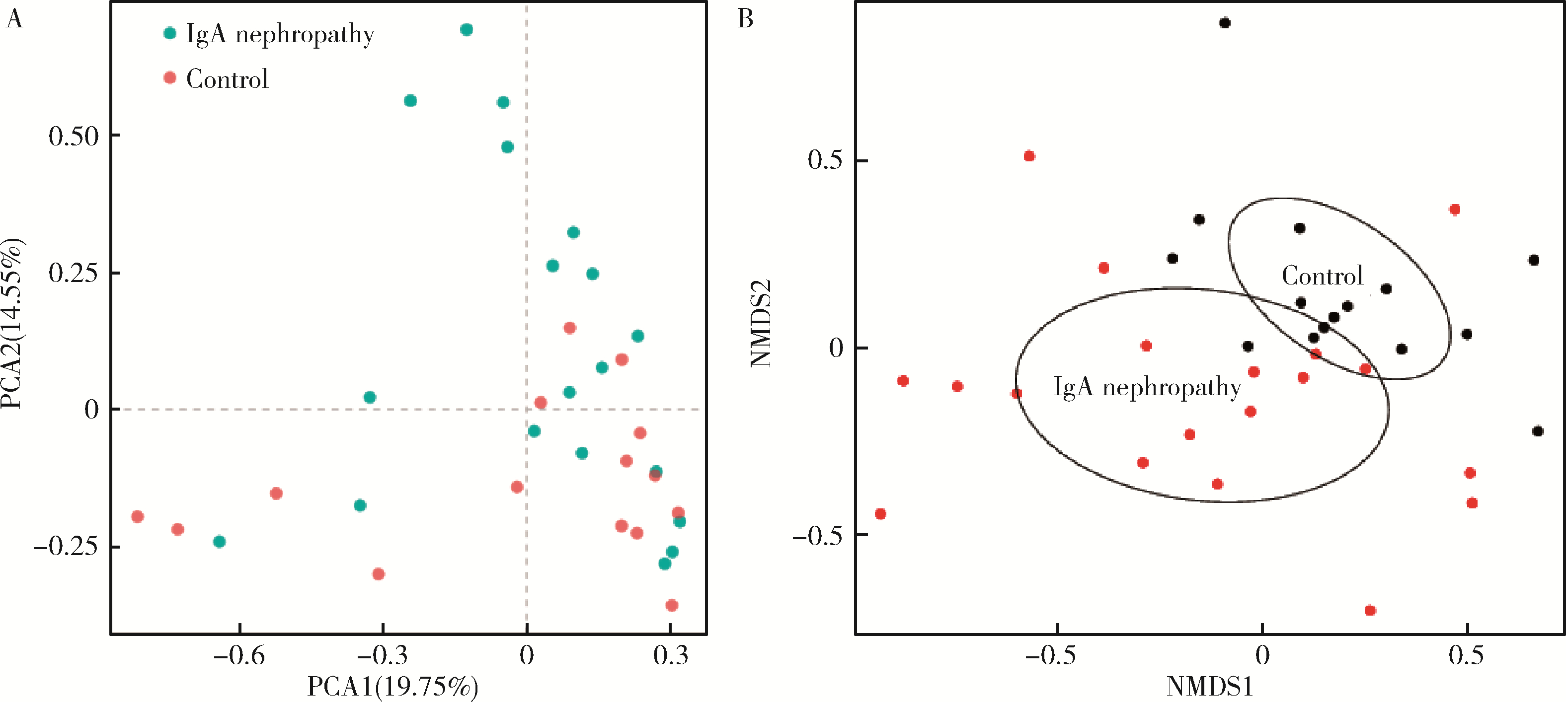
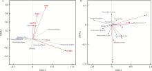
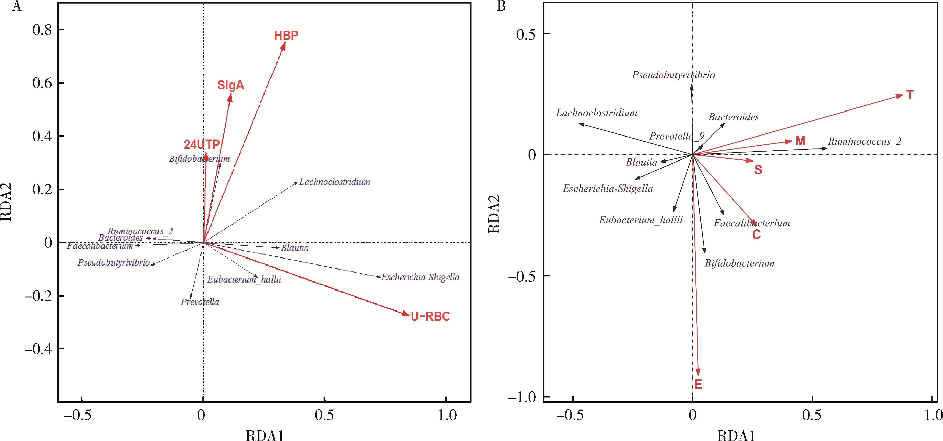
目的: 观察慢性肾脏病(chronic kidney disease,CKD)1~2期的初诊IgA肾病患者肠道微生物群的特征,进一步探索IgA肾病疾病进展因素与肠道微生物之间的关系。方法: 收集19例CKD 1~2期的初诊IgA肾病患者和15例年龄、性别相匹配的健康对照组的新鲜粪便样本,提取粪便细菌DNA,针对V3-V4区域进行16S核糖体RNA(16S ribosomal RNA,16S rRNA)高通量测序分析肠道菌群的组成,应用Illumina Miseq平台对粪便菌群测序结果进行分析。收集初诊IgA肾病患者的疾病进展因素,研究肠道菌群与IgA肾病疾病进展因素之间的相关性。结果: (1) 与健康对照组相比,在门水平上,拟杆菌门(Bacteroidetes)的丰度显著降低(P=0.046),放线菌门(Actinobacteria)的丰度显著升高(P=0.001)。在属水平上,埃希菌-志贺菌属(Escherichia-Shigella)、双歧杆菌属(Bifidobacterium)、Dorea等菌属的丰度显著升高(P < 0.05),毛螺菌属(Lachnospira)、粪球菌属_2(Coprococcus_2)、萨特氏菌属(Sutterella)等菌属的丰度显著降低(P < 0.05)。(2)初诊IgA肾病患者与健康对照组之间肠道菌群的丰度比较差异无统计学意义(P>0.05),但两组肠道菌群的组成存在差异。LEfSe分析结果显示,初诊IgA肾病患者和健康对照组比较有16个差异菌,其中,初诊IgA肾病患者肠杆菌目(Enterobacteriales)、放线菌门、埃希菌-志贺菌属等的丰度升高,健康对照组拟杆菌门、毛螺菌属的丰度升高。(3)冗余分析(redundancy analysis,RDA)的结果显示,初诊IgA肾病患者粪便中双歧杆菌属与血清IgA水平、24 h尿蛋白定量和合并高血压呈正相关,Lachnoclostridium菌属与合并高血压呈正相关,埃希菌-志贺菌属与尿红细胞数量呈正相关,双歧杆菌属与毛细血管内增殖呈正相关,粪杆菌属与细胞/纤维细胞性新月体呈正相关,瘤胃球菌属_2(Ruminococcus_2)与系膜细胞增殖、肾小球节段硬化、肾小管萎缩/间质纤维化呈正相关。结论: 初诊CKD 1~2期IgA肾病患者的肠道菌群与健康对照相比存在差异,肠道的某些菌属与IgA肾病疾病进展因素呈相关性,需要进一步的研究来了解这些菌属在IgA肾病中的潜在作用。

关键词: 肾小球肾炎, 免疫球蛋白A, 胃肠道微生物组, 疾病特征, 16S核糖体RNA

Abstract:

Objective: To investigate the gut microbiota in newly diagnosed IgA nephropathy patients with chronic kidney disease (CKD) stages 1-2 and the association between the gut microbiota and the clinical risk factors of IgA nephropathy. Methods: Fresh fecal samples were collected from nineteen newly diagnosed IgA nephropathy patients with CKD stages 1-2 and fifteen age- and sex-matched healthy controls. Fecal bacterial DNA was extracted and microbiota composition were characterized using 16S ribosomal RNA (16S rRNA) high-throughput sequencing for the V3-V4 region. The Illumina Miseq platform was used to analyze the results of 16S rRNA high-throughput sequencing of fecal flora. At the same time, the clinical risk factors of IgA nephropathy patients were collected to investigate the association between the gut microbiota and the clinical risk factors. Results: (1) At the phylum level, the abundance of Bacteroidetes was significantly reduced (P=0.046), and the abundance of Actinobacteria was significantly increased (P=0.001). At the genus level, the abundance of Escherichia-Shigella, Bifidobacte-rium, Dorea and others were significantly increased (P < 0.05). The abundance of Lachnospira, Coprococcus_2 and Sutterella was significantly reduced (P < 0.05). (2) There was no significant difference in the abundance of gut microbiota between the newly diagnosed IgA nephropathy patients and the healthy control group (P>0.05), but there were differences in the structure of the gut microbiota between the two groups. The results of LEfSe analysis showed that there were 16 differential bacteria in the newly diagnosed IgA nephropathy patients and healthy controls. Among them, the abundance of the newly diagnosed IgA nephropathy patients was increased in Enterobacteriales, Actinobacteria, Escherichia-Shigella, etc. The healthy control group was increased in Bacteroidetes and Lachnospira. (3) The result of redundancy analysis (RDA) showed that Bifidobacterium was positively correlated with serum IgA levels, 24-hour urinary protein levels and the presence of hypertension. Lachnoclostridium was positively correlated with the presence of hypertension. Escherichia-Shigella was positively correlated with urine red blood cells account. Bifidobacterium was positively correlated with the proliferation of capillaries. Faecalibacterium was positively correlated with cell/fibrocytic crescents. Ruminococcus_2 was positively correlated with mesangial cell proliferation, glomerular segmental sclerosis and renal tubular atrophy/interstitial fibrosis. Conclusion: The gut microbiota in the newly diagnosed IgA nephropathy patients with CKD stages 1-2 is different from that of the healthy controls. Most importantly, some gut bacteria are related to the clinical risk factors of IgA nephropathy. Further research is needed to understand the potential role of these bacteria in IgA nephropathy.

Key words: Glomerulonephritis, immunoglobulin A, Gastrointestinal microbiome, Disease attributes, RNA, ribosomal, 16S

中图分类号: 

  • R692.3

表1

IgA肾病和健康对照的基本特征"

Items IgA nephropathy (n=19) Health controls (n=15) P
Age/years, ˉx±s 33.53±3.96 34.95±8.58 0.529
Male 53% 47% 0.790
Serum creatinine/(μmol/L), ˉx±s 84.9±19.3 64.4±6.7 < 0.001
CKD-EPI eGFR/[mL/(min·1.73 m2)], ˉx±s 90.7±17.7 126.0±21.0 < 0.001
Pathological features of IgA nephropathy renal biopsy*
  Mesangial hypercellularity (M) M0=0%, M1=94.74%, M2=5.26%, M3=0%
  Endocapillary proliferation (E) E0=26.32%, E1=73.68%
  Segmental sclerosis (S) S0=68.42%, S1=31.58%
  Tubular atrophy/interstitial fibrosis (T) T0=73.68%, T1=26.32%, T2=0%
  Cellular/fibrocytic crescent (C) C0=84.21%, C1=15.79%, C2=0%

图1

样本稀释性曲线"

图2

IgA肾病患者和健康对照者肠道菌群门水平的对比"

表2

IgA肾病患者和健康对照者肠道菌群的相对丰度变化"

Items IgA nephropathy (n=19), M (min, max) Controls (n=15), M (min, max) P
Phylum
  Bacteroidetes 3.13×10⁻¹ (3.86×10⁻², 7.65×10⁻¹) 4.13×10⁻¹ (8.29×10⁻², 8.61×10⁻¹) 0.046
  Actinobacteria 2.47×10⁻² (6.84×10⁻⁴, 1.91×10⁻¹) 2.59×10⁻³ (2.63×10⁻⁴, 7.52×10⁻²) 0.001
  Lentisphaerae 0 (0, 2.93×10⁻⁵) 0 (0, 6.81×10⁻³) 0.011
  Saccharibacteria 5.07×10⁻⁵ (0, 6.40×10⁻⁴) 0 (0, 4.25×10⁻⁵) < 0.001
Genus
  Escherichia-Shigella 2.88×10⁻³ (7.79×10⁻⁵, 3.88×10⁻¹) 3.79×10⁻⁴ (0, 6.38×10⁻²) 0.025
  Bifidobacterium 1.17×10⁻² (2.409×10⁻⁵, 1.87×10⁻¹) 1.15×10⁻³ (5.42×10⁻⁵, 6.84×10⁻²) 0.036
  Lachnospira 1.43×10⁻³ (0, 1.82×10⁻²) 1.73×10⁻² (0, 1.25×10⁻¹) 0.002
  Dorea 8.84×10⁻³ (7.77×10⁻⁴, 1.03×10⁻¹) 4.67×10⁻³ (1.29×10⁻³, 1.07×10⁻²) 0.025
  Erysipelotrichaceae_UCG-003 7.42×10⁻³ (0, 1.65×10⁻¹) 1.75×10⁻³ (1.78×10⁻⁴, 3.03×10⁻²) 0.013
  Coprococcus_2 0 (0, 8.76×10⁻³) 3.97×10⁻³ (0, 4.59×10⁻²) 0.028
  Sutterella 0 (0, 1.87×10⁻³) 3.73×10⁻³ (0, 4.08×10⁻²) < 0.001
  Tyzzerella_3 0 (0, 2.52×10⁻²) 2.90×10⁻³ (0, 1.61×10⁻²) 0.012
  Thalassospira 0 (0, 1.73×10⁻³) 0 (0, 5.52×10⁻²) 0.015
  Lachnospiraceae_UCG-004 2.78×10⁻⁴ (0, 5.13×10⁻³) 2.50×10⁻³ (2.93×10⁻⁴, 1.27×10⁻²) 0.001
  Eubacterium_ventriosum_group 3.04×10⁻⁴ (0, 1.02×10⁻²) 1.31×10⁻³ (2.13×10⁻⁵, 1.27×10⁻²) 0.042
  Lachnospiraceae_ND3007_group 1.37×10⁻⁴ (0, 1.95×10⁻³) 1.68×10⁻³ (2.44×10⁻⁵, 1.08×10⁻²) 0.002
  Eggerthella 1.11×10⁻³ (0, 1.23×10⁻²) 0 (0, 6.58×10⁻⁵) < 0.001
  Ruminiclostridium_5 8.97×10⁻⁴ (7.78×10⁻⁵, 7.58×10⁻³) 4.14×10⁻⁴ (0, 1.34×10⁻³) 0.030
  Lachnospiraceae_UCG-010 5.06×10⁻⁵ (0, 3.48×10⁻³) 5.72×10⁻⁴ (0, 2.68×10⁻³) 0.049
  Butyricimonas 2.60×10⁻⁵ (0, 1.86×10⁻³) 3.73×10⁻⁴ (0, 8.27×10⁻³) 0.013
  Flavonifractor 2.63×10⁻⁴ (0, 5.19×10⁻³) 2.96×10⁻⁵ (0, 6.30×10⁻⁴) 0.035
  Desulfovibrio 0 (0, 1.82×10⁻³) 0 (0, 1.25×10⁻²) 0.045
  Ruminococcaceae_UCG-003 2.60×10⁻⁴ (0, 7.00×10⁻⁴) 4.44×10⁻⁴ (4.25×10⁻⁵, 3.12×10⁻³) 0.006
  Terrisporobacter 2.53×10⁻⁵ (0, 8.71×10⁻³) 0 (0, 6.58×10⁻⁴) 0.003
  Granulicatella 0.30×10⁻⁵ (0, 6.34×10⁻³) 0 (0, 5.72×10⁻⁵) 0.003
  Erysipelatoclostridium 4.05×10⁻⁵ (0, 3.34×10⁻³) 0 (0, 5.48×10⁻⁵) 0.009
  Ruminiclostridium_9 2.16×10⁻⁵ (0, 1.19×10⁻³) 2.36×10⁻⁴ (0, 1.14×10⁻³) 0.015
  Victivallis 0 (0, 2.93×10⁻⁵) 0 (0, 6.81×10⁻³) 0.011
  Cercis_gigantea 2.53×10⁻⁵ (0, 4.07×10⁻³) 0 (0, 3.33×10⁻⁵) 0.015
  Ezakiella 0 (0, 3.88×10⁻³) 0 (0, 2.74×10⁻⁵) 0.021
  Family_XIII_UCG-001 0 (0, 6.07×10⁻⁴) 6.91×10⁻⁵ (0, 3.52×10⁻⁴) 0.042
  Rothia 0 (0, 2.47×10⁻³) 0 (0, 8.91×10⁻⁵) 0.030
  Gemella 3.00×10⁻⁵ (0, 1.45×10⁻³) 0 (0, 8.26×10⁻⁵) 0.001
  Solobacterium 2.16×10⁻⁵ (0, 7.25×10⁻⁴) 0 (0, 3.33×10⁻⁵) 0.045
  Acinetobacter 0 (0, 2.13×10⁻⁴) 0 (0, 2.74×10⁻⁵) 0.028
  Corynebacterium 0 (0, 2.13×10⁻⁴) 0 (0, 0) 0.005
  Olsenella 2.16×10⁻⁵ (0, 8.09×10⁻⁵) 0 (0, 3.33×10⁻⁵) 0.007
  Atopobium 0 (0, 1.20×10⁻⁴) 0 (0, 0) 0.035
  Leptotrichia 0 (0, 1.20×10⁻⁴) 0 (0, 0) 0.010
  Methylobacterium 0 (0, 8.69×10⁻⁵) 0 (0, 0) 0.019

图3

IgA肾病患者和健康对照者肠道菌群属水平的对比"

表3

IgA肾病和健康对照肠道菌群α多样性分析"

Items IgA nephropathy (n=19), ˉx±s Controls (n=15), ˉx±s P
Shannon index 4.31±0.47 4.05±0.99 0.316
Chao1 index 158±32 157±35 0.942

图4

IgA肾病和健康对照间β多样性分析"

图5

IgA肾病和健康对照间LEfSe分析"

表4

IgA肾病患者的疾病进展因素"

Items IgA nephropathy (n=19)
Hypertention (yes/no) 7/12
Systolic pressure/mmHg 133 (121, 189)
Diastolic pressure/mmHg 88.32±11.56
Serum IgA levels/(mg/dL) 4.15 (1.78, 47.60)
Urine red blood cells account (/μL) 175 (12, 1 886)
24-hour urinary protein levels/g 1.86 (0.26, 9.37)

图6

肠道菌群与IgA肾病临床进展因素及肾脏病理相关性分析"

1 Schena FP , Nistor I . Epidemiology of IgA nephropathy: A global perspective[J]. Semin Nephrol, 2018, 38 (5): 435- 442.
doi: 10.1016/j.semnephrol.2018.05.013
2 Wang M , Lv J , Zhang X , et al. Secondary IgA nephropathy shares the same immune features with primary IgA nephropathy[J]. Kidney Int Rep, 2020, 5 (2): 165- 172.
doi: 10.1016/j.ekir.2019.10.012
3 Hu X , Du J , Xie Y , et al. Fecal microbiota characteristics of Chinese patients with primary IgA nephropathy: A cross-sectional study[J]. BMC Nephrol, 2020, 21 (1): 97.
doi: 10.1186/s12882-020-01741-9
4 De Angelis M , Montemurno E , Piccolo M , et al. Microbiota and metabolome associated with immunoglobulin A nephropathy (IgAN)[J]. PLoS One, 2014, 9 (6): e99006.
doi: 10.1371/journal.pone.0099006
5 Hobby GP , Karaduta O , Dusio GF , et al. Chronic kidney disease and the gut microbiome[J]. Am J Physiol Renal Physiol, 2019, 316 (6): F1211- F1217.
doi: 10.1152/ajprenal.00298.2018
6 Munyaka PM , Eissa N , Bernstein CN , et al. Antepartum antibio-tic treatment increases offspring susceptibility to experimental colitis: A role of the gut microbiota[J]. PLoS One, 2015, 10 (11): e0142536.
doi: 10.1371/journal.pone.0142536
7 Edgar RC . UPARSE: highly accurate OTU sequences from microbial amplicon reads[J]. Nat Methods, 2013, 10 (10): 996- 998.
doi: 10.1038/nmeth.2604
8 Cole JR , Wang Q , Cardenas E , et al. The ribosomal database project: Improved alignments and new tools for rRNA analysis[J]. Nucleic Acids Res, 2009, 37 (Suppl 1): D141- D145.
9 Segata N , Izard J , Waldron L , et al. Metagenomic biomarker discovery and explanation[J]. Genome Biol, 2011, 12 (6): R60.
doi: 10.1186/gb-2011-12-6-r60
10 Mitchell RJ , Hewison RL , Fielding DA , et al. Decline in atmospheric sulphur deposition and changes in climate are the major drivers of long-term change in grassland plant communities in Scotland[J]. Environ Pollut, 2018, 235, 956- 964.
doi: 10.1016/j.envpol.2017.12.086
11 Li H , Li T , Beasley DE , et al. Diet diversity is associated with beta but not alpha diversity of pika gut microbiota[J]. Front Microbiol, 2016, 7, 1169.
12 Suzuki H , Kiryluk K , Novak J , et al. The pathophysiology of IgA nephropathy[J]. J Am Soc Nephrol, 2011, 22 (10): 1795- 1803.
doi: 10.1681/ASN.2011050464
13 Nakajima A , Vogelzang A , Maruya M , et al. IgA regulates the composition and metabolic function of gut microbiota by promoting symbiosis between bacteria[J]. J Exp Med, 2018, 215 (8): 2019- 2034.
doi: 10.1084/jem.20180427
14 Floege J , Feehally J . The mucosa-kidney axis in IgA nephropathy[J]. Nat Rev Nephrol, 2016, 12 (3): 147- 156.
doi: 10.1038/nrneph.2015.208
15 Olive C , Allen AC , Harper SJ , et al. Expression of the mucosal T cell receptor V region repertoire in patients with IgA nephropathy[J]. Kidney Int, 1997, 52 (4): 1047- 1053.
doi: 10.1038/ki.1997.427
16 McCarthy DD , Kujawa J , Wilson C , et al. Mice overexpressing BAFF develop a commensal flora-dependent, IgA-associated nephropathy[J]. J Clin Invest, 2011, 121 (10): 3991- 4002.
doi: 10.1172/JCI45563
17 Kiryluk K , Li Y , Scolari F , et al. Discovery of new risk loci for IgA nephropathy implicates genes involved in immunity against intestinal pathogens[J]. Nat Genet, 2014, 46 (11): 1187- 1196.
doi: 10.1038/ng.3118
18 Grosserichter-Wagener C , Radjabzadeh D , van der Weide H , et al. Differences in systemic IgA reactivity and circulating Th subsets in healthy volunteers with specific microbiota enterotypes[J]. Front Immunol, 2019, 10, 341.
doi: 10.3389/fimmu.2019.00341
19 Barko PC , McMichael MA , Swanson KS , et al. The gastrointestinal microbiome: A review[J]. J Vet Intern Med, 2018, 32 (1): 9- 25.
doi: 10.1111/jvim.14875
20 Gomaa EZ . Human gut microbiota/microbiome in health and diseases: A review[J]. Antonie Van Leeuwenhoek, 2020, 113 (12): 2019- 2040.
doi: 10.1007/s10482-020-01474-7
21 包文晗, 王悦. 慢性肾脏病患者肠道菌群的变化及影响研究[J]. 中国全科医学, 2018, 21 (24): 2927- 2931.
doi: 10.3969/j.issn.1007-9572.2018.00.168
22 Gibiino G , Lopetuso LR , Scaldaferri F , et al. Exploring Bacteroidetes: Metabolic key points and immunological tricks of our gut commensals[J]. Dig Liver Dis, 2018, 50 (7): 635- 639.
doi: 10.1016/j.dld.2018.03.016
23 Lo Presti A , Zorzi F , Del Chierico F , et al. Fecal and mucosal microbiota profiling in irritable bowel syndrome and inflammatory bowel disease[J]. Front Microbiol, 2019, 10, 1655.
doi: 10.3389/fmicb.2019.01655
24 Amabebe E , Robert FO , Agbalalah T , et al. Microbial dysbiosis-induced obesity: Role of gut microbiota in homoeostasis of energy metabolism[J]. Br J Nutr, 2020, 123 (10): 1127- 1137.
doi: 10.1017/S0007114520000380
25 Coppo R . The gut-kidney axis in IgA nephropathy: Role of microbiota and diet on genetic predisposition[J]. Pediatr Nephrol, 2018, 33 (1): 53- 61.
doi: 10.1007/s00467-017-3652-1
26 Liong MT . Beneficial microorganisms in medical and health applications[M]. Switzerland: Springer International Publishing, 2015.
[1] 张家赫,史佳琪,陈章健,贾光. 基于人消化道微生态体外模拟系统观察纳米二氧化钛对肠道菌群的影响[J]. 北京大学学报(医学版), 2022, 54(3): 468-476.
[2] 王子靖,李在玲. 有幽门螺杆菌感染家族史儿童胃部菌群的特点[J]. 北京大学学报(医学版), 2021, 53(6): 1115-1121.
[3] 张梅香,史文芝,刘建新,王春键,李燕,王蔚,江滨. MLL-AF6融合基因阳性急性髓系白血病的临床特征及预后[J]. 北京大学学报(医学版), 2021, 53(5): 915-920.
[4] 颜勇卿, 张培训 , 王天兵 , 陈建海, 姜保国. 手术治疗的桡骨远端骨折多中心社会学与临床特点分析[J]. 北京大学学报(医学版), 2014, 46(2): 254-257.
Viewed
Full text
134


Abstract

Cited

  Shared   
  Discussed   
No Suggested Reading articles found!