北京大学学报(医学版) ›› 2023, Vol. 55 ›› Issue (2): 333-338. doi: 10.19723/j.issn.1671-167X.2023.02.019
不同根管内容物对口腔颌面锥形束CT诊断牙根纵裂准确性的影响
章锦花1,3,潘洁1,*( ),孙志鹏2,王霄3
- 1. 北京大学口腔医学院·口腔医院综合科,国家口腔医学中心,国家口腔疾病临床医学研究中心,口腔生物材料和数字诊疗装备国家工程研究中心,北京 100081
2. 北京大学口腔医学院·口腔医院放射科,北京 100081
3. 北京大学第三医院口腔科,北京 100191
Effect of various intracanal materials on the diagnostic accuracy of cone-beam computed tomography in vertical root fractures
Jin-hua ZHANG1,3,Jie PAN1,*(),Zhi-peng SUN2,Xiao WANG3
- 1. Department of Stomatology, Peking University School and Hospital of Stomatology & National Center for Stomatology & National Clinical Research Center for Oral Diseases & National Engineering Research Center of Oral Biomaterials and Digi-tal Medical Devices, Beijing 100081, China
2. Department of Radiology, Peking University School and Hospital of Stomatology, Beijing 100081, China
3. Department of Stomatology, Peking University Third Hospital, Beijing 100191, China
摘要:
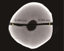
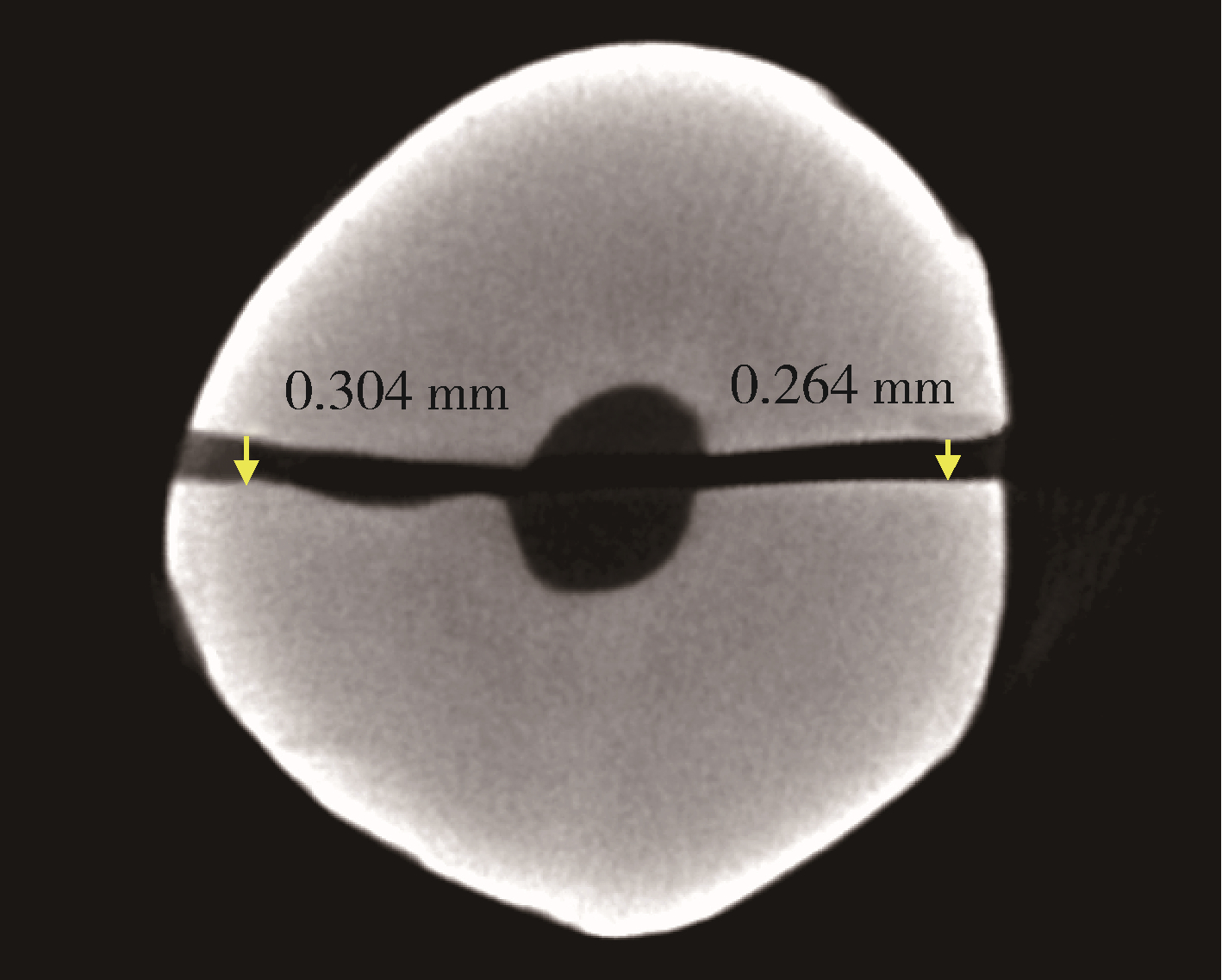
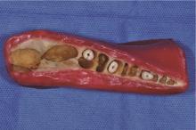
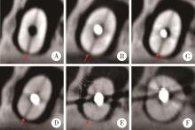
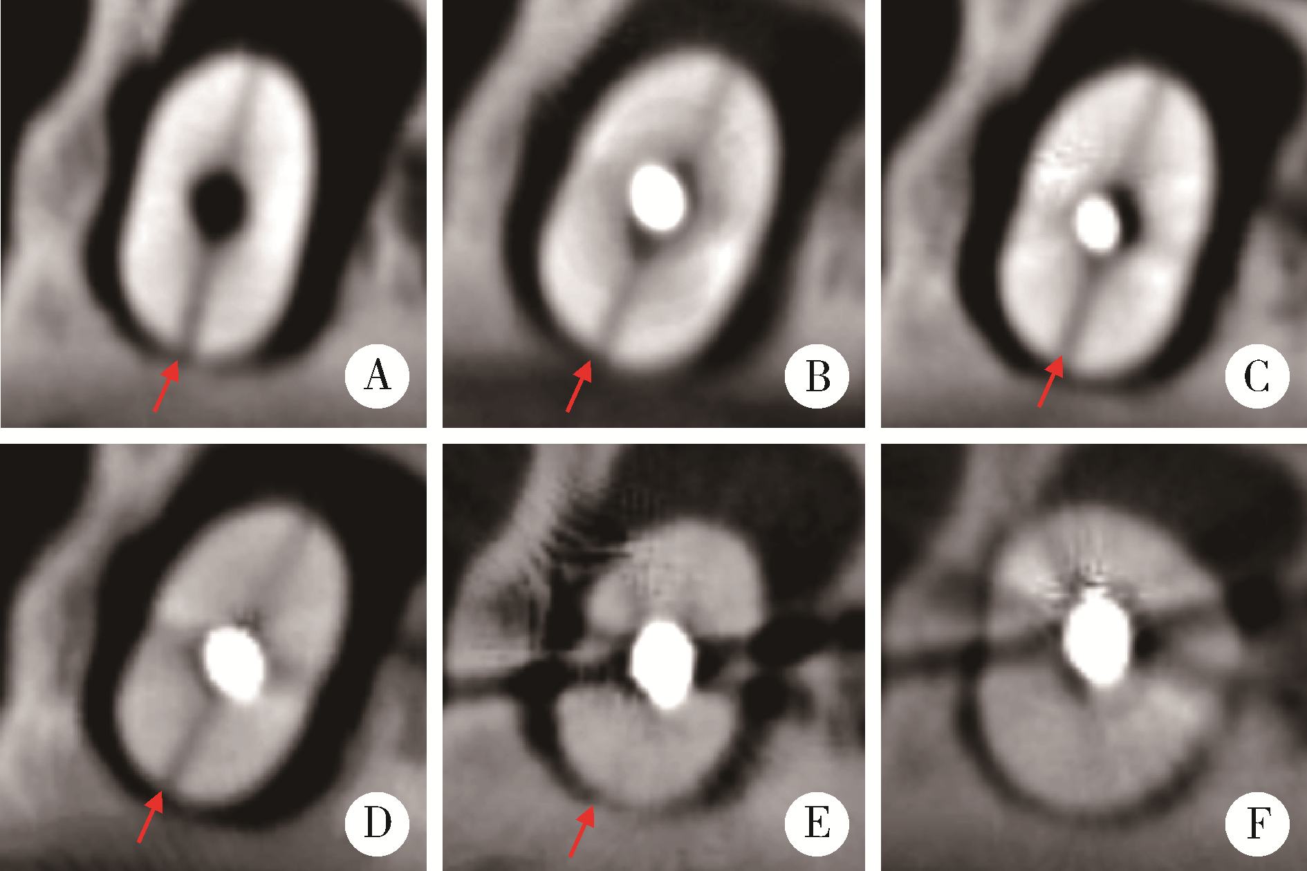
目的: 研究不同根管内容物对口腔颌面锥形束CT(cone-beam computed tomography,CBCT)诊断牙根纵裂(vertical root fracture,VRF)准确性的影响。方法: 选择因正畸治疗或牙周病而拔除的结构完整的单根管干燥离体牙24颗,沿釉牙骨质界去除牙冠,进行根管和桩道预备后制备为研究样本。将24个样本分为两组,每组12个。A组为对照组(无VRF组),根据根管内容物分为5个亚组:空白组、纤维桩组、牙胶尖组、钛桩组和金钯桩组;B组为实验组(VRF组),亚组分组同上。VRF组使用统一方法制备为VRF模型,以专用根管钉使牙根发生颊舌向完全裂开后再粘接复位;对照组不进行模拟VRF的操作。钛桩和金钯桩根据根管预备情况个性制作,通过X线检查确认桩与根管壁的密合性。所有样本均置于猪下颌骨牙槽窝中进行CBCT扫描,由经验丰富的牙体牙髓病学专科医师和口腔颌面医学影像学专科医师进行盲法判读,统计分析诊断准确率。结果: 空白组、纤维桩组、牙胶尖组、钛桩组、金钯桩组中VRF的诊断准确率依次为95.83%、91.67%、87.50%、79.17%和45.83%,与空白组相比,纤维桩组(P>0.999)、牙胶尖组(P=0.500)和钛桩组(P=0.125)的诊断准确率差异均无统计学意义。金钯桩组中VRF诊断准确率最低,与其他各组相比差异均有统计学意义(P < 0.001)。结论: 不同根管内容物在CBCT中的伪影对VRF诊断的准确性产生不同程度的影响,纤维桩、牙胶尖和钛桩的影响较小,而金钯桩影响较显著。
中图分类号:
- R781

| 1 |
Moule AJ , Kahler B . Diagnosis and management of teeth with vertical root fractures[J]. Aust Dent J, 1999, 44 (2): 75- 87.
doi: 10.1111/j.1834-7819.1999.tb00205.x |
| 2 |
Chen SC , Chueh LH , Hsiao CK , et al. First untoward events and reasons for tooth extraction after nonsurgical endodontic treatment in Taiwan[J]. J Endod, 2008, 34 (6): 671- 674.
doi: 10.1016/j.joen.2008.03.016 |
| 3 |
Walton RE . Vertical root fracture: Factors related to identification[J]. J Am Dent Assoc, 2017, 148 (2): 100- 105.
doi: 10.1016/j.adaj.2016.11.014 |
| 4 |
Mohammadpour M , Bakhshalian N , Shahab S , et al. Effect of titanium and stainless steel posts in detection of vertical root fractures using NewTom VG cone beam computed tomography system[J]. Imaging Sci Dent, 2014, 44 (2): 89- 94.
doi: 10.5624/isd.2014.44.2.89 |
| 5 |
Bernardes RA , de Moraes IG , Húngaro Duarte MA , et al. Use of cone-beam volumetric tomography in the diagnosis of root fractures[J]. Oral Surg Oral Med Oral Pathol Oral Radiol Endod, 2009, 108 (2): 270- 277.
doi: 10.1016/j.tripleo.2009.01.017 |
| 6 |
Brito-Júnior M , Santos LA , Faria-e-Silva AL , et al. Ex vivo eva-luation of artifacts mimicking fracture lines on cone-beam computed tomography produced by different root canal sealers[J]. Int Endod J, 2014, 47 (1): 26- 31.
doi: 10.1111/iej.12121 |
| 7 |
Hassan B , Metska ME , Ozok AR , et al. Detection of vertical root fractures in endodontically treated teeth by a cone beam computed tomography scan[J]. J Endod, 2009, 35 (5): 719- 722.
doi: 10.1016/j.joen.2009.01.022 |
| 8 |
Guo XL , Li G , Zheng JQ , et al. Accuracy of detecting vertical root fractures in non-root filled teeth using cone beam computed tomography: Effect of voxel size and fracture width[J]. Int Endod J, 2019, 52 (6): 887- 898.
doi: 10.1111/iej.13076 |
| 9 |
Nikbin A , Dalili Kajan Z , Taramsari M , et al. Effect of object position in the field of view and application of a metal artifact reduction algorithm on the detection of vertical root fractures on cone-beam computed tomography scans: An in vitro study[J]. Imaging Sci Dent, 2018, 48 (4): 245- 254.
doi: 10.5624/isd.2018.48.4.245 |
| 10 |
Fox A , Basrani B , Lam E . the performance of a zirconium-based root filling material with artifact reduction properties in the detection of artificially induced root fractures using cone-beam computed tomographic imaging[J]. J Endod, 2018, 44 (5): 828- 833.
doi: 10.1016/j.joen.2018.02.007 |
| 11 |
Saati S , Eskandarloo A , Falahi A , et al. Evaluation of the efficacy of the metal artifact reduction algorithm in the detection of a vertical root fracture in endodontically treated teeth in cone-beam computed tomography images: An in vitro study[J]. Dent Med Probl, 2019, 56 (4): 357- 363.
doi: 10.17219/dmp/109902 |
| 12 |
刘玉, 杨洁, 廖倬逸, 等. CBCT在牙根纵折诊断中的作用及影响因素分析[J]. 临床口腔医学杂志, 2018, 34 (11): 663- 666.
doi: 10.3969/j.issn.1003-1634.2018.11.006 |
| 13 |
Mozzo P , Procacci C , Tacconi A , et al. A new volumetric CT machine for dental imaging based on the cone-beam technique: Preliminary results[J]. Eur Radiol, 1998, 8 (9): 1558- 1564.
doi: 10.1007/s003300050586 |
| 14 |
Patel S , Durack C , Abella F , et al. Cone beam computed tomography in endodontics: A review[J]. Int Endod J, 2015, 48 (1): 3- 15.
doi: 10.1111/iej.12270 |
| 15 | Paul RA , Tamse A , Rosenberg E . Cracked and broken teeth: Definitions, differential diagnosis and treatment[J]. Refuat Hapeh Vehashinayim (1993), 2007, 24 (2): 7- 12. |
| 16 |
Schulze R , Heil U , Gross D , et al. Artefacts in CBCT: A review[J]. Dentomaxillofac Radiol, 2011, 40 (5): 265- 273.
doi: 10.1259/dmfr/30642039 |
| 17 |
Neves FS , Freitas DQ , Campos PS , et al. Evaluation of cone-beam computed tomography in the diagnosis of vertical root fractures: The influence of imaging modes and root canal materials[J]. J Endod, 2014, 40 (10): 1530- 1536.
doi: 10.1016/j.joen.2014.06.012 |
| 18 |
Gaêta-Araujo H , Silva de Souza GQ , Freitas DQ , et al. Optimization of tube current in cone-beam computed tomography for the detection of vertical root fractures with different intracanal materials[J]. J Endod, 2017, 43 (10): 1668- 1673.
doi: 10.1016/j.joen.2017.04.003 |
| 19 |
Rabelo KA , Cavalcanti YW , de Oliveira Pinto MG , et al. Quantitative assessment of image artifacts from root filling materials on CBCT scans made using several exposure parameters[J]. Imaging Sci Dent, 2017, 47 (3): 189- 197.
doi: 10.5624/isd.2017.47.3.189 |
| 20 |
Makeeva IM , Byakova SF , Novozhilova NE , et al. Detection of artificially induced vertical root fractures of different widths by cone beam computed tomography in vitro and in vivo[J]. Int Endod J, 2016, 49 (10): 980- 989.
doi: 10.1111/iej.12549 |
| 21 | 古丽比热·艾合买提, 曹雅, 谢思静, 等. 根管治疗后隐匿性VRF裂纹宽度对CBCT检出率的影响[J]. 口腔医学研究, 2020, 36 (2): 172- 176. |
| 22 |
Brady E , Mannocci F , Brown J , et al. A comparison of cone beam computed tomography and periapical radiography for the detection of vertical root fractures in nonendodontically treated teeth[J]. Int Endod J, 2014, 47 (8): 735- 746.
doi: 10.1111/iej.12209 |
| 23 |
Jakobson SJ , Westphalen VP , Silva Neto UX , et al. The influence of metallic posts in the detection of vertical root fractures using different imaging examinations[J]. Dentomaxillofac Radiol, 2014, 43 (1): 20130287.
doi: 10.1259/dmfr.20130287 |
| 24 | 曾艳, 王嘉德, 周书敏. 牙根纵裂患者的咬合应力分析[J]. 中华口腔医学杂志, 2000, 35 (2): 142- 143. |
| 25 |
Özer SY . Detection of vertical root fractures by using cone beam computed tomography with variable voxel sizes in an in vitro model[J]. J Endod, 2011, 37 (1): 75- 79.
doi: 10.1016/j.joen.2010.04.021 |
| 26 |
Melo SL , Haiter-Neto F , Correa LR , et al. Comparative diagnostic yield of cone beam CT reconstruction using various software programs on the detection of vertical root fractures[J]. Dentomaxillofac Radiol, 2013, 42 (9): 20120459.
doi: 10.1259/dmfr.20120459 |
| 27 |
Pinto M , Rabelo KA , Sousa Melo SL , et al. Influence of exposure parameters on the detection of simulated root fractures in the pre-sence of various intracanal materials[J]. Int Endod J, 2017, 50 (6): 586- 594.
doi: 10.1111/iej.12655 |
| 28 |
Queiroz PM , Santaella GM , Capelozza A , et al. Zoom reconstruction tool: Evaluation of image quality and influence on the diagnosis of root fracture[J]. J Endod, 2018, 44 (4): 621- 625.
doi: 10.1016/j.joen.2017.10.011 |
| 29 | 吴秦, 白石柱, 谢瑞, 等. 口腔常用修复材料的CBCT影像灰度值差异的探索[J]. 实用口腔医学杂志, 2016, 32 (1): 5- 9. |
| 30 |
Bragatto FP , Iwaki Filho L , Kasuya AV , et al. Accuracy in the diagnosis of vertical root fractures, external root resorptions, and root perforations using cone-beam computed tomography with different voxel sizes of acquisition[J]. J Conserv Dent, 2016, 19 (6): 573- 577.
doi: 10.4103/0972-0707.194029 |
| 31 |
Menezes RF , Araújo NC , Santa Rosa JM , et al. Detection of vertical root fractures in endodontically treated teeth in the absence and in the presence of metal post by cone-beam computed tomography[J]. BMC Oral Health, 2016, 16, 48.
doi: 10.1186/s12903-016-0207-y |
| 32 |
Ferreira RI , Bahrami G , Isidor F , et al. Detection of vertical root fractures by cone-beam computerized tomography in endodontically treated teeth with fiber-resin and titanium posts: An in vitro study[J]. Oral Surg Oral Med Oral Pathol Oral Radiol, 2013, 115 (1): e49- e57.
doi: 10.1016/j.oooo.2012.06.012 |
| 33 |
Melo SL , Bortoluzzi EA , Abreu M Jr , et al. Diagnostic ability of a cone-beam computed tomography scan to assess longitudinal root fractures in prosthetically treated teeth[J]. J Endod, 2010, 36 (11): 1879- 1882.
doi: 10.1016/j.joen.2010.08.025 |
| 34 |
Salineiro F , Kobayashi-Velasco S , Braga MM , et al. Radiographic diagnosis of root fractures: A systematic review, meta-analyses and sources of heterogeneity[J]. Dentomaxillofac Radiol, 2017, 46 (8): 20170400.
doi: 10.1259/dmfr.20170400 |
| 35 |
Elsaltani MH , Farid MM , Eldin Ashmawy MS . Detection of simulated vertical root fractures: Which cone-beam computed tomographic system is the most accurate[J]. J Endod, 2016, 42 (6): 972- 977.
doi: 10.1016/j.joen.2016.03.013 |
| 36 |
Huang CC , Lee BS . Diagnosis of vertical root fracture in endodontically treated teeth using computed tomography[J]. J Dental Sciences, 2015, 10 (3): 227- 232.
doi: 10.1016/j.jds.2015.01.002 |
| 37 |
Patel S , Brady E , Wilson R , et al. The detection of vertical root fractures in root filled teeth with periapical radiographs and CBCT scans[J]. Int Endod J, 2013, 46 (12): 1140- 1152.
doi: 10.1111/iej.12109 |
| 38 |
Chambrone L , Chambrone LA , Lima LA . Effects of occlusal overload on peri-implant tissue health: A systematic review of animal-model studies[J]. J Periodontol, 2010, 81 (10): 1367- 1378.
doi: 10.1902/jop.2010.100176 |
| 39 | 洪虹, 徐圆, 齐宏亮, 等. 基于数学形态学的CT图像金属伪影消除算法[J]. 计算机应用与软件, 2013, 30 (6): 29- 32. |
| 40 | 越野, 王敏, 张春宝, 等. 纯钛固定修复体铸件缺陷原因分析及预防措施[J]. 牙体牙髓牙周病学杂志, 2009, 19 (10): 608. |
| [1] | 薄士仕,高承志. 基于卷积神经网络实现锥形束CT牙齿分割及牙位标定[J]. 北京大学学报(医学版), 2024, 56(4): 735-740. |
| [2] | 潘孟乔,刘建,徐莉,徐筱,侯建霞,李小彤,王晓霞. 牙周-正畸-正颌联合治疗骨性安氏Ⅲ类错 |
| [3] | 叶佳学,梁宇红. 牙髓专科医师应用锥形束CT的现况调查[J]. 北京大学学报(医学版), 2023, 55(1): 114-119. |
| [4] | 高娟,吕航苗,马慧敏,赵一姣,李小彤. 锥形束CT三维体积测量评估骨性Ⅲ类错 |
| [5] | 刘伟涛,王怡然,王雪东,周彦恒. 锥形束CT研究上颌反复扩缩前方牵引后上颌骨缝的三维变化[J]. 北京大学学报(医学版), 2022, 54(2): 346-355. |
| [6] | 孟圆,张丽琪,赵雅宁,柳登高,张祖燕,高岩. 67例上颌根尖周囊肿的三维影像特点分析[J]. 北京大学学报(医学版), 2021, 53(2): 396-401. |
| [7] | 曹畅,王菲,王恩博,刘宇. β-磷酸三钙用于下颌第三磨牙拔除术后骨缺损修复的自身对照研究[J]. 北京大学学报(医学版), 2020, 52(1): 97-102. |
| [8] | 谢晓艳,贾淑梅,孙志辉,张祖燕. 分辨率设置与锥形束CT检测牙根外吸收的可靠性[J]. 北京大学学报(医学版), 2019, 51(1): 75-79. |
| [9] | 赵一姣,刘怡,孙玉春,王勇. 一种基于曲率连续算法的冠、根三维数据融合方法[J]. 北京大学学报(医学版), 2017, 49(4): 719-723. |
| [10] | 钟金晟, 欧阳翔英, 柳登高, 曹采方. 锥形束CT测量离体下颌磨牙Ⅱ°根分叉病变效果的评价[J]. 北京大学学报(医学版), 2010, 42(1): 41-45. |
| [11] | 刘怡, James MAH, 许天民. 锥形束计算机断层扫描中牙齿的分割精度[J]. 北京大学学报(医学版), 2010, 42(1): 98-102. |
| [12] | 王瑞永, 马绪臣, 张万林, 柳登高. 健康成年人颞下颌关节间隙锥形束计算机体层摄影术测量分析[J]. 北京大学学报(医学版), 2007, 39(5): 503-506. |
|
||
