北京大学学报(医学版) ›› 2019, Vol. 51 ›› Issue (1): 75-79. doi: 10.19723/j.issn.1671-167X.2019.01.014
分辨率设置与锥形束CT检测牙根外吸收的可靠性
谢晓艳1,贾淑梅2,孙志辉3,张祖燕1,△( )
)
- 1. 北京大学口腔医学院·口腔医院,放射科 国家口腔疾病临床医学研究中心 口腔数字化医疗技术和材料国家工程实验室 口腔数字医学北京市重点实验室,北京 100081
2. 清华大学工程物理系,粒子信息获取与处理教育部重点实验室,北京 100084
3. 北京大学口腔医学院?口腔医院,口腔材料研究室,北京 100081
Diagnostic accuracy of cone beam computed tomography with different resolution settings for external root resorption
Xiao-yan XIE1,Shu-mei JIA2,Zhi-hui SUN3,Zu-yan ZHANG1,△()
- 1. Department of Oral and Maxillofacial Radiology, Peking University School and Hospital of Stomatology & National Clinical Research Center for Oral Diseases & National Engineering Laboratory for Digital and Material Technology of Stomatology & Beijing Key Laboratory of Digital Stomatology, Beijing 100081, China
2. Department of Engineering Physics, Tsinghua University & Key Laboratory of Particle and Radiation Imaging, Ministry of Education, Beijing 100084, China
3. Department of Dental Materials, Peking University School and Hospital of Stomatology, Beijing 100081, China
摘要:
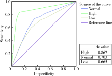
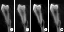
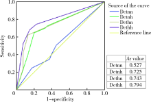
目的:评价锥形束CT(cone beam computed tomography,CBCT)在不同分辨率设置下检测牙根外吸收的可靠性,为临床选择恰当的CBCT扫描参数设置提供依据。方法:收集51颗离体单根前磨牙,分别在其舌面的根颈、根中和根尖部随机制备洞型,直径为1 mm,深度分别为0.1、0.2和0.3 mm,共153个位点(其中随机选择51个位点未制备洞型)用以模拟牙根外吸收的微小缺损。将离体牙置于人干下颌骨内,采用ProMax 3D和 DCT PRO型CBCT机分别对样本进行容积扫描;两名观察者评估图像,进行受试者工作特征(receiver operating characteristics,ROC)曲线分析,得到曲线下面积(Az值),用于评估不同分辨率设置下该设备检测牙根外吸收的可靠性。结果:ProMax 3D CBCT可提供高(high)、正常(normal)和低(low)3种分辨率模式扫描,其Az值分别为0.867、0.703和0.665(P<0.05)。DCT PRO CBCT提供了两种分辨率参数设置,组合可得到4种曝光模式,分别为正常质量+正常分辨率(normal quality + normal resolution)、正常质量+高分辨率(normal quality + high resolution)、高质量+正常分辨率(high quality + normal resolution)和高质量+高分辨率(high quality + high resolution), 其Az值分别为0.527、0.725、0.743和0.794(P<0.05)。结论:分辨率设置可显著影响锥形束CT检测牙根外吸收的可靠性,在不增加患者辐射剂量的前提下,采用计算机辅助后处理的方法可显著提高锥形束CT的诊断能力。
中图分类号:
- R782


| [1] |
Iglesias-Linares A, Hartsfield JK Jr . Cellular and molecular pathways leading to external root resorption[J]. J Dent Res, 2017,96(2):145-152.
doi: 10.1177/0022034516677539 pmid: 27811065 |
| [2] |
Fuss Z, Tsesis I, Lin S . Root resorption-diagnosis, classification and treatment choices based on stimulation factors[J]. Dent Traumatol, 2003,19(4):175-182.
doi: 10.1034/j.1600-9657.2003.00192.x |
| [3] |
Li J, Wang X, Li N , et al. Short-term effects of nicotine on orthodontically induced root resorption in rats[J]. Angle Orthod, 2016,86(2):199-205.
doi: 10.2319/101014-727.1 pmid: 26083055 |
| [4] |
Makedonas D, Lund H, Hansen K . Root resorption diagnosed with cone beam computed tomography after 6 months and at the end of orthodontic treatment with fixed appliances[J]. Angle Orthod, 2013,83(3):389-393.
doi: 10.2319/042012-332.1 pmid: 23092202 |
| [5] |
Khojastepour L, Moazami F, Babaei M , et al. Assessment of root perforation within simulated internal resorption cavities using cone-beam computed tomography[J]. J Endod, 2015,41(9):1520-1523.
doi: 10.1016/j.joen.2015.04.015 pmid: 26025347 |
| [6] |
Madani Z, Moudi E, Bijani A , et al. Diagnostic accuracy of cone-beam computed tomography and periapical radiography in internal root resorption[J]. Iran Endod J, 2016,11(1):51-56.
doi: 10.7508/iej.2016.01.010 pmid: 26843878 |
| [7] |
Durack C, Patel S, Davies J , et al. Diagnostic accuracy of small volume cone beam computed tomography and intraoral periapical radiography for the detection of simulated external inflammatory root resorption[J]. Int Endod J, 2011,44(2):136-147.
doi: 10.1111/j.1365-2591.2010.01819.x pmid: 21083575 |
| [8] |
Nasseh I, Al-Rawi W . Cone beam computed tomography[J]. Dent Clin North Am, 2018,62(3):361-391.
doi: 10.1016/j.cden.2018.03.002 |
| [9] |
Aminoshariae A, Kulild JC, Syed A . Cone-beam computed tomography compared with intraoral radiographic lesions in endodontic outcome studies: a systematic review[J]. J Endod, 2018,44(11):1626-1631.
doi: 10.1016/j.joen.2018.08.006 |
| [10] |
谢晓艳, 张祖燕 . 锥形束CT与8层螺旋CT检测牙根外吸收的可靠性[J]. 北京大学学报(医学版), 2012,44(4):628-632.
doi: 10.3969/j.issn.1671-167X.2012.04.030 |
| [11] |
Qu XM, Li G, Ludlow JB , et al. Effective radiation dose of ProMax 3D cone-beam computerized tomography scanner with different dental protocols[J]. Oral Surg Oral Med Oral Pathol Oral Radiol Endod, 2010,110(6):770-776.
doi: 10.1016/j.tripleo.2010.06.013 pmid: 20952220 |
| [12] |
Bernardes RA, de Paulo RS, Pereira LO , et al. Comparative study of cone beam computed tomography and intraoral periapical radiographs in diagnosis of lingual-simulated external root resorptions[J]. Dent Traumatol, 2012,28(4):268-272.
doi: 10.1111/j.1600-9657.2011.01113.x pmid: 22233265 |
| [13] |
Andreasen FM, Sewerin I, Mandel U , et al. Radiographic assessment of simulated root resorption cavities[J]. Endod Dent Traumatol, 1987,3(1):21-27.
doi: 10.1111/j.1600-9657.1987.tb00167.x pmid: 3471513 |
| [14] |
Haghanifar S, Moudi E, Mesgarani A , et al. A comparative study of cone-beam computed tomography and digital periapical radiography in detecting mandibular molars root perforations[J]. Imaging Sci Dent, 2014,44(2):115-119.
doi: 10.5624/isd.2014.44.2.115 pmid: 4061294 |
| [15] | 马绪臣 . 口腔颌面锥形束CT的临床应用 [M]. 北京: 人民卫生出版社, 2011: 5. |
| [16] |
van Erkel AR, Pattynama PM . Receiver operating characteristic (ROC) analysis: basic principles and applications in radiology[J]. Eur J Radiol, 1998,27(2):88-94.
doi: 10.1016/S0720-048X(97)00157-5 pmid: 9639133 |
| [17] |
Deliga Schröder ÂG, Westphalen FH, Schröder JC , et al. Accuracy of digital periapical radiography and cone-beam computed tomography for diagnosis of natural and simulated external root resorption[J]. J Endod, 2018,44(7):1151-1158.
doi: 10.1016/j.joen.2018.03.011 |
| [1] | 薄士仕,高承志. 基于卷积神经网络实现锥形束CT牙齿分割及牙位标定[J]. 北京大学学报(医学版), 2024, 56(4): 735-740. |
| [2] | 章锦花,潘洁,孙志鹏,王霄. 不同根管内容物对口腔颌面锥形束CT诊断牙根纵裂准确性的影响[J]. 北京大学学报(医学版), 2023, 55(2): 333-338. |
| [3] | 潘孟乔,刘建,徐莉,徐筱,侯建霞,李小彤,王晓霞. 牙周-正畸-正颌联合治疗骨性安氏Ⅲ类错 |
| [4] | 叶佳学,梁宇红. 牙髓专科医师应用锥形束CT的现况调查[J]. 北京大学学报(医学版), 2023, 55(1): 114-119. |
| [5] | 高娟,吕航苗,马慧敏,赵一姣,李小彤. 锥形束CT三维体积测量评估骨性Ⅲ类错 |
| [6] | 刘伟涛,王怡然,王雪东,周彦恒. 锥形束CT研究上颌反复扩缩前方牵引后上颌骨缝的三维变化[J]. 北京大学学报(医学版), 2022, 54(2): 346-355. |
| [7] | 杨雨卉,黄一平,李巍然. 骨皮质切开加速正畸牙齿移动对牙根吸收的影响[J]. 北京大学学报(医学版), 2021, 53(2): 434-437. |
| [8] | 孟圆,张丽琪,赵雅宁,柳登高,张祖燕,高岩. 67例上颌根尖周囊肿的三维影像特点分析[J]. 北京大学学报(医学版), 2021, 53(2): 396-401. |
| [9] | 曹畅,王菲,王恩博,刘宇. β-磷酸三钙用于下颌第三磨牙拔除术后骨缺损修复的自身对照研究[J]. 北京大学学报(医学版), 2020, 52(1): 97-102. |
| [10] | 赵一姣,刘怡,孙玉春,王勇. 一种基于曲率连续算法的冠、根三维数据融合方法[J]. 北京大学学报(医学版), 2017, 49(4): 719-723. |
| [11] | 沈潇,施捷,徐莉,焦剑,路瑞芳,孟焕新. 伴错牙合畸形的侵袭性牙周炎患者牙周-正畸联合治疗的临床评价[J]. 北京大学学报(医学版), 2017, 49(1): 60-066. |
| [12] | 陈小贤, 林碧琛, 钟洁, 葛立宏. 改良乳牙根管充填材料体内降解匹配性与临床效果[J]. 北京大学学报(医学版), 2015, 47(3): 529-535. |
| [13] | 白洁, 赵玉鸣, 秦满. 儿童恒牙全脱出牙周组织预后的回顾性研究[J]. 北京大学学报(医学版), 2015, 47(2): 312-316. |
| [14] | 杨祥, 张趁英, 郑树国. 颅骨锁骨发育不良患者乳牙牙根吸收特点及牙齿结构分析[J]. 北京大学学报(医学版), 2011, 43(1): 98-101. |
| [15] | 钟金晟, 欧阳翔英, 柳登高, 曹采方. 锥形束CT测量离体下颌磨牙Ⅱ°根分叉病变效果的评价[J]. 北京大学学报(医学版), 2010, 42(1): 41-45. |
|
||
