北京大学学报(医学版) ›› 2019, Vol. 51 ›› Issue (5): 919-924. doi: 10.19723/j.issn.1671-167X.2019.05.021

• 论著 • 上一篇    下一篇

侵袭性牙周炎患者正畸前后的咬合变化

杜仁杰1,焦剑2,周彦恒1,施捷1,()   

  1. 1. 北京大学口腔医学院·口腔医院,正畸科 国家口腔疾病临床研究中心 口腔数字化医疗技术和材料国家工程试验室 口腔数字医学北京市重点实验室,北京 100081
    2. 北京大学口腔医学院·口腔医院,牙周科 国家口腔疾病临床研究中心 口腔数字化医疗技术和材料国家工程试验室 口腔数字医学北京市重点实验室,北京 100081
  • 收稿日期:2018-10-08 出版日期:2019-10-18 发布日期:2019-10-23
  • 通讯作者: 施捷 E-mail:sjlily@yeah.net
  • 基金资助:
    北京大学口腔医院临床新技术新疗法项目(PKUSSNCT-16A03);北京大学口腔医院临床新技术新疗法项目(YS010118)

Occlusal changes before and after orthodontic treatment in patients with aggressive periodontitis

Ren-jie DU1,Jian JIAO2,Yan-heng ZHOU1,Jie SHI1,()   

  1. 1. Department of Orthodontics, Peking University School and Hospital of Stomatology & National Clinical Research Center for Oral Diseases & National Engineering Laboratory for Digital and Material Technology of Stomatology & Beijing Key Laboratory of Digital Stomatology, Beijing 100081, China
    2. Department of Periodontology, Peking University School and Hospital of Stomatology & National Clinical Research Center for Oral Diseases & National Engineering Laboratory for Digital and Material Technology of Stomatology & Beijing Key Laboratory of Digital Stomatology, Beijing 100081, China
  • Received:2018-10-08 Online:2019-10-18 Published:2019-10-23
  • Contact: Jie SHI E-mail:sjlily@yeah.net
  • Supported by:
    Supported by the New Clinical Techniques and Therapies of Peking University School and Hospital of Stomatology(PKUSSNCT-16A03);Supported by the New Clinical Techniques and Therapies of Peking University School and Hospital of Stomatology(YS010118)

摘要:

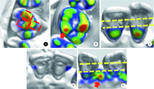
目的:评价侵袭性牙周炎患者行唇侧固定矫治后咬合改善的有效性,探究咬合改善与炎症控制的关系。方法:将纳入的22例牙周-正畸联合治疗患者的牙合相与正畸模型匹配,使用3Shape R700激光扫描仪获得数字化三维模型,在OrthoAnalyzer 软件中获得咬合分布图。从咬合分布与邻面接触两方面,对患者治疗前后的咬合变化进行评估。建立治疗后牙周探诊深度降低的多水平线性回归模型,筛选出对炎症控制有利的正畸方法。结果:在全牙列水平上,咬合分布评分在正畸治疗后显著提升(84.5±20.9 vs.105.3±22.6, P <0.001), 邻面接触评分在正畸治疗后显著提升(68.9±9.1 vs. 83.7±6.3,P <0.001)。在牙位水平上,正畸治疗后的咬合分布评分在上颌前牙区有显著提升(P <0.001), 正畸治疗后的邻面接触评分在上下前牙区均有显著提升(P <0.01)。多水平线性回归模型表明年龄和性别与治疗后牙周探诊深度降低未见显著的相关关系(P >0.05)。初始牙周探诊深度、咬合分布评分改善值和邻面接触评分改善值与治疗后牙周探诊深度降低均呈正相关(P <0.001)。结论:正畸治疗后,侵袭性牙周炎患者的咬合力分布、邻面接触均有显著改善。咬合分布评分改善值及邻面接触评分改善值与牙周探诊深度降低呈正相关,提示侵袭性牙周炎患者的牙周-正畸联合治疗中,应改善牙齿的咬合力分布与邻面接触,以促进牙周炎症控制。

关键词: 侵袭性牙周炎, 错牙合畸形, 探诊深度, 咬合力分布

Abstract:

Objective: To evaluate the efficacy of occlusal improvement in the labial fixed orthodontic treatment in aggressive periodontitis patients and to explore the relationship between occlusal improvement and inflammation control. Methods: Twenty-two aggressive periodontitis patients who underwent combined periodontal-orthodontic treatment were included in this study. The patient’s photos were matched to the dental models and digital three dimentional models were acquired using 3Shape R700 laser scanner. The occlusal force distribution maps were generated in the OrthoAnalyzer software. The newly established occlusal force distribution score (OFDS) and proximal contact score (PCS) were used to evaluate the occlusal distribution changes before and after labial fixed orthodontic treatment for assessing the effectiveness of orthodontic treatment. The multi-level linear regression analysis was used to explore the relationship between the probing depth changes and OFDS or PCS changes to screen out the favorable orthodontic strategy for inflammation control, which would provide clinical strategy for combined periodontal-orthodontic treatment in aggressive periodontitis patients. Results: At the patient level, OFDS was improved significantly after orthodontic treatment compared with the score before orthodontic treatment(84.5±20.9 vs.105.3±22.6, P <0.001) and PCS was improved significantly after orthodontic treatment compared with the score before orthodontic treatment(68.9±9.1 vs. 83.7±6.3, P <0.001).At the tooth level, the OFDS was significantly increased in the maxillary anterior teeth (P <0.001) while the PCS of the anterior teeth in both maxillary and mandible arches were significantly increased significantly (P <0.01). No significant changes were found in other tooth positions. The multi-level linear regression model showed that no significant correlation was found between age and gender and probing depth decrease (P >0.05). The baseline probing depth,OFDS improvements and PCS improvements (P <0.001) were positively correlated with probing depth decrease. Conclusion: This study showed that the distribution of occlusal force was more reasonable and the proximal contacts were more ideal in aggressive periodontitis patients. Orthodontic treatment was effective in improving occlusal force distribution by the above two ways. Especially, the OFDS and PCS improvements were both positively correlated with probing depth decrease, indicating that in the combined periodontal-orthodontic treatment for aggressive periodontitis patients, occlusal force distribution and proximal contact should be improved in order to facilitate periodontal improvement.

Key words: Aggressive periodontitis, Malocclusion, Probing depth, Occlusal force distribution

中图分类号: 

  • R783.5

图1

咬合分布评分积分规则"

表1

22例侵袭性牙周炎患者的临床特点"

Variebles n(%)
Gender
Male 6(27.3)
Female 16(72.7)
Angle classification
4(18.2)
16(72.7)
2(9.1)
Classification of periodontitis
Local 1(4.5)
Generalized 21(95.5)
Extraction or not
Extraction 6(27.3)
Non-extraction 16(72.7)
Total 22(100)

表2

正畸前后咬合分布、邻面接触评分在全牙列水平上的配对t检验结果"

Items Pre-treatment Post-treatment t P
OFDS 84.5±20.9 105.3±22.6 -9.028 <0.001
PCS 68.9±9.1 83.7±6.3 -8.671 <0.001

表3

正畸前后咬合分布在牙位水平上的配对t检验结果"

OFDS of different tooth position Pre-treatment Post-treatment t P
Maxillary
Left
Central incisor 2.2±1.8 5.2±1.9 -6.890 <0.001
Lateral incisor 2.4±2.0 5.2±1.8 -7.738 <0.001
Canine 3.0±1.7 5.6±1.2 -6.411 <0.001
Second premolar 2.9±1.6 3.2±1.6 -0.646 0.525
First molar 3.6±3.3 4.0±2.5 -0.509 0.616
Second molar 2.3±2.0 2.8±1.6 -1.779 0.090
Right
Central incisor 1.9±1.3 5.5±1.4 -9.279 <0.001
Lateral incisor 2.7±1.5 5.4±1.5 -7.286 <0.001
Canine 3.1±1.6 5.5±0.9 -5.786 <0.001
Second premolar 2.9±1.7 3.5±1.6 -1.204 0.242
First molar 4.0±3.0 4.9±2.4 -1.870 0.076
Second molar 2.6±1.8 3.1±2.1 -0.917 0.369
Mandible
Left
Central incisor 4.8±2.4 5.7±1.3 -2.084 0.050
Lateral incisor 5.0±2.1 5.0±2.1 <0.001 1.000
Canine 5.7±1.3 5.7±1.3 <0.001 1.000
Second premolar 3.5±1.2 3.3±1.5 0.576 0.571
First molar 4.7±2.5 4.8±2.8 -0.234 0.817
Second molar 2.0±1.8 2.1±1.5 -0.568 0.576
Right
Central incisor 4.9±2.4 5.4±1.8 -1.451 0.162
Lateral incisor 5.5±1.8 5.3±1.8 1.000 0.329
Canine 5.6±1.4 5.7±1.3 -1.000 0.329
Second premolar 2.8±1.7 3.1±1.8 -0.819 0.422
First molar 5.0±2.6 3.9±3.2 1.827 0.082
Second molar 2.0±1.5 2.1±1.7 -0.311 0.759

表4

邻面接触评分在牙位水平上的对比结果"

PCS of different tooth position Pre-treatment Post-treatment P
Maxillary
Left
Central incisor 2.1±1.5 4.0(4.0, 4.0) <0.001
Lateral incisor 2.6±1.4 3.9±0.4 <0.001
Canine 3.2±1.0 4.0(4.0, 4.0) 0.001
Second premolar 3.8±0.6 3.7±0.7 0.665
First molar 3.8±0.6 3.9±0.5 0.331
Second molar 1.7±0.9 1.9±0.8 0.481
Right
Central incisor 1.7±0.8 3.9±0.4 <0.001
Lateral incisor 2.0±1.6 4.0(4.0, 4.0) <0.001
Canine 2.5±1.6 4.0(4.0, 4.0) <0.001
Second premolar 3.8±0.5 3.9±0.4 0.162
First molar 4.0(4.0, 4.0) 4.0±0.2 0.329
Second molar 1.9±0.4 2.0(2.0, 2.0) 0.329
Mandible
Left
Central incisor 2.8±1.2 3.9±0.4 <0.001
Lateral incisor 2.9±1.2 3.8±0.9 0.001
Canine 3.5±0.7 4.0(4.0, 4.0) 0.009
Second premolar 3.7±0.5 3.9±0.4 0.083
First molar 3.9±0.4 3.9±0.4 1.000
Second molar 1.9±0.4 1.9±0.4 1.000
Right
Central incisor 2.7±1.2 3.9±0.4 <0.001
Lateral incisor 2.9±1.2 3.8±0.9 0.001
Canine 3.3±0.7 4.0(4.0, 4.0) <0.001
Second premolar 4.0±0.2 3.9±0.6 0.540
First molar 4.0±0.2 4.0(4.0, 4.0) 0.329
Second molar 2.0±0.2 2.0(2.0, 2.0) 0.329

表5

牙周探诊深度降低的多水平线性回归结果"

Items 95%CI P
Patient level
Age 0.006 (-0.042, 0.030) 0.744
Gender -0.072 (-0.390, 0.533) 0.760
Tooth level
OFDS(T2-T1) 0.050(0.024, 0.077) <0.001
PCS(T2-T1) 0.244(0.182,0.305) <0.001
Baseline PD(T0) 0.363(0.286,0.440) <0.001
[1] 孟焕新 . 临床牙周病学[M]. 北京: 北京大学医学出版社, 2014: 185-195.
[2] Azuma S, Kohzuki M, Saeki S , et al. Beneficial effects of orthodontic treatment on quality of life in patients with malocclusion[J]. Tohoku J Exp Med, 2008,214(1):39-50.
[3] Bernabé E, Tsakos G, Messias d OC, et al. Impacts on daily performances attributed to malocclusions using the condition-specific feature of the oral impacts on daily performances index[J]. Angle Orthod, 2008,78(2):241-247.
[4] 施捷 . 牙周炎患者的正畸减数治疗及其远期疗效观察[J]. 中华口腔正畸学杂志, 2010,17(4):181-187.
[5] Armitage GC . Development of a classification system for periodontal diseases and conditions[J]. Ann Periodontol, 2005,4(1):1-6.
[6] Dawson P . Functional occlusion: from TMJ to smile design[M]. Missouri: Mosby Elsevier, 2008: 404-405.
[7] 徐莉, 孟焕新, 田雨 , 等侵袭性牙周炎患者牙根形态异常的观察[J]. 中华口腔医学杂志, 2009,44(5):266-269.
[8] 乔敏, 徐莉, 孟焕新 , 等. 侵袭性牙周炎核心家系牙槽骨吸收和牙根形态的遗传度分析[J]. 中华口腔医学杂志, 2013,48(10):577-580.
[9] Slot J, Rosling BG . Suppression of the periodontopathic microflora in localized juvenile periodontitis by systemic tetracycline[J]. Journal Clin Periodontol, 1983,10(5):465-486.
[10] Lindhe J, Hamp SE, Loe H . Experimental periodontitis in the beagle dog[J]. J Periodontal Res, 1973,8(1):1-10.
[11] Shi J, Liu Z, Kawai T , et al. Antibiotic administration alleviates the aggravating effect of orthodontic force on ligature-induced experimental periodontitis bone loss in mice[J]. J Periodontal Res, 2017,52(4):725-733.
[1] 朱小玲,李文静,王宪娥,宋文莉,徐莉,张立,冯向辉,路瑞芳,释栋,孟焕新. 细胞色素B-245α链及胆固醇酯转运蛋白基因多态性与广泛型侵袭性牙周炎易感性的关系[J]. 北京大学学报(医学版), 2022, 54(1): 18-22.
[2] 刘建,王宪娥,吕达,乔敏,张立,孟焕新,徐莉,毛铭馨. 广泛型侵袭性牙周炎患者牙根形态异常与相关致病基因的关联[J]. 北京大学学报(医学版), 2021, 53(1): 16-23.
[3] 王秀婧,张怡美,周彦恒. 骨性Ⅲ类错牙合畸形患者正畸-正颌联合治疗的稳定性[J]. 北京大学学报(医学版), 2019, 51(1): 86-92.
[4] 张又文,辛天艺,焦剑,周彦恒,施捷. 慢性牙周炎的减数正畸治疗[J]. 北京大学学报(医学版), 2018, 50(2): 308-313.
[5] 张海东,张立,释栋,韩劼,闫夏,谢也斯,孟焕新. 锥形锁柱种植体用于因牙周炎缺牙患者修复的临床观察[J]. 北京大学学报(医学版), 2018, 50(2): 300-307.
[6] 郑旭,胡兴学,马宁,陈晓红. 正畸矫治牙性牙合平面倾斜的新方法——波浪形弓[J]. 北京大学学报(医学版), 2017, 49(1): 176-180.
[7] 沈潇,施捷,徐莉,焦剑,路瑞芳,孟焕新. 伴错牙合畸形的侵袭性牙周炎患者牙周-正畸联合治疗的临床评价[J]. 北京大学学报(医学版), 2017, 49(1): 60-066.
[8] 宋文莉, 田雨, 王宪娥, 张立, 徐莉, 释栋, 冯向辉, 路瑞芳, 陈智滨, 孟焕新. FADS1 rs174537基因多态性与侵袭性牙周炎患者血清蛋白的相关性[J]. 北京大学学报(医学版), 2016, 48(1): 10-15.
[9] 冯向辉, 张立, 徐莉, 孟焕新, 陈智滨, 释栋, 路瑞芳. 侵袭性牙周炎患者抗伴放线聚集杆菌血清c型IgG滴度分析[J]. 北京大学学报(医学版), 2015, 47(5): 820-824.
[10] 路瑞芳, 冯向辉, 徐莉, 孟焕新. 侵袭性牙周炎在非手术治疗后不同治疗反应位点的临床和可疑致病微生物特性[J]. 北京大学学报(医学版), 2015, 47(1): 13-18.
[11] 路瑞芳, 冯琳, 高学军, 孟焕新, 冯向辉. 侵袭性牙周炎龈沟液中有机酸与牙龈卟啉单胞菌和齿垢密螺旋体的关系[J]. 北京大学学报(医学版), 2013, 45(1): 12-16.
[12] 吕达, 孟焕新, 徐莉, 张立, 陈智滨, 冯向辉, 释栋, 路瑞芳, 王宪娥. 侵袭性牙周炎患者治疗后失牙的预测模型[J]. 北京大学学报(医学版), 2013, 45(03): 480-483.
Viewed
Full text


Abstract

Cited

  Shared   
  Discussed   
[1] 田增民, 陈涛, Nanbert ZHONG, 李志超, 尹丰, 刘爽. 神经干细胞移植治疗遗传性小脑萎缩的临床研究(英文稿)[J]. 北京大学学报(医学版), 2009, 41(4): 456 -458 .
[2] 卢恬, 朱晓辉, 柳世庆, 郑杰, 邱晓彦. 白细胞介素2促进宫颈癌细胞系HeLaS3免疫球蛋白G的表达[J]. 北京大学学报(医学版), 2009, 41(2): 158 -161 .
[3] 袁惠燕, 张苑, 范田园. 离子交换型栓塞微球及其载平阳霉素的制备与性质研究[J]. 北京大学学报(医学版), 2009, 41(2): 217 -220 .
[4] 祁琨, 邓芙蓉, 郭新彪. 纳米二氧化钛颗粒对人肺成纤维细胞缝隙连接通讯的影响[J]. 北京大学学报(医学版), 2009, 41(3): 297 -301 .
[5] 丰雷, 王玉凤, 曹庆久. 哌甲酯对注意缺陷多动障碍儿童平衡功能影响的开放性研究[J]. 北京大学学报(医学版), 2007, 39(3): 304 -309 .
[6] 钱秋谨, 杨莉, 王玉凤. 儿童注意缺陷多动障碍的研究进展[J]. 北京大学学报(医学版), 2007, 39(3): 323 -328 .
[7] 王海燕. 办好病例分析类栏目 为提高临床诊疗水平提供新平台[J]. 北京大学学报(医学版), 2007, 39(4): 341 .
[8] 马明信. 重视并提高疑难/罕见淋巴瘤的诊治[J]. 北京大学学报(医学版), 2007, 39(4): 342 .
[9] 田雨, 刘建湘, 王化虹. 经皮肝穿刺活检确诊无症状原发性肝结核一例[J]. 北京大学学报(医学版), 2007, 39(4): 365 -368 .
[10] 李淳德, 李宏, 刘宪义. 后路脊柱截骨矫形治疗重度僵硬后凸型脊柱侧凸[J]. 北京大学学报(医学版), 2007, 39(4): 399 -402 .