北京大学学报(医学版) ›› 2020, Vol. 52 ›› Issue (2): 362-367. doi: 10.19723/j.issn.1671-167X.2020.02.026
脑对流增强给药对老年大鼠脑细胞外间隙微观结构的影响
宋宇1,2,韩鸿宾2,3,4,△( ),杨军5,王艾博3,4,和清源3,4,李媛媛3,4,赵国梅3,4,高亚娟3,4,王睿3,4,韩易兴3,4,刘爱连1,△(),宋清伟1,△()
- 1. 大连医科大学附属第一医院放射科, 辽宁大连 116011
2. 北京大学医学部医学技术研究院, 北京 100191
3. 北京市磁共振成像设备与技术重点实验室, 北京 100191
4. 北京大学第三医院放射科, 北京 100191
5. 北京大学第三医院神经外科, 北京 100191
Effect of convection enhanced delivery on the microstructure of brain extracellular space in aged rats
Yu SONG1,2,Hong-bin HAN2,3,4,△(),Jun YANG5,Ai-bo WANG3,4,Qing-yuan HE3,4,Yuan-yuan LI3,4,Guo-mei ZHAO3,4,Ya-juan GAO3,4,Rui WANG3,4,Yi-xing HAN3,4,Ai-lian LIU1,△(),Qing-wei SONG1,△()
- 1. Department of Radiology, the First Affiliated Hospital of Dalian Medical University, Dalian 116011, Liaoning, China
2. Institute of Medical Technology, Peking University Health Science Center, Beijing 100191, China
3. Beijing Key Lab of Magnetic Resonance Imaging Device and Technique, Beijing 100191, China
4. Department of Radiology, Peking University Third Hospital, Beijing 100191, China
5. Department of Neurosurgery, Peking University Third Hospital, Beijing 100191, China
摘要:
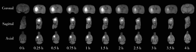
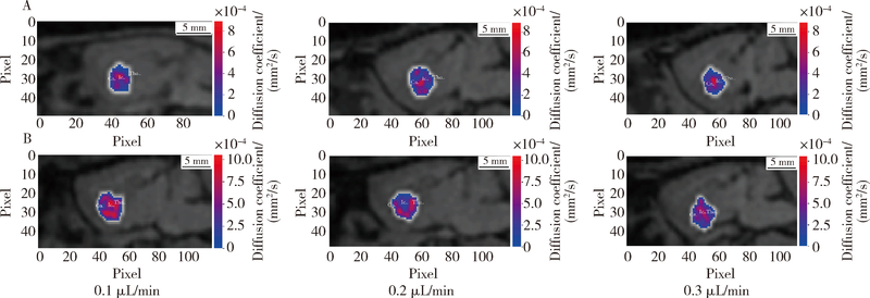
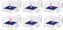
目的 对比研究经细胞外间隙(extracellular space,ECS)途径的脑对流增强给药(convection enhanced delivery, CED)在进行脑病微创治疗时,不同给药速率下成年大鼠与老年大鼠脑ECS结构参数及局部药物分布的改变.方法: 36只SD雄性大鼠按照月龄分为成年大鼠组(2~8月龄,18只)和老年大鼠组(18~24月龄,18只),每组再按照不同的给药速率(0.1 μL/min,0.2 μL/min,0.3 μL/min)随机分为3个亚组,每亚组6只.采用立体定位注射法分别在各组鼠脑尾状核区导入浓度为10 mmol/L的磁示踪剂钆-二乙三胺五乙酸(gadolinium-diethylene triamine pentaacetic acid,Gd-DTPA)后,应用磁示踪法动态采集Gd-DTPA在脑间质系统(brain interstitial system, ISS)中的扩散和分布图像.利用自主研发的MRI影像测量分析系统软件对所获得的图像进行处理和分析,可获得各组大鼠脑尾状核区ECS内的有效扩散系数(DECS),清除率,容积占比和半衰期(T1/2)等参数.比较分析在不同给药速率下,老年大鼠与成年大鼠脑ECS内药物清除以及ECS结构功能的影响和差异.应用磁示踪DECS-mapping技术观察示踪剂在尾状核区的分布引流情况.结果: 0.1 μL/min注药速率下,与成年大鼠相比,老年大鼠的容积占比增加(18.20%±0.04% vs. 17.20%±0.03%,t=3.752,P=0.004),迂曲度下降(1.63±0.04 vs. 1.78±0.09, t=-3.680,P=0.004),药物清除速率下降[(1.94±0.68) mm 2/s vs. (3.25±0.43) mm 2/s,t=-3.971,P=0.003],ECS内分子扩散速率增快[(3.99±0.21)×10 -4mm 2/s vs. (3.36±0.37)×10 -4mm 2/s,t=3.663,P=0.004].注药速率增加到0.2 μL/min时,老年大鼠ECS内药物清除减慢[(2.53±0.45) mmol/L vs. (3.37±0.72) mmol/L,t=-1.828,P=0.021],但容积占比,ECS内分子扩散和宏观药物代谢参数无明显差异.注药速率增加到0.3 μL/min时,老年大鼠容积占比减小(17.20%±0.03% vs. 18.20%±0.05%,t=-0.869,P=0.045), ECS内药物清除明显加快[(4.04±0.76) mmol/L vs. (3.26±0.55) mmol/L,t=1.786,P=0.014],迂曲度和ECS内分子扩散速率无明显差异.结论: 老年脑CED给药在不同速率时ECS内药物清除及ECS结构参数发生改变,0.2 μL/min速率下CED给药对老年脑ECS影响最小.应用CED进行脑病治疗时应综合考虑年龄和注药速率等因素的影响,经ECS途径给药进行脑病微创治疗时应制定个体化临床治疗方案.
中图分类号:
- R814.4


| [1] | Mehta AM, Sonabend AM, Bruce JN . Convection-enhanced deli-very[J]. Neurotherapeutics, 2017,14(2):358-371. |
| [2] | Souweidane MM, Singh R, Zhou Z . Convection-enhanced delivery for diffuse intrinsic pontine glioma treatment[J]. Curr Neuropharmacol, 2017,15(1):116-128. |
| [3] | Lei Y, Han H, Yuan F , et al. Brain interstitial system: anatomy, modeling, in vivo measurement, and application[J]. Prog Neurobiol, 2016,157:230-246. |
| [4] | Nicholson C, Hrabětová S . Brain extracellular space: the final frontier of neuroscience[J]. Biophys J, 2017,113(10):1-10. |
| [5] | Himes BT, Zhang L, Daniels DJ . Treatment strategies in diffuse midline gliomas with the H3K27M mutation: the role of convection-enhanced delivery in overcoming anatomic challenges[J]. Front Oncol, 2019,9:31. |
| [6] | Oertel W, Schulz JB . Current and experimental treatments of Parkinson disease: A guide for neuroscientists[J]. J Neurochem, 2016,139(Suppl 1):325-337. |
| [7] | Chen PY, Yeh CK, Hsu PH , et al. Drug-carrying microbubbles as a theranostic tool in convection-enhanced delivery for brain tumor therapy[J]. Oncotarget, 2017,8(26):42359-42371. |
| [8] | Fan X, Nelson BD, Ai Y , et al. Continuous intraputamenal convection-enhanced delivery in adult rhesus macaques[J]. J Neurosurg, 2015,123(6):1569-1577. |
| [9] | Sugiyama SI, Saito R, Nakamura T , et al. Safety and feasibility of convection-enhanced delivery of nimustine hydrochloride co-infused with free gadolinium for real-time monitoring in the primate brain[J]. Neurol Res, 2012,34(6):581-587. |
| [10] | Hou J, Wang W, Quan X , et al. Quantitative visualization of dynamic tracer transportation in the extracellular space of deep brain regions using tracer-based magnetic resonance imaging[J]. Med Sci Monit, 2017,23:4260-4268. |
| [11] | Han H, Shi C, Fu Y , et al. A novel mri tracer-based method for measuring water diffusion in the extracellular space of the rat brain[J]. IEEE J Biomed Health Inform, 2014,18(3):978-983. |
| [12] | Daneman R, Prat A . The blood-brain barrier[J]. Cold Spring Harb Perspect Biol, 2015,7(1):a020412. |
| [13] | Zhan W, Alamer M, Xu XY . Computational modelling of drug delivery to solid tumour: Understanding the interplay between chemotherapeutics and biological system for optimised delivery systems[J]. Adv Drug Deliv Rev, 2018,132:81-103. |
| [14] | Miners JS, Barua N, Kehoe PG , et al. Aβ-degrading enzymes: potential for treatment of Alzheimer disease[J]. J Neuropathol Exp Neurol, 2011,70(11):944-959. |
| [15] | Han HB, Xia ZL, Chen H , et al. Simple diffusion delivery via brain interstitial route for the treatment of cerebral ischemia[J]. Sci China Life Sci, 2011,54(3):235-239. |
| [16] | Xu F, Han H, Yan J , et al. Greatly improved neuroprotective efficiency of citicoline by stereotactic delivery in treatment of ischemic injury[J]. Drug Deliv, 2011,18(7):461-467. |
| [17] | Bobo RH, Laske DW, Akbasak A , et al. Convection-enhanced delivery of macromolecules in the brain[J]. Proc Natl Acad Sci USA, 1994,91(6):2076-2080. |
| [18] | Chittiboina P, Heiss JD, Warren KE , et al. Magnetic resonance imaging properties of convective delivery in diffuse intrinsic pontine gliomas[J]. J Neurosurg Pediatr, 2014,13(3):276-282. |
| [19] | Miranpuri GS, Kumbier L, Hinchman A , et al. Gene-based therapy of Parkinson's disease: Translation from animal model to human clinical trial employing convection enhanced delivery[J]. Ann of Neurosci, 2012,19(3):133-146. |
| [20] | Whone A, Luz M, Boca M , et al. Randomized trial of intermittent intraputamenal glial cell line-derived neurotrophic factor in Parkinson's disease[J]. Brain, 2019,142(3):512-525. |
| [21] | Souweidane MM, Kim K, Neeta PT , et al. Convection-enhanced delivery for diffuse intrinsic pontine glioma: a single-centre, dose-escalation, phase 1 trial[J]. Lancet Oncol, 2018,19(8):1040-1050. |
| [22] | Bishop NA, Lu T, Yankner BA . Neural mechanisms of ageing and cognitive decline[J]. Nature, 2010,464(7288):529-535. |
| [23] | Aibo W, Rui W, Dehua C , et al. The drainage of interstitial fluid in the deep brain is controlled by the integrity of myelination[J]. Aging Dis, 2019,10(5):937-948. |
| [24] | Syková E, Nicholson C . Diffusion in brain extracellular space[J]. Physiol Rev, 2008,88(4):1277-1340. |
| [1] | 孟庆伟, 范梦, 郭煌达, 章涵宇, 王梦莹, 王斯悦, 彭和香, 王雪珩, 侯天姣, 秦雪英, 陈大方, 李劲, 武轶群, 吴涛, 陈洪波, 胡永华. 老年人心源性卒中抗凝治疗的预后[J]. 北京大学学报(医学版), 2026, 58(3): 536-542. |
| [2] | 王欧洋, 朱鹏磊, 林杰, 吴昊. LncRNA DANCR调节miR-656/BMPR1A轴对脑胶质瘤细胞恶性行为的影响[J]. 北京大学学报(医学版), 2026, 58(3): 616-623. |
| [3] | 王泽远, 于栓宝, 郑浩轲, 陶金, 范雅峰, 张雪培. 基于临床特征和多参数MRI的前列腺癌盆腔淋巴结转移的术前预测模型[J]. 北京大学学报(医学版), 2025, 57(4): 684-691. |
| [4] | 邹晶, 高天姿, 汪洋, 任蒙蒙, 刘东阳, 龙仁, 成雨萌, 刘萌, 徐正仁, 谢肇恒, 吕鹏宇, 袁兰, 韩鸿宾. 99mTc-DTPA经脑细胞外间隙给药后的动态分布及消除规律[J]. 北京大学学报(医学版), 2025, 57(3): 562-568. |
| [5] | 钟鹏, 胡晓丹, 王振洲. 大鼠脑创伤半暗带光学相干断层血管造影及微血管密度定量[J]. 北京大学学报(医学版), 2025, 57(2): 262-266. |
| [6] | 孙建军, 马千权, 尹晓亮, 杨辰龙, 张嘉, 陈素华, 吴超, 谢京城, 韩芸峰, 林国中, 司雨, 杨军, 邬海博, 赵强. 任意维度重建磁共振对骶管囊肿进行精准分型对于指导微创手术和康复的意义[J]. 北京大学学报(医学版), 2025, 57(2): 303-308. |
| [7] | 邢念增,王明帅,杨飞亚,尹路,韩苏军. 前列腺免活检创新理念的临床实践及其应用前景[J]. 北京大学学报(医学版), 2024, 56(4): 565-566. |
| [8] | 田宇轩,阮明健,刘毅,李德润,吴静云,沈棋,范宇,金杰. 双参数MRI改良PI-RADS评分4分和5分病灶的最大径对临床有意义前列腺癌的预测效果[J]. 北京大学学报(医学版), 2024, 56(4): 567-574. |
| [9] | 李晋娜,许丽娜,李敏,宋怡,张静,贾龙斌. 急性脑梗死患者血清BDNF、IL-18、hs-CRP水平与血管性认知障碍的相关性[J]. 北京大学学报(医学版), 2024, 56(4): 708-714. |
| [10] | 龙仁, 毛鑫, 高天姿, 解倩, 谈瀚博, 李子寅, 韩鸿宾, 袁兰. 熊果酸改善精神分裂症小鼠脱髓鞘和脑组织间液引流紊乱[J]. 北京大学学报(医学版), 2024, 56(3): 487-494. |
| [11] | 刘毅,袁昌巍,吴静云,沈棋,肖江喜,赵峥,王霄英,李学松,何志嵩,周利群. 靶向穿刺+6针系统穿刺对PI-RADS 5分患者的前列腺癌诊断效能[J]. 北京大学学报(医学版), 2023, 55(5): 812-817. |
| [12] | 袁昌巍,李德润,李志华,刘毅,山刚志,李学松,周利群. 多参数磁共振成像中动态对比增强状态在诊断PI-RADS 4分前列腺癌中的应用[J]. 北京大学学报(医学版), 2023, 55(5): 838-842. |
| [13] | 刘颖,霍然,徐慧敏,王筝,王涛,袁慧书. 磁共振血管壁成像评估颈动脉中重度狭窄患者斑块特征与脑血流灌注的相关性[J]. 北京大学学报(医学版), 2023, 55(4): 646-651. |
| [14] | 傅强,高冠英,徐雁,林卓华,孙由静,崔立刚. 无症状髋关节前上盂唇撕裂超声与磁共振检查的对比研究[J]. 北京大学学报(医学版), 2023, 55(4): 665-669. |
| [15] | 范常锋,黄亚平,李霞,陈芸,李真,乔淑冬. 以发作性体位性视物双影为前期症状的后循环卒中1例[J]. 北京大学学报(医学版), 2023, 55(4): 762-765. |
|
||
