北京大学学报(医学版) ›› 2021, Vol. 53 ›› Issue (1): 88-94. doi: 10.19723/j.issn.1671-167X.2021.01.014
应用超声器械改善预备体肩台的效果
李思雨1,段雪飞2,曹烨2,Δ( )
)
- 1.北京大学口腔医学院·口腔医院,综合科, 国家口腔疾病临床医学研究中心 口腔数字化医疗技术和材料国家工程实验室 口腔数字医学北京市重点实验室,北京 100081
2.修复科 国家口腔疾病临床医学研究中心 口腔数字化医疗技术和材料国家工程实验室 口腔数字医学北京市重点实验室,北京 100081
Evaluation of the effect of using ultrasonic instruments to improve the shoulder of the preparations
LI Si-yu1,DUAN Xue-fei2,CAO Ye2,Δ()
- 1. Department of General Dentistry, Peking University School and Hospital of Stomatology & National Clinical Research Center for Oral Diseases & National Engineering Laboratory for Digital and Material Technology of Stomatology & Beijing Key Laboratory of Digital Stomatology, Beijing 100081, China
2. Department of Prosthodontics, Peking University School and Hospital of Stomatology & National Clinical Research Center for Oral Diseases & National Engineering Laboratory for Digital and Material Technology of Stomatology & Beijing Key Laboratory of Digital Stomatology, Beijing 100081, China
摘要:
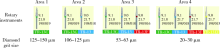
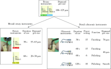
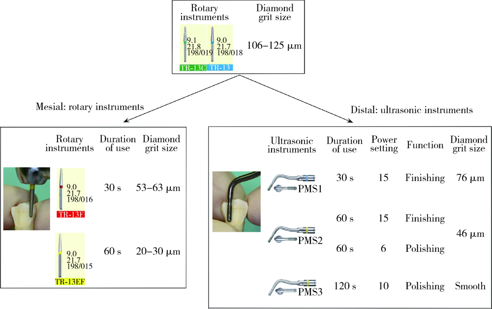
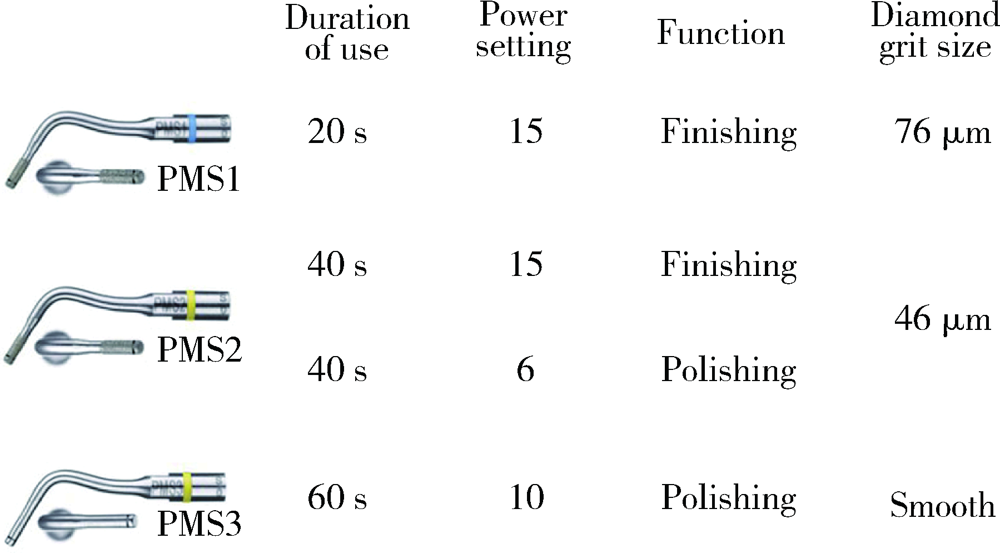
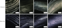
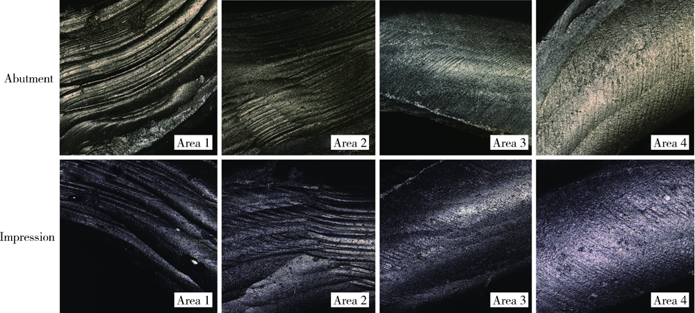
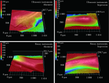
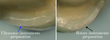
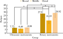
目的: 比较超声器械与金刚砂车针预备所形成的肩台表面粗糙度的差异,为超声肩台预备的临床开展提供依据。方法: (1)6颗离体前磨牙颊舌侧的不同区域分别使用不同粗糙度的金刚砂车针进行预备,聚醚橡胶制取肩台区印模,三维形貌测量激光显微镜(three-dimensional topography measurement laser microscope, 3-D TMLM)扫描印模并比较表面粗糙度。(2)6颗离体前磨牙颊侧的近、远中分别使用金刚砂车针和超声器械进行预备,聚醚橡胶制取肩台区印模,3-D TMLM扫描印模并比较表面粗糙度,牙科显微镜(×25)观察肩台表面形态。(3)上颌对称同名离体前牙20颗,灌注标准模型后随机分为超声器械组和金刚砂车针组,于仿头模上牙体预备后聚醚橡胶制取肩台印模;扫描并对比肩台印模近中、中央、远中区域表面粗糙度。用SPSS 25.0软件进行统计学分析。结果: (1)不同区域内预备体肩台和印模肩台的Ra和Rz差异均无统计学意义。(2)超声器械预备后肩台印模表面粗糙度[Ra:(6.59±2.33) μm,Rz:(34.69±7.29) μm]明显小于金刚砂车针[Ra:(21.79±4.89) μm,Rz:(91.69±14.82) μm](P<0.05);牙科显微镜下可见超声预备的肩台形态清晰连续。(3)超声器械预备后肩台印模各区域的表面粗糙度Ra均小于金刚砂车针,其差异有统计学意义(P<0.001);超声器械预备后肩台印模各区域表面粗糙度Ra的差异无统计学意义,而金刚砂车针预备后肩台印模在近中及远中区域的表面粗糙度Ra均大于中央区域,其差异有统计学意义(P<0.001)。结论: 超声器械可获得更光滑的肩台表面,尤其可以显著改善肩台近邻面处的预备效果。
中图分类号:
- R783.4


| [1] |
Blair FM, Wassell RW, Steele JG. Crowns and other extra-coronal restorations: Preparations for full veneer crowns[J]. Br Dent J, 2002,192(10):561-571.
doi: 10.1038/sj.bdj.4801428 pmid: 12075956 |
| [2] |
Douglass CW, Watson AJ. Future needs for fixed and removable partial dentures in the United States[J]. J Prosthet Dent, 2002,87(1):9-14.
doi: 10.1067/mpr.2002.121204 pmid: 11807477 |
| [3] |
Kronström M, Palmqvist S, Eriksson T, et al. Practice profile differences among Swedish dentists. A questionnaire study with special reference to prosthodontics[J]. Acta Odontol Scand, 1997,55(5):265-269.
doi: 10.3109/00016359709114962 pmid: 9370022 |
| [4] |
Ranjitha K, Jamie ADS. Ultrasonic vs. hand instrumentation in periodontal therapy: clinical outcomes[J]. Periodontol 2000, 2016,71(1):113-127.
doi: 10.1111/prd.12119 pmid: 27045433 |
| [5] |
Galler KM, Grubmüller V, Schlichting R, et al. Penetration depth of irrigants into root dentine after sonic, ultrasonic and photoacoustic activation[J]. Int Endod J, 2019,52(8):1210-1217.
doi: 10.1111/iej.13108 pmid: 30828819 |
| [6] |
Ju-Hyoung L. Guided tooth preparation for a pediatric zirconia crown[J]. J Am Dent Assoc, 2018,149(3):202-208.
doi: 10.1016/j.adaj.2017.08.048 pmid: 29395008 |
| [7] | Jamal MS, Christian H, Sareh SM. Combination of ultrasonic decontamination, soft tissue curettage, and submucosal air poli-shing with povidone-iodine application for non-surgical therapy of peri-implantitis: 12 month clinical outcomes[J]. J Periodontol, 2018,89:139-147. |
| [8] |
Atieh MA, Alsabeeha NHM, Tawse-Smith A, et al. Piezoelectric versus conventional implant site preparation: A systematic review and meta-analysis[J]. Clin Implant Dent Relat Res, 2018,20(2):261-270.
doi: 10.1111/cid.12555 pmid: 29148161 |
| [9] |
Ella AN, Fabian S, Wolfgang HA, et al. Marginal quality of ceramic inlays after three different instrumental cavity preparation methods of the proximal boxes[J]. Clin Oral Investig, 2019,23(2):793-803.
doi: 10.1007/s00784-018-2492-0 pmid: 29862414 |
| [10] |
Sous M, Lepetitcorps Y, Lasserre JF, et al. Ultrasonic sulcus preparation: a new approach for full crown preparations[J]. Int J Periodontics Restorative Dent, 2009,29(3):277-287.
pmid: 19537467 |
| [11] |
Horne P, Bennani V, Chandler N, et al. Ultrasonic margin pre-paration for fixed prosthodontics: a pilot study[J]. J Esthet Restor Dent, 2012,24(3):201-209.
doi: 10.1111/j.1708-8240.2011.00477.x |
| [12] |
Faus-Matoses I, Solá-Ruiz F. Dental preparation with sonic vs. high-speed finishing: analysis of microleakage in bonded veneer restorations[J]. J Adhes Dent, 2014,16(1):29-34.
doi: 10.3290/j.jad.a30754 |
| [13] | 许立侠, 郭红延, 郭红梅, 等. 三维形貌测量激光显微镜下对早期釉质龋的纳米结构分析[J]. 中国现代医学杂志, 2015,25(36):16-20. |
| [14] | 许晓波, 侯永福, 边华琴, 等. 超声肩台预备对全冠边缘密合性影响的随机对照研究[J]. 上海口腔医学, 2018,27(3):318-321. |
| [15] | 侯永福, 边华琴, 张磊, 等. 超声预备牙体表面的三维形貌分析[J]. 上海口腔医学, 2018,25(3):288-291. |
| [1] | 杨咏涛, 田淯文, 单珅瑶, 李文博, 商相宜, 王艺蓁, 郭殊玮, 高梓翔, 温奥楠, 赵一姣, 王勇. 基于多视图立体视觉的无牙颌种植固定修复软组织数字印模的方法[J]. 北京大学学报(医学版), 2026, 58(1): 126-132. |
| [2] | 钱锟, 刘亦洪. 基于直接法和间接法数字印模制作的高嵌体适合性评价的体外研究[J]. 北京大学学报(医学版), 2025, 57(3): 604-609. |
| [3] | 罗昊,田福聪,王晓燕. 不同椅旁可切削修复材料序列抛光时间及表面粗糙度与光泽度的比较[J]. 北京大学学报(医学版), 2022, 54(3): 565-571. |
| [4] | 王鹃,尉华杰,孙井德,邱立新. 预成刚性连接杆用于无牙颌种植即刻印模制取的应用评价[J]. 北京大学学报(医学版), 2022, 54(1): 187-192. |
| [5] | 徐啸翔,曹烨,赵一姣,贾璐,谢秋菲. 数字化个齿托盘制取下颌全牙列全冠预备体印模的体外评价[J]. 北京大学学报(医学版), 2021, 53(1): 54-61. |
| [6] | 徐迪,魏冬豪,张亚池,邸萍,林野. 以苄索氯胺和异丙醇为主要有效成分的消毒剂对牙科印模精度的影响[J]. 北京大学学报(医学版), 2020, 52(6): 1112-1116. |
| [7] | 孙玉春,王勇,邓珂慧,陈虎,李伟伟,赵一姣,潘韶霞,叶红强,周永胜. 功能易适数字化全口义齿的自主创新研发[J]. 北京大学学报(医学版), 2020, 52(2): 390-394. |
| [8] | 曹悦,陈俊锴,邓珂慧,王勇,孙玉春,赵一姣. 三款口内三维扫描仪获取无牙颌红膏初印模精度的对比评价[J]. 北京大学学报(医学版), 2020, 52(1): 129-137. |
| [9] | 胡菁颖,李莉,周倩妹,丁瑞宇,尚冉,白伟. 不同调拌板对玻璃离子水门汀物理性能的影响[J]. 北京大学学报(医学版), 2019, 51(5): 964-967. |
| [10] | 程明轩,姜婷,孙玉春,张皓羽. 比较口内扫描和模型扫描对数字化牙列模型咬合定量分析的影响[J]. 北京大学学报(医学版), 2018, 50(1): 136-140. |
| [11] | 张皓羽,姜婷,程明轩,张玉玮. 类瓷树脂及玻璃陶瓷牙合贴面疲劳实验前后的磨耗及表面粗糙度的变化[J]. 北京大学学报(医学版), 2018, 50(1): 73-77. |
| [12] | 曲春娜1,2,康军1△,栾庆先1. Er, Cr:YSGG激光对牙周炎患牙及健康牙根面的影响[J]. 北京大学学报(医学版), 2016, 48(1): 71-75. |
| [13] | 杨鑫, 孙一飞, 田雷, 司文捷, 冯海兰, 刘亦洪. 模拟口腔取模环境下TRIOS数字印模的精密度[J]. 北京大学学报(医学版), 2015, 47(1): 85-89. |
| [14] | 邢燕西, 彭东, 王宇光. Triple-tray和常规印模技术修复全冠所需的临床与工艺操作时间的对比研究[J]. 北京大学学报(医学版), 2012, 44(1): 151-156. |
| [15] | 张津京, 刘玉华, 吕培军, 赵一姣. 三维模型分析法评价不同排龈时间下的龈沟宽度[J]. 北京大学学报(医学版), 2011, 43(1): 73-76. |
|
||
